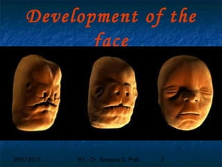
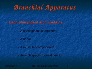
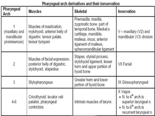
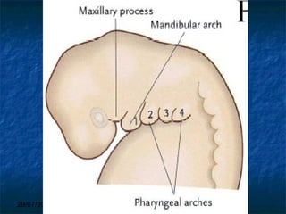
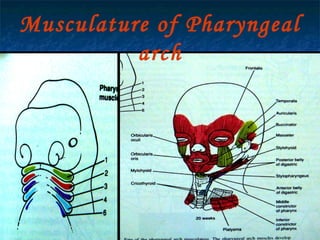
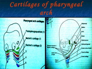
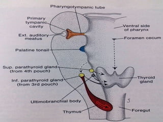
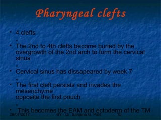
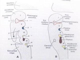
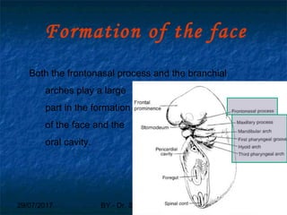
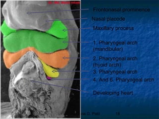
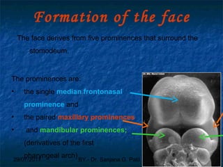
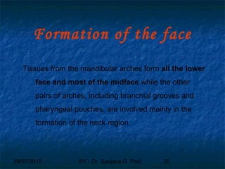
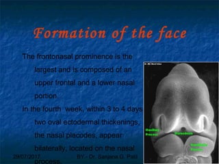
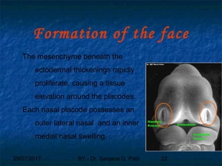
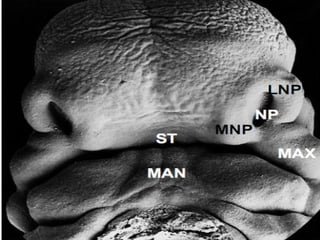
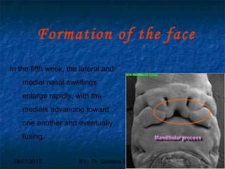
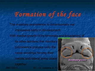
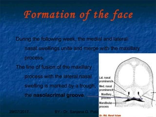
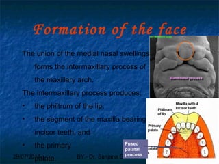
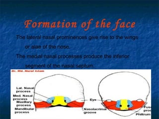
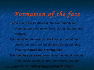
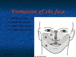
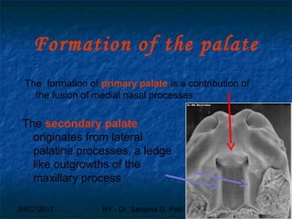
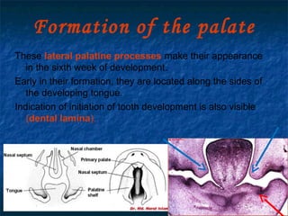
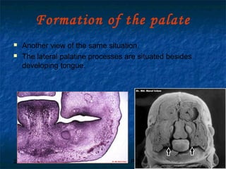
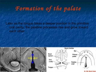
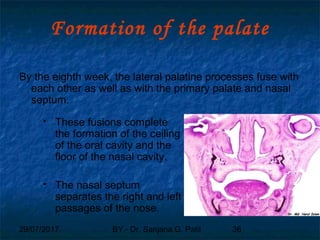
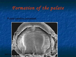
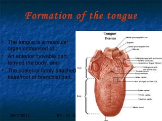
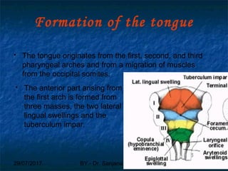
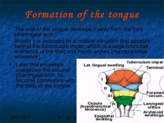
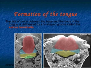
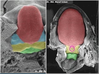
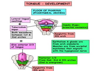
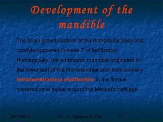
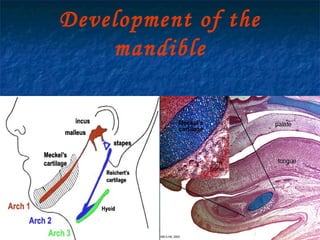
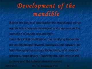
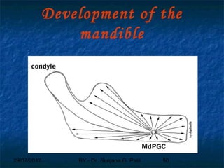
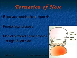
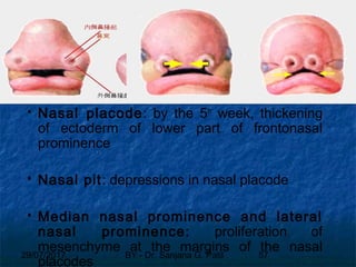
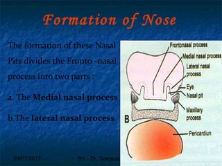
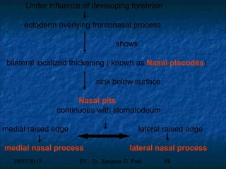
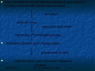
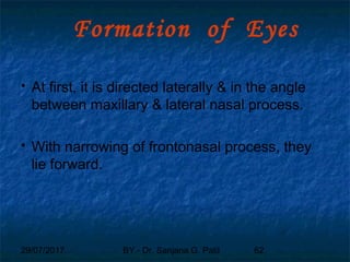
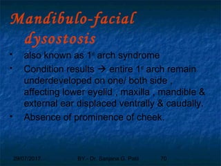
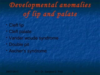
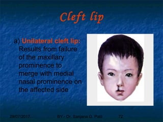
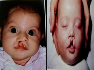
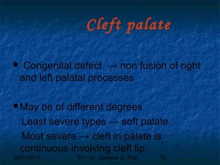
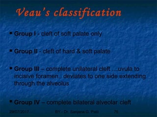
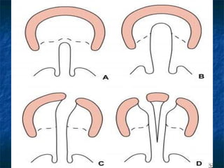
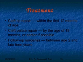
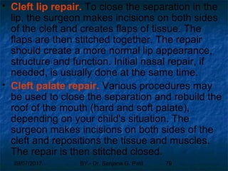
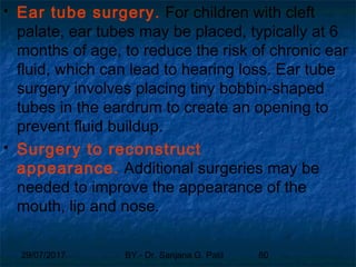
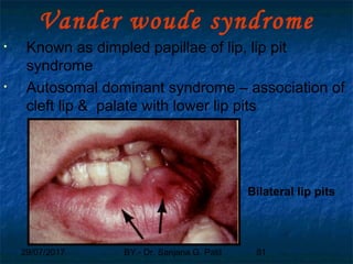
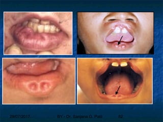
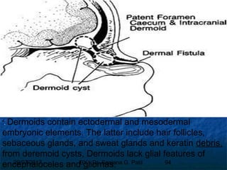
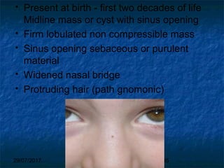
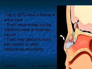
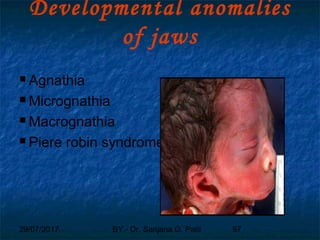
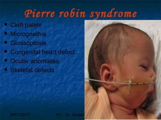
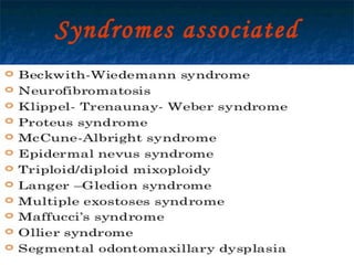
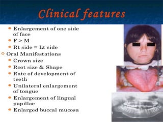
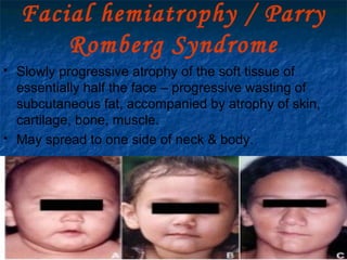
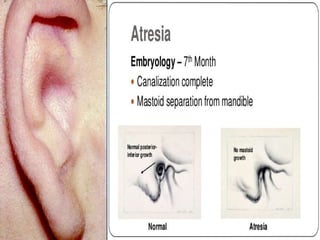
29/07/2017 BY - Dr. Sanjana G. Patil 1
29/07/2017 BY - Dr. Sanjana G. Patil 2
Development of the
face
29/07/201729/07/2017 BY - Dr. Sanjana G. PatilBY - Dr. Sanjana G. Patil 33
Development
Definition: “Development is a progress
towards maturity” – Todd (1931)
According to Moyers Development refers to
all the naturally occurring unidirectional
changes in the life of an individual from its
existence as a single cell to its elaboration as
a multifunctional unit terminating in death..
29/07/2017 BY - Dr. Sanjana G. Patil 4
Branchial Apparatus
 Arches
– mesenchymal tissue surrounded by ectoderm
and endoderm
 „ Clefts (or grooves)
– Separate adjacent arches along ectodermal
surface
 „ Pouches
– Outpouching of endoderm from foregut
– Penetrate adjacent mesenchyme
29/07/2017 BY - Dr. Sanjana G. Patil 5
Branchial Apparatus
29/07/2017 BY - Dr. Sanjana G. Patil 6
Branchial Apparatus
 Each pharyngeal arch contains :
- A cartilaginous component.
- A nerve.
- A muscular component &
- An arch specific cranial nerve.
29/07/2017 BY - Dr. Sanjana G. Patil 7
29/07/2017 BY - Dr. Sanjana G. Patil 8
29/07/2017 BY - Dr. Sanjana G. Patil 9
Musculature of Pharyngeal
arch
29/07/2017 BY - Dr. Sanjana G. Patil 10
Cartilages of pharyngeal
arch
29/07/2017 BY - Dr. Sanjana G. Patil 11
Cranial nerves of pharyngeal
arch
29/07/2017 BY - Dr. Sanjana G. Patil 12
Arterial supply of pharyngeal
arch
29/07/2017 BY - Dr. Sanjana G. Patil 13
Pharyngeal
Pouch
Derivative
1 Epithelium of tympanic cavity
and auditory tube
2 Epithelium of tonsil
3 Inferior parathyroid glands and
epithelium of thymus
4 Superior parathyroid glands.
5 Ultimobranchial body.
29/07/2017 BY - Dr. Sanjana G. Patil 14
29/07/2017 BY - Dr. Sanjana G. Patil 15
Pharyngeal clefts
 4 clefts
„
 The 2nd to 4th clefts become buried by the
overgrowth of the 2nd arch to form the cervical
sinus
„
 Cervical sinus has dissapeared by week 7
„
 The first cleft persists and invades the
mesenchyme
opposite the first pouch
„
 This becomes the EAM and ectoderm of the TM
29/07/2017 BY - Dr. Sanjana G. Patil 16
29/07/2017 BY - Dr. Sanjana G. Patil 17
Formation of the face
Both the frontonasal process and the branchial
arches play a large
part in the formation
of the face and the
oral cavity.
29/07/2017 BY - Dr. Sanjana G. Patil 18
Maxillary process
1. Pharyngeal arch
(mandibular)
2. Pharyngeal arch
(hyoid arch)
3. Pharyngeal arch
4. And 6. Pharyngeal arch
Developing heart
Nasal placode
Frontonasal prominence
29/07/2017 BY - Dr. Sanjana G. Patil 19
Formation of the face
The face derives from five prominences that surround the
stomodeum.
The prominences are:
• the single median frontonasal
prominence and
• the paired maxillary prominences
• and mandibular prominences;
(derivatives of the first
pharyngeal arch)
29/07/2017 BY - Dr. Sanjana G. Patil 20
Formation of the face
Tissues from the mandibular arches form all the lower
face and most of the midface while the other
pairs of arches, including branchial grooves and
pharyngeal pouches, are involved mainly in the
formation of the neck region.
29/07/2017 BY - Dr. Sanjana G. Patil 21
Formation of the face
The frontonasal prominence is the
largest and is composed of an
upper frontal and a lower nasal
portion.
In the fourth week, within 3 to 4 days,
two oval ectodermal thickenings,
the nasal placodes, appear
bilaterally, located on the nasal
process.
29/07/2017 BY - Dr. Sanjana G. Patil 22
Formation of the face
The mesenchyme beneath the
ectodermal thickenings rapidly
proliferate, causing a tissue
elevation around the placodes.
Each nasal placode possesses an
outer lateral nasal and an inner
medial nasal swelling.
29/07/2017 BY - Dr. Sanjana G. Patil 23
29/07/2017 BY - Dr. Sanjana G. Patil 24
Formation of the face
In the fifth week, the lateral and
medial nasal swellings
enlarge rapidly, with the
medials advancing toward
one another and eventually
fusing.
29/07/2017 BY - Dr. Sanjana G. Patil 25
Formation of the face
The maxillary prominence is dimensionally not
impressive early in development.
With medial growth of its terminals and broadening of
its other borders, the maxillary
prominence presses onto the
nasal swellings forcing their
medial and lateral arms closer
together.
29/07/2017 BY - Dr. Sanjana G. Patil 26
Formation of the face
During the following week, the medial and lateral
nasal swellings unite and merge with the maxillary
process.
The line of fusion of the maxillary
process with the lateral nasal
swelling is marked by a trough,
the nasolacrimal groove.
29/07/2017 BY - Dr. Sanjana G. Patil 27
Formation of the face
The union of the medial nasal swellings
forms the intermaxillary process of
the maxillary arch.
The intermaxillary process produces:
• the philtrum of the lip,
• the segment of the maxilla bearing the
incisor teeth, and
• the primary
palate.
Fused
palatal
process
29/07/2017 BY - Dr. Sanjana G. Patil 28
Formation of the face
The lateral nasal prominences give rise to the wings
or alae of the nose.
The medial nasal processes produce the inferior
segment of the nasal septum.
29/07/2017 BY - Dr. Sanjana G. Patil 29
Formation of the face
By the end of the fourth week, the two mandibular
prominences have grown towards one another and
merged.
The mandible, the lower lip, the lower portion of the
cheek, the chin, and the gingiva take their origins
from the mandibular prominences.
The maxillary process gives rise to the lateral portions
of the upper lip and cheeks, the maxilla, and the
secondary palate with its associated gingiva.
29/07/2017 BY - Dr. Sanjana G. Patil 30
Formation of the face
A = Maxillary process
B = Mandibular process
C = Medial nasal process
D = Lateral nasal process
29/07/2017 BY - Dr. Sanjana G. Patil 31
Development of the
palate
29/07/2017 BY - Dr. Sanjana G. Patil 32
Formation of the palate
The formation of primary palate is a contribution of
the fusion of medial nasal processes.
The secondary palate
originates from lateral
palatine processes, a ledge
like outgrowths of the
maxillary process
29/07/2017 BY - Dr. Sanjana G. Patil 33
Formation of the palate
These lateral palatine processes make their appearance
in the sixth week of development.
Early in their formation, they are located along the sides of
the developing tongue.
Indication of initiation of tooth development is also visible
(dental lamina).
29/07/2017 BY - Dr. Sanjana G. Patil 34
Formation of the palate
 Another view of the same situation.
 The lateral palatine processes are situated besides
developing tongue.
29/07/2017 BY - Dr. Sanjana G. Patil 35
Formation of the palate
Later as the tongue takes a deeper position in the primitive
oral cavity, the palatine processes rise and grow toward
each other.
29/07/2017 BY - Dr. Sanjana G. Patil 36
Formation of the palate
By the eighth week, the lateral palatine processes fuse with
each other as well as with the primary palate and nasal
septum.
 These fusions complete
the formation of the ceiling
of the oral cavity and the
floor of the nasal cavity.
 The nasal septum
separates the right and left
passages of the nose.
29/07/2017 BY - Dr. Sanjana G. Patil 37
Formation of the palate
 Fused palatine processes.
29/07/2017 BY - Dr. Sanjana G. Patil 38
Development of face and oral
Cavity
Development of the tongue
29/07/2017 BY - Dr. Sanjana G. Patil 39
Formation of the tongue
 The tongue is a muscular
organ composed of :
 An anterior movable part,
termed the body, and
 The posterior firmly attached
base/root or branchial part.
29/07/2017 BY - Dr. Sanjana G. Patil 40
Formation of the tongue
 The tongue originates from the first, second, and third
pharyngeal arches and from a migration of muscles
from the occipital somites.
 The anterior part arising from
the first arch is formed from
three masses, the two lateral
lingual swellings and the
tuberculum impar.
29/07/2017 BY - Dr. Sanjana G. Patil 41
Formation of the tongue
 These lateral lingual swellings rapidly enlarge, merge
with each other, and overgrow the tuberculum impar to
form the oral part of the tongue.
 An U-shaped sulcus develops in front of and on both
sides of this oral part, which allows it to be free and
highly mobile, except at the region of the lingual
frenulum where it remains attached to the floor of the
mouth.
29/07/2017 BY - Dr. Sanjana G. Patil 42
Formation of the tongue
 The root of the tongue develops mainly from the third
pharyngeal arch.
 Initially it is indicated by a midline elevation that appears
behind the tuberculum imper, which is a large branchial
eminence of the third and fourth arches (hypobranchial
eminence).
 Later this eminence
overgrows the second
pharyngeal arch, to
become continuous with
the body of the tongue.
29/07/2017 BY - Dr. Sanjana G. Patil 43
Formation of the tongue
The site of union between the base and the body of the
tongue is delineated by a V-shaped groove called the
sulcus terminalis.
29/07/2017 BY - Dr. Sanjana G. Patil 44
29/07/2017 BY - Dr. Sanjana G. Patil 45
29/07/2017 BY - Dr. Sanjana G. Patil 46
Development of face and oral
Cavity
Development of the mandible
29/07/2017 BY - Dr. Sanjana G. Patil 47
Development of the
mandible
The basic growth pattern of the mandibular body and
condyle appeared in week 7 of fertilization.
Histologically, the embryonic mandible originated in
the lower part of the first branchial arch from primary
intramembranous ossification in the fibrous
mesenchymal tissue around the Meckel's cartilage.
29/07/2017 BY - Dr. Sanjana G. Patil 48
Development of the
mandible
29/07/2017 BY - Dr. Sanjana G. Patil 49
Development of the
mandible
Before the begin of ossification the mandibular nerve
and its branches are developed and they ensure the
formation of canals around them.
From this initial ossification, the ramifying trabecular
bones developed forward, backward and upward, to
form the symphysis, mandibular body, and coronoid
process, respectively. (following the path way of the
incisive and the inferior alveolar nerve)
29/07/2017 BY - Dr. Sanjana G. Patil 50
Development of the
mandible
29/07/2017 BY - Dr. Sanjana G. Patil 51
Development of the
mandible
The accessory cartilages are:
• the condylar cartilage
• the coronoid cartilage
• the symphysial cartilage
29/07/2017 BY - Dr. Sanjana G. Patil 52
Development of the
mandible
The condylar cartilage is the largest and most
important one. It appears in the 12th
week i. u. and is
quickly replaced by enchondral ossification.
A thin layer of cartilage remains in the condylar head
and persists until the 20
years of life providing
a mechanism of mandibular
growth.
29/07/2017 BY - Dr. Sanjana G. Patil 53
Growth of the mandible
The shape and size of the mandible undergo
considerable transformation from embryonic to adult
mandible.
29/07/2017 BY - Dr. Sanjana G. Patil 54
Growth of the mandible
The mandible grows in all directions:
• Anterio-posterior by bone deposition along the
posterior border of the ramus.
• Vertical by growth of the condyle, along the upper
border of the ramus and the formation of the alveolar
process.
• Transverse by bone deposition on the external
surface and bone resorption at the inner surface.
29/07/2017 BY - Dr. Sanjana G. Patil 55
Development of the
mandible
The alveolar bone formation begins as the teeth
reach the early bell stage.
The bone begins to grow around the tooth germs and
upward in occlusal direction.
Later this area is called alveolar bone and is divided
into alveoli by septa.
29/07/2017 BY - Dr. Sanjana G. Patil 56
Formation of Nose
 Receives contributions from 
• Frontonasal process
• Medial & lateral nasal process
of right & left side
56
29/07/2017 BY - Dr. Sanjana G. Patil 57
 Nasal placode: by the 5th
week, thickening
of ectoderm of lower part of frontonasal
prominence
 Nasal pit: depressions in nasal placode
 Median nasal prominence and lateral
nasal prominence: proliferation of
mesenchyme at the margins of the nasal
placodes
29/07/2017 BY - Dr. Sanjana G. Patil 58
The formation of these Nasal
Pits divides the Fronto -nasal
process into two parts :
a. The Medial nasal process
b.The lateral nasal process
Formation of Nose
29/07/2017 BY - Dr. Sanjana G. Patil 59
Under influence of developing forebrain
ectoderm overlying frontonasal process
shows
bilateral localized thickening ( known as Nasal placodes)
sink below surface
Nasal pits
continuous with stomatodeum
medial raised edge lateral raised edge
medial nasal process lateral nasal process
29/07/2017 BY - Dr. Sanjana G. Patil 60
 Nasal pits later on cut off from stomatodeum and fusion of
maxillary process with medial nasal process
formation
external nares
approach each other
narrowing of frontonasal process
 mesoderm heaped up in median plane
prominence of nose
 external nares open downward instead of
forward
29/07/2017 BY - Dr. Sanjana G. Patil 61
Formation of Eyes
 Formation starts – 8th
week in utero
 Firstly, ectodermal thickening known as
lens placode appears on ventrolateral
side of developing forebrain, lateral &
cranial – nasal placode
29/07/2017 BY - Dr. Sanjana G. Patil 62
Formation of Eyes
 At first, it is directed laterally & in the angle
between maxillary & lateral nasal process.
 With narrowing of frontonasal process, they
lie forward.
29/07/2017 BY - Dr. Sanjana G. Patil 63
Formation of
External Ear
 External acoustic meatus –
 dorsal part of 1st
ectodermal cleft
 deeper part  proliferation of its lining epithelium
which grows towards middle ear.
 Firstly this proliferation – solid , later on canalized.
29/07/2017 BY - Dr. Sanjana G. Patil 64
 Auricle ( pinna )
• Series of 6 mesodermal thickenings appear –
mandibular & hyoid arch around opening of
external acoustic meatus
• Fusion of these thickenings leads to pinna
• Firstly it lies caudal – developing jaw , later on
pushed upward & backward to its definitive
position because of great enlargement of
mandibular process
29/07/2017 BY - Dr. Sanjana G. Patil 65
 Mandibular arch forms only tragus & small
area around it.
 Rest of auricle formed from hyoid arch.
29/07/2017 BY - Dr. Sanjana G. Patil 66
References
 J.W Larsen, 1998. Essentials of Human
Embryology. 2nd
Ed Churchill Livingstone
publisher, New York. Pg-345-370.
 K L Moore, and T. V. N. Persaud. 1998. The
Developing Human, 6th Ed. Saunders,
Philadelphia. Pg- 236-254
 T W Sadler,1985. Langman’s Medical
Embryology, 5th Ed. Williams and Wilkins,
Baltimore. Pg-345-390
29/07/2017 BY - Dr. Sanjana G. Patil 67
Developmental Anomalies
of Face
 Formation of various part of Face 
fusion of diverse components
 This fusion occasionally incomplete 
various anomalies
 Formation of various part of Face 
fusion of diverse components
 This fusion occasionally incomplete 
various anomalies
29/07/2017 BY - Dr. Sanjana G. Patil 68
Developmental anomalies
of face
 Oblique facial cleft
 Manidibulofacial dysostosis
29/07/2017 BY - Dr. Sanjana G. Patil 69
Oblique facial
cleft
• Non fusion of the maxillary and lateral
nasal process  cleft running from the
medial angle of the eye to the mouth.
• Nasolacrimal duct not formed.
29/07/2017 BY - Dr. Sanjana G. Patil 70
Mandibulo-facial
dysostosis
 also known as 1st
arch syndrome
 Condition results  entire 1st
arch remain
underdeveloped on one/ both side ,
affecting lower eyelid , maxilla , mandible &
external ear displaced ventrally & caudally.
 Absence of prominence of cheek.
29/07/2017 BY - Dr. Sanjana G. Patil 71
Developmental anomalies
of lip and palate
 Cleft lip
 Cleft palate
 Vander woude syndrome
 Double pit
 Ascher’s syndrome
29/07/2017 BY - Dr. Sanjana G. Patil 72
Cleft lip
a) Unilateral cleft lip:
Results from failure
of the maxillary
prominence to
merge with medial
nasal prominence on
the affected side
29/07/2017 BY - Dr. Sanjana G. Patil 73
b. Bilateral cleft lip : Results from failure
of the maxillary prominences to merge
with the medial nasal prominence on
both sides
c. Median cleft lip: Results from failure of
the medial nasal prominences to merge
and form the intermaxillary segments
29/07/2017 BY - Dr. Sanjana G. Patil 74
29/07/2017 BY - Dr. Sanjana G. Patil 75
Cleft palate
 Congenital defect → non fusion of right
and left palatal processes
 May be of different degrees
Least severe types → soft palate.
Most severe → cleft in palate is
continuous involving cleft lip.
29/07/2017 BY - Dr. Sanjana G. Patil 76
Veau’s classification
 Group I - cleft of soft palate only
 Group II - cleft of hard & soft palate
 Group III – complete unilateral cleft …uvula to
incisive foramen.. deviates to one side extending
through the alveolus
 Group IV – complete bilateral alveolar cleft
29/07/2017 BY - Dr. Sanjana G. Patil 77 77
29/07/2017 BY - Dr. Sanjana G. Patil 78
Treatment
 Cleft lip repair — within the first 12 months
of age
 Cleft palate repair — by the age of 18
months, or earlier if possible
 Follow-up surgeries — between age 2 and
late teen years
29/07/2017 BY - Dr. Sanjana G. Patil 79
 Cleft lip repair. To close the separation in the
lip, the surgeon makes incisions on both sides
of the cleft and creates flaps of tissue. The
flaps are then stitched together. The repair
should create a more normal lip appearance,
structure and function. Initial nasal repair, if
needed, is usually done at the same time.
 Cleft palate repair. Various procedures may
be used to close the separation and rebuild the
roof of the mouth (hard and soft palate),
depending on your child's situation. The
surgeon makes incisions on both sides of the
cleft and repositions the tissue and muscles.
The repair is then stitched closed.
29/07/2017 BY - Dr. Sanjana G. Patil 80
 Ear tube surgery. For children with cleft
palate, ear tubes may be placed, typically at 6
months of age, to reduce the risk of chronic ear
fluid, which can lead to hearing loss. Ear tube
surgery involves placing tiny bobbin-shaped
tubes in the eardrum to create an opening to
prevent fluid buildup.
 Surgery to reconstruct
appearance. Additional surgeries may be
needed to improve the appearance of the
mouth, lip and nose.
29/07/2017 BY - Dr. Sanjana G. Patil 81
Vander woude syndrome
• Known as dimpled papillae of lip, lip pit
syndrome
• Autosomal dominant syndrome – association of
cleft lip & palate with lower lip pits
Bilateral lip pits
29/07/2017 BY - Dr. Sanjana G. Patil 82
29/07/2017 BY - Dr. Sanjana G. Patil 83
• Diagnosis – hyper nasal voice & bifid
uvula.
• May have maxillary
hypodontia( missing incisors /
premolars) ,ankyloglossia , high arched
palate.
29/07/2017 BY - Dr. Sanjana G. Patil 84
Double Lip
 A redundant fold of tissue on the mucosa
side of the lip.
 Usually congenital but can be acquired.
 Acquired may be associated with Ascher’s
Syndrome (double lip, blepharochalasis,
non toxic thyroid enlargement)
 Treatment – Removal of accessory lip.
29/07/2017 BY - Dr. Sanjana G. Patil 85
Ascher’s Syndrome
Blepharochalasis
Double Lip
Thyroid Enlargement
29/07/2017 BY - Dr. Sanjana G. Patil 86
Developmental anomalies
of nose
 Encephalocele
 Glioma
 Dermal fistula ( Nasal dermoid)
29/07/2017 BY - Dr. Sanjana G. Patil 87
29/07/2017 BY - Dr. Sanjana G. Patil 88
Encephalocele
Extra cranial herniation of meninges and brain tissue
through a defect in the skull.
Meningocele presents similarly without herniation of
brain tissue
a) Sincipital Encephalocele b) Basal Encephalocele.
29/07/2017 BY - Dr. Sanjana G. Patil 89
 A) Sincipital encephaloceles are also
known as Frontoethmoidal
encephaloceles. They occur between the
frontal and ethmoid bones at the foramen
cecum immediately anterior to the cribiform
plate. They may be further subdivided as
nasofrontal, nasoethmoidal and naso-orbital
and manifest as external nasal masses.
 B) Generally Basal Encephaloceles arise
through the cribiform plate or through the
superior orbital fissure and manifest as an
intranasal mass.
29/07/2017 BY - Dr. Sanjana G. Patil 90
29/07/2017 BY - Dr. Sanjana G. Patil 91
Nasal Glioma
 Heterotopic glial tissue that lacks a patent
CSF communication to subarachnoid
space
 Abnormal closure of the fonticulus frontalis
 During retraction of the
embryonic Dural
diverticulum, rests of glial
tissue become sequeatered
29/07/2017 BY - Dr. Sanjana G. Patil 92
Extra nasal (60%)
 Smooth, firm, non-compressible masses,
skin telangiectasia
 Glabella (most common), nasomaxillary
suture line
Intranasal (30%)
 Polypoid, pale masses
 Arise in the lateral nasal wall near the
middle turbinate
 Nasal septum (rare)
29/07/2017 BY - Dr. Sanjana G. Patil 93
 During development, dura projects
through the foramen cecum and attaches
to skin
 Separates from the nasal skin and retracts
through foramen cecum
 If there is a persistent attachment to
underling fibrous tissue, nasal capsule or
dura, epithelial elements are trapped in
the prenasal space
Nasal dermoids
29/07/2017 BY - Dr. Sanjana G. Patil 94

. Dermoids contain ectodermal and mesodermal
embryonic elements. The latter include hair follicles,
sebaceous glands, and sweat glands and keratin debris.
from deremoid cysts, Dermoids lack glial features of
encephaloceles and gliomas.
29/07/2017 BY - Dr. Sanjana G. Patil 95
 Present at birth - first two decades of life
Midline mass or cyst with sinus opening
 Firm lobulated non compressible mass
 Sinus opening sebaceous or purulent
material
 Widened nasal bridge
 Protruding hair (path gnomonic)
29/07/2017 BY - Dr. Sanjana G. Patil 96
 Up to 50% have a fistula or
sinus tract
 Tract transverses via the
cribiform plate or foramen
cecum
 Tract may attach to dura,
falx cerebri or other
intracranial structures
29/07/2017 BY - Dr. Sanjana G. Patil 97
Developmental anomalies
of jaws
 Agnathia
 Micrognathia
 Macrognathia
 Piere robin syndrome
29/07/2017 BY - Dr. Sanjana G. Patil 98
Agnathia
29/07/2017 BY - Dr. Sanjana G. Patil 99
Micrognathia
29/07/2017 BY - Dr. Sanjana G. Patil 100
 Congenital –
1) It is associated with congenital heart disease &
Pierre Robin syndrome
2) Maxillary Micrognathia due to deficiency in
premaxillary area.
3) Retracted middle third of face.
4) Etiology – mouth breathing.
 Mandibular micrognathia may be due to
posterior positioning of mandible, steep
mandibular angle, agenesis of condyle.
29/07/2017 BY - Dr. Sanjana G. Patil 101
 Acquired type is post natal –
 Due to disturbance in TMJ region.
 Ankylosis due to trauma or infection of mastoid.
Clinical features
 Severe retrusion of chin
 Steep mandibular angle
 Deficient chin button.
29/07/2017 BY - Dr. Sanjana G. Patil 102
Pierre robin syndrome
 Cleft palate
 Micrognathia
 Glossoptosis
 Congenital heart defect
 Ocular anomalies
 Skeletal defects
29/07/2017 BY - Dr. Sanjana G. Patil 103
Macrognathia
 Abnormally large jaws
 Pituitary gigantism – both jaws
 Maxillary enlargement – Paget’s disease, Leontiasis
ossea, Cherubism.
 Mandibular enlargement – Acromegaly.
Treatment
 Surgical correction of defect.
29/07/2017 BY - Dr. Sanjana G. Patil 104
29/07/2017 BY - Dr. Sanjana G. Patil 105
Facial hemihypertrophy /
hyperplasia
 Characterized by asymmetric overgrowth of one or
more body parts.
 Etiology
 Hormonal imbalance
 Incomplete twinning
 Chromosal abnormalities
 Lymphatic abnormalities
 Vascular abnormalities
 Neurogenic abnormalities.
29/07/2017 BY - Dr. Sanjana G. Patil 106
Syndromes associated
29/07/2017 BY - Dr. Sanjana G. Patil 107
Clinical features
29/07/2017 BY - Dr. Sanjana G. Patil 108
 Treatment
• Cosmetic repair
 Differential diagnosis
• Neurofibromatosis
• Fibrous dysplasia
29/07/2017 BY - Dr. Sanjana G. Patil 109
Facial hemiatrophy / Parry
Romberg Syndrome
 Slowly progressive atrophy of the soft tissue of
essentially half the face – progressive wasting of
subcutaneous fat, accompanied by atrophy of skin,
cartilage, bone, muscle.
 May spread to one side of neck & body.
29/07/2017 BY - Dr. Sanjana G. Patil 110
 Etiology
 Cerebral disturbances leading to increased &
unregulated activity of sympathetic nervous system.
 Trauma
 Infection
 Hereditary
 Peripheral trigeminal neuritis
 Localized scleroderma
29/07/2017 BY - Dr. Sanjana G. Patil 111
Clinical features
 Atrophy of skin, subcutaneous tissue, muscle,
bone & cartilage on affected side.
 Begins in 1st
decade & lasts for 3years before
becomes quiscent.
 Basilar kyphosis.
 Misshapen ear or small ear.
 Dark pigmentation of affected side.
 Trigeminal neuralgia, facial parathesia.
29/07/2017 BY - Dr. Sanjana G. Patil 112
oral manifestations
 Atrophy of half lip & tongue
 Shorter ramus & body of mandible resulting in
malocclusion.
 Delayed eruption of teeth on affected side.
Treatment
 A new technic for the treatment of facial hemiatrophy
(Romberg's disease) in children with multiple
subcutaneous injections of dimethylpolysiloxane has
been highly satisfactory. The technic is investigative in
nature and will remain so for several years.
 Surgical corrections.
29/07/2017 BY - Dr. Sanjana G. Patil 113
Developmental anomalies
of tongue
• Microglossia
• Macroglossia
• Tongue tie
29/07/2017 BY - Dr. Sanjana G. Patil 114
Microglossia
 May be mild and difficult to detect to
aglossia
(very rare).
 May be a presence of rudimentary or small
tongue
29/07/2017 BY - Dr. Sanjana G. Patil 115
Macroglossia
 Caused by a wide variety of conditions.
 Congenital and hereditary: vascular
malformations (lymphangioma and
hemangioma), hemihyperplasia, cretinism,
Beckwith-hypogycemic syndrome, down
syndrome,
 Acquired; amyloidosis, myxedema, acromegaly,
angioedema, tumors.
29/07/2017 BY - Dr. Sanjana G. Patil 116
Tongue tie
 Tongue -Tie (Ankyloglossia) is a problem caused
by abnormal development of the tongue.It is a
congenital condition, in which the tongue is tied
down to the floor of the mouth. The tongue is
normally connected to the floor of the mouth by a
thin cord, called a frenum.
 With tongue-tie the frenum is too short and thick. It
prevents the tongue from moving freely.
 Effect of Tongue-Tie (Ankyloglossia)
 Ankyloglossia in breastfeeding infants can cause
ineffective latch, inadequate milk transfer, and
maternal nipple pain, resulting in ultimately weaning.
It also causes the speech problem and there is
difficulty in swallowing.
29/07/2017 BY - Dr. Sanjana G. Patil 117
Treatment
 Frenotomy – The lingual frenum is given a cut to
release the tongue
 Frenectomy – Removal of frenum
 Revision of the frenum by laser without a
general anaesthetic.
 Revision by electrocautery using a local
anaesthetic.
29/07/2017 BY - Dr. Sanjana G. Patil 118
Developmental anomalies of
ear
 Microtia
 Auditory canal atresia.
29/07/2017 BY - Dr. Sanjana G. Patil 119
Microtia
 Microtia is a developmental abnormality in which the
external part of the ear (the pinna or auricle) has not
fully developed. There are varying degrees of microtia
depending on how well developed the auricle is. The
degree of auricle development can range from simply
a small ear that has all the proper cartilage (grade I) to
a small "nubbin" or peanut ear that has no cartilage
and only a mound of skin and soft tissue (grade III).
Rarely, people are born without an auricle, a condition
called anotia.
29/07/2017 BY - Dr. Sanjana G. Patil 120
Treatment
A plastic surgeon skilled in microtia repair can often
rebuild the auricle to a very favorable appearance using
a prosthesis, the person's own rib cartilage, or an
implant.
29/07/2017 BY - Dr. Sanjana G. Patil 121
Auditory canal atresia
 Auditory canal atresia is the partial or total failure of
the ear canal to develop. Absence of the ear canal is
usually associated with absence of the eardrum
(tympanic membrane) and underdevelopment of the
middle ear and middle ear bones (hammer, anvil, and
stirrup). Children with auditory canal atresia have
hearing loss because sound is not efficiently carried,
or conducted, into the middle and inner ear. Usually
the inner ear (cochlea) is healthy and normally
developed in these children.
29/07/2017 BY - Dr. Sanjana G. Patil 122
29/07/2017 BY - Dr. Sanjana G. Patil 123
Treatment
 In some children, doctors can
surgically open the ear canal, build an
eardrum, and restore the natural
sound-conducting pathway of the ear
canal and middle ear to the healthy
inner ear. Although surgical correction
of auditory canal atresia, called
canalplasty, can improve hearing, it
seldom results in normal hearing at all
pitches (frequencies) of sound.
29/07/2017 BY - Dr. Sanjana G. Patil 124
Developmental anomalies
of eye
 Anopthalmia & Micropthalmia.
 Colombomas.
29/07/2017 BY - Dr. Sanjana G. Patil 125
Anopthalmia &
Micropthalmos Complete failure of eye development is
extremely rare. Only histologic examination can
differentiate between true anophthalmia and
severe microphthalmia. In cases of
microphthalmia, ocular structures are identified,
whereas they are absent in primary
anophthalmia. Microphthalmos is considered
simple or pure when the eye is small but
anatomically correct, and complex when the eye
is malformed
29/07/2017 BY - Dr. Sanjana G. Patil 126
Treatment (anopthalmia)
 Inflatable expander
 Self expanding hydrophilic, osmotic expanders.
 Injectable calcium hydroxylapatite
 Eyelid surgery
29/07/2017 BY - Dr. Sanjana G. Patil 127
Treatment (micropthalmia)
Conformer expansion therapy
29/07/2017 BY - Dr. Sanjana G. Patil 128
Colombomas
 Colobomas The term coloboma is of Greek origin
meaning curtailment and is used in ophthalmology to
refer to defects in ocular structures. Those colobomas
that result from failure of the embryonic.
 choroidal fissure to close properly are classified as
typical colobomas. Colobomas are common
malformations, constituting about 2% of all congenital
abnormalities. They are usually bilateral. A variant of
typical coloboma of the disc is designated the morning
glory syndrome, because in this malformation the optic
disc is posteriorly displaced at the apex of a funnel-
shaped staphylomatous excavation. This configuration
is thought to resemble the flower.
29/07/2017 BY - Dr. Sanjana G. Patil 129
Treatment
 There is no proper treatment .
 Use of contact lens can be done.
29/07/2017 BY - Dr. Sanjana G. Patil 130

Development of the face

  • 1.
    29/07/2017 BY -Dr. Sanjana G. Patil 1
  • 2.
    29/07/2017 BY -Dr. Sanjana G. Patil 2 Development of the face
  • 3.
    29/07/201729/07/2017 BY -Dr. Sanjana G. PatilBY - Dr. Sanjana G. Patil 33 Development Definition: “Development is a progress towards maturity” – Todd (1931) According to Moyers Development refers to all the naturally occurring unidirectional changes in the life of an individual from its existence as a single cell to its elaboration as a multifunctional unit terminating in death..
  • 4.
    29/07/2017 BY -Dr. Sanjana G. Patil 4 Branchial Apparatus  Arches – mesenchymal tissue surrounded by ectoderm and endoderm  „ Clefts (or grooves) – Separate adjacent arches along ectodermal surface  „ Pouches – Outpouching of endoderm from foregut – Penetrate adjacent mesenchyme
  • 5.
    29/07/2017 BY -Dr. Sanjana G. Patil 5 Branchial Apparatus
  • 6.
    29/07/2017 BY -Dr. Sanjana G. Patil 6 Branchial Apparatus  Each pharyngeal arch contains : - A cartilaginous component. - A nerve. - A muscular component & - An arch specific cranial nerve.
  • 7.
    29/07/2017 BY -Dr. Sanjana G. Patil 7
  • 8.
    29/07/2017 BY -Dr. Sanjana G. Patil 8
  • 9.
    29/07/2017 BY -Dr. Sanjana G. Patil 9 Musculature of Pharyngeal arch
  • 10.
    29/07/2017 BY -Dr. Sanjana G. Patil 10 Cartilages of pharyngeal arch
  • 11.
    29/07/2017 BY -Dr. Sanjana G. Patil 11 Cranial nerves of pharyngeal arch
  • 12.
    29/07/2017 BY -Dr. Sanjana G. Patil 12 Arterial supply of pharyngeal arch
  • 13.
    29/07/2017 BY -Dr. Sanjana G. Patil 13 Pharyngeal Pouch Derivative 1 Epithelium of tympanic cavity and auditory tube 2 Epithelium of tonsil 3 Inferior parathyroid glands and epithelium of thymus 4 Superior parathyroid glands. 5 Ultimobranchial body.
  • 14.
    29/07/2017 BY -Dr. Sanjana G. Patil 14
  • 15.
    29/07/2017 BY -Dr. Sanjana G. Patil 15 Pharyngeal clefts  4 clefts „  The 2nd to 4th clefts become buried by the overgrowth of the 2nd arch to form the cervical sinus „  Cervical sinus has dissapeared by week 7 „  The first cleft persists and invades the mesenchyme opposite the first pouch „  This becomes the EAM and ectoderm of the TM
  • 16.
    29/07/2017 BY -Dr. Sanjana G. Patil 16
  • 17.
    29/07/2017 BY -Dr. Sanjana G. Patil 17 Formation of the face Both the frontonasal process and the branchial arches play a large part in the formation of the face and the oral cavity.
  • 18.
    29/07/2017 BY -Dr. Sanjana G. Patil 18 Maxillary process 1. Pharyngeal arch (mandibular) 2. Pharyngeal arch (hyoid arch) 3. Pharyngeal arch 4. And 6. Pharyngeal arch Developing heart Nasal placode Frontonasal prominence
  • 19.
    29/07/2017 BY -Dr. Sanjana G. Patil 19 Formation of the face The face derives from five prominences that surround the stomodeum. The prominences are: • the single median frontonasal prominence and • the paired maxillary prominences • and mandibular prominences; (derivatives of the first pharyngeal arch)
  • 20.
    29/07/2017 BY -Dr. Sanjana G. Patil 20 Formation of the face Tissues from the mandibular arches form all the lower face and most of the midface while the other pairs of arches, including branchial grooves and pharyngeal pouches, are involved mainly in the formation of the neck region.
  • 21.
    29/07/2017 BY -Dr. Sanjana G. Patil 21 Formation of the face The frontonasal prominence is the largest and is composed of an upper frontal and a lower nasal portion. In the fourth week, within 3 to 4 days, two oval ectodermal thickenings, the nasal placodes, appear bilaterally, located on the nasal process.
  • 22.
    29/07/2017 BY -Dr. Sanjana G. Patil 22 Formation of the face The mesenchyme beneath the ectodermal thickenings rapidly proliferate, causing a tissue elevation around the placodes. Each nasal placode possesses an outer lateral nasal and an inner medial nasal swelling.
  • 23.
    29/07/2017 BY -Dr. Sanjana G. Patil 23
  • 24.
    29/07/2017 BY -Dr. Sanjana G. Patil 24 Formation of the face In the fifth week, the lateral and medial nasal swellings enlarge rapidly, with the medials advancing toward one another and eventually fusing.
  • 25.
    29/07/2017 BY -Dr. Sanjana G. Patil 25 Formation of the face The maxillary prominence is dimensionally not impressive early in development. With medial growth of its terminals and broadening of its other borders, the maxillary prominence presses onto the nasal swellings forcing their medial and lateral arms closer together.
  • 26.
    29/07/2017 BY -Dr. Sanjana G. Patil 26 Formation of the face During the following week, the medial and lateral nasal swellings unite and merge with the maxillary process. The line of fusion of the maxillary process with the lateral nasal swelling is marked by a trough, the nasolacrimal groove.
  • 27.
    29/07/2017 BY -Dr. Sanjana G. Patil 27 Formation of the face The union of the medial nasal swellings forms the intermaxillary process of the maxillary arch. The intermaxillary process produces: • the philtrum of the lip, • the segment of the maxilla bearing the incisor teeth, and • the primary palate. Fused palatal process
  • 28.
    29/07/2017 BY -Dr. Sanjana G. Patil 28 Formation of the face The lateral nasal prominences give rise to the wings or alae of the nose. The medial nasal processes produce the inferior segment of the nasal septum.
  • 29.
    29/07/2017 BY -Dr. Sanjana G. Patil 29 Formation of the face By the end of the fourth week, the two mandibular prominences have grown towards one another and merged. The mandible, the lower lip, the lower portion of the cheek, the chin, and the gingiva take their origins from the mandibular prominences. The maxillary process gives rise to the lateral portions of the upper lip and cheeks, the maxilla, and the secondary palate with its associated gingiva.
  • 30.
    29/07/2017 BY -Dr. Sanjana G. Patil 30 Formation of the face A = Maxillary process B = Mandibular process C = Medial nasal process D = Lateral nasal process
  • 31.
    29/07/2017 BY -Dr. Sanjana G. Patil 31 Development of the palate
  • 32.
    29/07/2017 BY -Dr. Sanjana G. Patil 32 Formation of the palate The formation of primary palate is a contribution of the fusion of medial nasal processes. The secondary palate originates from lateral palatine processes, a ledge like outgrowths of the maxillary process
  • 33.
    29/07/2017 BY -Dr. Sanjana G. Patil 33 Formation of the palate These lateral palatine processes make their appearance in the sixth week of development. Early in their formation, they are located along the sides of the developing tongue. Indication of initiation of tooth development is also visible (dental lamina).
  • 34.
    29/07/2017 BY -Dr. Sanjana G. Patil 34 Formation of the palate  Another view of the same situation.  The lateral palatine processes are situated besides developing tongue.
  • 35.
    29/07/2017 BY -Dr. Sanjana G. Patil 35 Formation of the palate Later as the tongue takes a deeper position in the primitive oral cavity, the palatine processes rise and grow toward each other.
  • 36.
    29/07/2017 BY -Dr. Sanjana G. Patil 36 Formation of the palate By the eighth week, the lateral palatine processes fuse with each other as well as with the primary palate and nasal septum.  These fusions complete the formation of the ceiling of the oral cavity and the floor of the nasal cavity.  The nasal septum separates the right and left passages of the nose.
  • 37.
    29/07/2017 BY -Dr. Sanjana G. Patil 37 Formation of the palate  Fused palatine processes.
  • 38.
    29/07/2017 BY -Dr. Sanjana G. Patil 38 Development of face and oral Cavity Development of the tongue
  • 39.
    29/07/2017 BY -Dr. Sanjana G. Patil 39 Formation of the tongue  The tongue is a muscular organ composed of :  An anterior movable part, termed the body, and  The posterior firmly attached base/root or branchial part.
  • 40.
    29/07/2017 BY -Dr. Sanjana G. Patil 40 Formation of the tongue  The tongue originates from the first, second, and third pharyngeal arches and from a migration of muscles from the occipital somites.  The anterior part arising from the first arch is formed from three masses, the two lateral lingual swellings and the tuberculum impar.
  • 41.
    29/07/2017 BY -Dr. Sanjana G. Patil 41 Formation of the tongue  These lateral lingual swellings rapidly enlarge, merge with each other, and overgrow the tuberculum impar to form the oral part of the tongue.  An U-shaped sulcus develops in front of and on both sides of this oral part, which allows it to be free and highly mobile, except at the region of the lingual frenulum where it remains attached to the floor of the mouth.
  • 42.
    29/07/2017 BY -Dr. Sanjana G. Patil 42 Formation of the tongue  The root of the tongue develops mainly from the third pharyngeal arch.  Initially it is indicated by a midline elevation that appears behind the tuberculum imper, which is a large branchial eminence of the third and fourth arches (hypobranchial eminence).  Later this eminence overgrows the second pharyngeal arch, to become continuous with the body of the tongue.
  • 43.
    29/07/2017 BY -Dr. Sanjana G. Patil 43 Formation of the tongue The site of union between the base and the body of the tongue is delineated by a V-shaped groove called the sulcus terminalis.
  • 44.
    29/07/2017 BY -Dr. Sanjana G. Patil 44
  • 45.
    29/07/2017 BY -Dr. Sanjana G. Patil 45
  • 46.
    29/07/2017 BY -Dr. Sanjana G. Patil 46 Development of face and oral Cavity Development of the mandible
  • 47.
    29/07/2017 BY -Dr. Sanjana G. Patil 47 Development of the mandible The basic growth pattern of the mandibular body and condyle appeared in week 7 of fertilization. Histologically, the embryonic mandible originated in the lower part of the first branchial arch from primary intramembranous ossification in the fibrous mesenchymal tissue around the Meckel's cartilage.
  • 48.
    29/07/2017 BY -Dr. Sanjana G. Patil 48 Development of the mandible
  • 49.
    29/07/2017 BY -Dr. Sanjana G. Patil 49 Development of the mandible Before the begin of ossification the mandibular nerve and its branches are developed and they ensure the formation of canals around them. From this initial ossification, the ramifying trabecular bones developed forward, backward and upward, to form the symphysis, mandibular body, and coronoid process, respectively. (following the path way of the incisive and the inferior alveolar nerve)
  • 50.
    29/07/2017 BY -Dr. Sanjana G. Patil 50 Development of the mandible
  • 51.
    29/07/2017 BY -Dr. Sanjana G. Patil 51 Development of the mandible The accessory cartilages are: • the condylar cartilage • the coronoid cartilage • the symphysial cartilage
  • 52.
    29/07/2017 BY -Dr. Sanjana G. Patil 52 Development of the mandible The condylar cartilage is the largest and most important one. It appears in the 12th week i. u. and is quickly replaced by enchondral ossification. A thin layer of cartilage remains in the condylar head and persists until the 20 years of life providing a mechanism of mandibular growth.
  • 53.
    29/07/2017 BY -Dr. Sanjana G. Patil 53 Growth of the mandible The shape and size of the mandible undergo considerable transformation from embryonic to adult mandible.
  • 54.
    29/07/2017 BY -Dr. Sanjana G. Patil 54 Growth of the mandible The mandible grows in all directions: • Anterio-posterior by bone deposition along the posterior border of the ramus. • Vertical by growth of the condyle, along the upper border of the ramus and the formation of the alveolar process. • Transverse by bone deposition on the external surface and bone resorption at the inner surface.
  • 55.
    29/07/2017 BY -Dr. Sanjana G. Patil 55 Development of the mandible The alveolar bone formation begins as the teeth reach the early bell stage. The bone begins to grow around the tooth germs and upward in occlusal direction. Later this area is called alveolar bone and is divided into alveoli by septa.
  • 56.
    29/07/2017 BY -Dr. Sanjana G. Patil 56 Formation of Nose  Receives contributions from  • Frontonasal process • Medial & lateral nasal process of right & left side 56
  • 57.
    29/07/2017 BY -Dr. Sanjana G. Patil 57  Nasal placode: by the 5th week, thickening of ectoderm of lower part of frontonasal prominence  Nasal pit: depressions in nasal placode  Median nasal prominence and lateral nasal prominence: proliferation of mesenchyme at the margins of the nasal placodes
  • 58.
    29/07/2017 BY -Dr. Sanjana G. Patil 58 The formation of these Nasal Pits divides the Fronto -nasal process into two parts : a. The Medial nasal process b.The lateral nasal process Formation of Nose
  • 59.
    29/07/2017 BY -Dr. Sanjana G. Patil 59 Under influence of developing forebrain ectoderm overlying frontonasal process shows bilateral localized thickening ( known as Nasal placodes) sink below surface Nasal pits continuous with stomatodeum medial raised edge lateral raised edge medial nasal process lateral nasal process
  • 60.
    29/07/2017 BY -Dr. Sanjana G. Patil 60  Nasal pits later on cut off from stomatodeum and fusion of maxillary process with medial nasal process formation external nares approach each other narrowing of frontonasal process  mesoderm heaped up in median plane prominence of nose  external nares open downward instead of forward
  • 61.
    29/07/2017 BY -Dr. Sanjana G. Patil 61 Formation of Eyes  Formation starts – 8th week in utero  Firstly, ectodermal thickening known as lens placode appears on ventrolateral side of developing forebrain, lateral & cranial – nasal placode
  • 62.
    29/07/2017 BY -Dr. Sanjana G. Patil 62 Formation of Eyes  At first, it is directed laterally & in the angle between maxillary & lateral nasal process.  With narrowing of frontonasal process, they lie forward.
  • 63.
    29/07/2017 BY -Dr. Sanjana G. Patil 63 Formation of External Ear  External acoustic meatus –  dorsal part of 1st ectodermal cleft  deeper part  proliferation of its lining epithelium which grows towards middle ear.  Firstly this proliferation – solid , later on canalized.
  • 64.
    29/07/2017 BY -Dr. Sanjana G. Patil 64  Auricle ( pinna ) • Series of 6 mesodermal thickenings appear – mandibular & hyoid arch around opening of external acoustic meatus • Fusion of these thickenings leads to pinna • Firstly it lies caudal – developing jaw , later on pushed upward & backward to its definitive position because of great enlargement of mandibular process
  • 65.
    29/07/2017 BY -Dr. Sanjana G. Patil 65  Mandibular arch forms only tragus & small area around it.  Rest of auricle formed from hyoid arch.
  • 66.
    29/07/2017 BY -Dr. Sanjana G. Patil 66 References  J.W Larsen, 1998. Essentials of Human Embryology. 2nd Ed Churchill Livingstone publisher, New York. Pg-345-370.  K L Moore, and T. V. N. Persaud. 1998. The Developing Human, 6th Ed. Saunders, Philadelphia. Pg- 236-254  T W Sadler,1985. Langman’s Medical Embryology, 5th Ed. Williams and Wilkins, Baltimore. Pg-345-390
  • 67.
    29/07/2017 BY -Dr. Sanjana G. Patil 67 Developmental Anomalies of Face  Formation of various part of Face  fusion of diverse components  This fusion occasionally incomplete  various anomalies  Formation of various part of Face  fusion of diverse components  This fusion occasionally incomplete  various anomalies
  • 68.
    29/07/2017 BY -Dr. Sanjana G. Patil 68 Developmental anomalies of face  Oblique facial cleft  Manidibulofacial dysostosis
  • 69.
    29/07/2017 BY -Dr. Sanjana G. Patil 69 Oblique facial cleft • Non fusion of the maxillary and lateral nasal process  cleft running from the medial angle of the eye to the mouth. • Nasolacrimal duct not formed.
  • 70.
    29/07/2017 BY -Dr. Sanjana G. Patil 70 Mandibulo-facial dysostosis  also known as 1st arch syndrome  Condition results  entire 1st arch remain underdeveloped on one/ both side , affecting lower eyelid , maxilla , mandible & external ear displaced ventrally & caudally.  Absence of prominence of cheek.
  • 71.
    29/07/2017 BY -Dr. Sanjana G. Patil 71 Developmental anomalies of lip and palate  Cleft lip  Cleft palate  Vander woude syndrome  Double pit  Ascher’s syndrome
  • 72.
    29/07/2017 BY -Dr. Sanjana G. Patil 72 Cleft lip a) Unilateral cleft lip: Results from failure of the maxillary prominence to merge with medial nasal prominence on the affected side
  • 73.
    29/07/2017 BY -Dr. Sanjana G. Patil 73 b. Bilateral cleft lip : Results from failure of the maxillary prominences to merge with the medial nasal prominence on both sides c. Median cleft lip: Results from failure of the medial nasal prominences to merge and form the intermaxillary segments
  • 74.
    29/07/2017 BY -Dr. Sanjana G. Patil 74
  • 75.
    29/07/2017 BY -Dr. Sanjana G. Patil 75 Cleft palate  Congenital defect → non fusion of right and left palatal processes  May be of different degrees Least severe types → soft palate. Most severe → cleft in palate is continuous involving cleft lip.
  • 76.
    29/07/2017 BY -Dr. Sanjana G. Patil 76 Veau’s classification  Group I - cleft of soft palate only  Group II - cleft of hard & soft palate  Group III – complete unilateral cleft …uvula to incisive foramen.. deviates to one side extending through the alveolus  Group IV – complete bilateral alveolar cleft
  • 77.
    29/07/2017 BY -Dr. Sanjana G. Patil 77 77
  • 78.
    29/07/2017 BY -Dr. Sanjana G. Patil 78 Treatment  Cleft lip repair — within the first 12 months of age  Cleft palate repair — by the age of 18 months, or earlier if possible  Follow-up surgeries — between age 2 and late teen years
  • 79.
    29/07/2017 BY -Dr. Sanjana G. Patil 79  Cleft lip repair. To close the separation in the lip, the surgeon makes incisions on both sides of the cleft and creates flaps of tissue. The flaps are then stitched together. The repair should create a more normal lip appearance, structure and function. Initial nasal repair, if needed, is usually done at the same time.  Cleft palate repair. Various procedures may be used to close the separation and rebuild the roof of the mouth (hard and soft palate), depending on your child's situation. The surgeon makes incisions on both sides of the cleft and repositions the tissue and muscles. The repair is then stitched closed.
  • 80.
    29/07/2017 BY -Dr. Sanjana G. Patil 80  Ear tube surgery. For children with cleft palate, ear tubes may be placed, typically at 6 months of age, to reduce the risk of chronic ear fluid, which can lead to hearing loss. Ear tube surgery involves placing tiny bobbin-shaped tubes in the eardrum to create an opening to prevent fluid buildup.  Surgery to reconstruct appearance. Additional surgeries may be needed to improve the appearance of the mouth, lip and nose.
  • 81.
    29/07/2017 BY -Dr. Sanjana G. Patil 81 Vander woude syndrome • Known as dimpled papillae of lip, lip pit syndrome • Autosomal dominant syndrome – association of cleft lip & palate with lower lip pits Bilateral lip pits
  • 82.
    29/07/2017 BY -Dr. Sanjana G. Patil 82
  • 83.
    29/07/2017 BY -Dr. Sanjana G. Patil 83 • Diagnosis – hyper nasal voice & bifid uvula. • May have maxillary hypodontia( missing incisors / premolars) ,ankyloglossia , high arched palate.
  • 84.
    29/07/2017 BY -Dr. Sanjana G. Patil 84 Double Lip  A redundant fold of tissue on the mucosa side of the lip.  Usually congenital but can be acquired.  Acquired may be associated with Ascher’s Syndrome (double lip, blepharochalasis, non toxic thyroid enlargement)  Treatment – Removal of accessory lip.
  • 85.
    29/07/2017 BY -Dr. Sanjana G. Patil 85 Ascher’s Syndrome Blepharochalasis Double Lip Thyroid Enlargement
  • 86.
    29/07/2017 BY -Dr. Sanjana G. Patil 86 Developmental anomalies of nose  Encephalocele  Glioma  Dermal fistula ( Nasal dermoid)
  • 87.
    29/07/2017 BY -Dr. Sanjana G. Patil 87
  • 88.
    29/07/2017 BY -Dr. Sanjana G. Patil 88 Encephalocele Extra cranial herniation of meninges and brain tissue through a defect in the skull. Meningocele presents similarly without herniation of brain tissue a) Sincipital Encephalocele b) Basal Encephalocele.
  • 89.
    29/07/2017 BY -Dr. Sanjana G. Patil 89  A) Sincipital encephaloceles are also known as Frontoethmoidal encephaloceles. They occur between the frontal and ethmoid bones at the foramen cecum immediately anterior to the cribiform plate. They may be further subdivided as nasofrontal, nasoethmoidal and naso-orbital and manifest as external nasal masses.  B) Generally Basal Encephaloceles arise through the cribiform plate or through the superior orbital fissure and manifest as an intranasal mass.
  • 90.
    29/07/2017 BY -Dr. Sanjana G. Patil 90
  • 91.
    29/07/2017 BY -Dr. Sanjana G. Patil 91 Nasal Glioma  Heterotopic glial tissue that lacks a patent CSF communication to subarachnoid space  Abnormal closure of the fonticulus frontalis  During retraction of the embryonic Dural diverticulum, rests of glial tissue become sequeatered
  • 92.
    29/07/2017 BY -Dr. Sanjana G. Patil 92 Extra nasal (60%)  Smooth, firm, non-compressible masses, skin telangiectasia  Glabella (most common), nasomaxillary suture line Intranasal (30%)  Polypoid, pale masses  Arise in the lateral nasal wall near the middle turbinate  Nasal septum (rare)
  • 93.
    29/07/2017 BY -Dr. Sanjana G. Patil 93  During development, dura projects through the foramen cecum and attaches to skin  Separates from the nasal skin and retracts through foramen cecum  If there is a persistent attachment to underling fibrous tissue, nasal capsule or dura, epithelial elements are trapped in the prenasal space Nasal dermoids
  • 94.
    29/07/2017 BY -Dr. Sanjana G. Patil 94  . Dermoids contain ectodermal and mesodermal embryonic elements. The latter include hair follicles, sebaceous glands, and sweat glands and keratin debris. from deremoid cysts, Dermoids lack glial features of encephaloceles and gliomas.
  • 95.
    29/07/2017 BY -Dr. Sanjana G. Patil 95  Present at birth - first two decades of life Midline mass or cyst with sinus opening  Firm lobulated non compressible mass  Sinus opening sebaceous or purulent material  Widened nasal bridge  Protruding hair (path gnomonic)
  • 96.
    29/07/2017 BY -Dr. Sanjana G. Patil 96  Up to 50% have a fistula or sinus tract  Tract transverses via the cribiform plate or foramen cecum  Tract may attach to dura, falx cerebri or other intracranial structures
  • 97.
    29/07/2017 BY -Dr. Sanjana G. Patil 97 Developmental anomalies of jaws  Agnathia  Micrognathia  Macrognathia  Piere robin syndrome
  • 98.
    29/07/2017 BY -Dr. Sanjana G. Patil 98 Agnathia
  • 99.
    29/07/2017 BY -Dr. Sanjana G. Patil 99 Micrognathia
  • 100.
    29/07/2017 BY -Dr. Sanjana G. Patil 100  Congenital – 1) It is associated with congenital heart disease & Pierre Robin syndrome 2) Maxillary Micrognathia due to deficiency in premaxillary area. 3) Retracted middle third of face. 4) Etiology – mouth breathing.  Mandibular micrognathia may be due to posterior positioning of mandible, steep mandibular angle, agenesis of condyle.
  • 101.
    29/07/2017 BY -Dr. Sanjana G. Patil 101  Acquired type is post natal –  Due to disturbance in TMJ region.  Ankylosis due to trauma or infection of mastoid. Clinical features  Severe retrusion of chin  Steep mandibular angle  Deficient chin button.
  • 102.
    29/07/2017 BY -Dr. Sanjana G. Patil 102 Pierre robin syndrome  Cleft palate  Micrognathia  Glossoptosis  Congenital heart defect  Ocular anomalies  Skeletal defects
  • 103.
    29/07/2017 BY -Dr. Sanjana G. Patil 103 Macrognathia  Abnormally large jaws  Pituitary gigantism – both jaws  Maxillary enlargement – Paget’s disease, Leontiasis ossea, Cherubism.  Mandibular enlargement – Acromegaly. Treatment  Surgical correction of defect.
  • 104.
    29/07/2017 BY -Dr. Sanjana G. Patil 104
  • 105.
    29/07/2017 BY -Dr. Sanjana G. Patil 105 Facial hemihypertrophy / hyperplasia  Characterized by asymmetric overgrowth of one or more body parts.  Etiology  Hormonal imbalance  Incomplete twinning  Chromosal abnormalities  Lymphatic abnormalities  Vascular abnormalities  Neurogenic abnormalities.
  • 106.
    29/07/2017 BY -Dr. Sanjana G. Patil 106 Syndromes associated
  • 107.
    29/07/2017 BY -Dr. Sanjana G. Patil 107 Clinical features
  • 108.
    29/07/2017 BY -Dr. Sanjana G. Patil 108  Treatment • Cosmetic repair  Differential diagnosis • Neurofibromatosis • Fibrous dysplasia
  • 109.
    29/07/2017 BY -Dr. Sanjana G. Patil 109 Facial hemiatrophy / Parry Romberg Syndrome  Slowly progressive atrophy of the soft tissue of essentially half the face – progressive wasting of subcutaneous fat, accompanied by atrophy of skin, cartilage, bone, muscle.  May spread to one side of neck & body.
  • 110.
    29/07/2017 BY -Dr. Sanjana G. Patil 110  Etiology  Cerebral disturbances leading to increased & unregulated activity of sympathetic nervous system.  Trauma  Infection  Hereditary  Peripheral trigeminal neuritis  Localized scleroderma
  • 111.
    29/07/2017 BY -Dr. Sanjana G. Patil 111 Clinical features  Atrophy of skin, subcutaneous tissue, muscle, bone & cartilage on affected side.  Begins in 1st decade & lasts for 3years before becomes quiscent.  Basilar kyphosis.  Misshapen ear or small ear.  Dark pigmentation of affected side.  Trigeminal neuralgia, facial parathesia.
  • 112.
    29/07/2017 BY -Dr. Sanjana G. Patil 112 oral manifestations  Atrophy of half lip & tongue  Shorter ramus & body of mandible resulting in malocclusion.  Delayed eruption of teeth on affected side. Treatment  A new technic for the treatment of facial hemiatrophy (Romberg's disease) in children with multiple subcutaneous injections of dimethylpolysiloxane has been highly satisfactory. The technic is investigative in nature and will remain so for several years.  Surgical corrections.
  • 113.
    29/07/2017 BY -Dr. Sanjana G. Patil 113 Developmental anomalies of tongue • Microglossia • Macroglossia • Tongue tie
  • 114.
    29/07/2017 BY -Dr. Sanjana G. Patil 114 Microglossia  May be mild and difficult to detect to aglossia (very rare).  May be a presence of rudimentary or small tongue
  • 115.
    29/07/2017 BY -Dr. Sanjana G. Patil 115 Macroglossia  Caused by a wide variety of conditions.  Congenital and hereditary: vascular malformations (lymphangioma and hemangioma), hemihyperplasia, cretinism, Beckwith-hypogycemic syndrome, down syndrome,  Acquired; amyloidosis, myxedema, acromegaly, angioedema, tumors.
  • 116.
    29/07/2017 BY -Dr. Sanjana G. Patil 116 Tongue tie  Tongue -Tie (Ankyloglossia) is a problem caused by abnormal development of the tongue.It is a congenital condition, in which the tongue is tied down to the floor of the mouth. The tongue is normally connected to the floor of the mouth by a thin cord, called a frenum.  With tongue-tie the frenum is too short and thick. It prevents the tongue from moving freely.  Effect of Tongue-Tie (Ankyloglossia)  Ankyloglossia in breastfeeding infants can cause ineffective latch, inadequate milk transfer, and maternal nipple pain, resulting in ultimately weaning. It also causes the speech problem and there is difficulty in swallowing.
  • 117.
    29/07/2017 BY -Dr. Sanjana G. Patil 117 Treatment  Frenotomy – The lingual frenum is given a cut to release the tongue  Frenectomy – Removal of frenum  Revision of the frenum by laser without a general anaesthetic.  Revision by electrocautery using a local anaesthetic.
  • 118.
    29/07/2017 BY -Dr. Sanjana G. Patil 118 Developmental anomalies of ear  Microtia  Auditory canal atresia.
  • 119.
    29/07/2017 BY -Dr. Sanjana G. Patil 119 Microtia  Microtia is a developmental abnormality in which the external part of the ear (the pinna or auricle) has not fully developed. There are varying degrees of microtia depending on how well developed the auricle is. The degree of auricle development can range from simply a small ear that has all the proper cartilage (grade I) to a small "nubbin" or peanut ear that has no cartilage and only a mound of skin and soft tissue (grade III). Rarely, people are born without an auricle, a condition called anotia.
  • 120.
    29/07/2017 BY -Dr. Sanjana G. Patil 120 Treatment A plastic surgeon skilled in microtia repair can often rebuild the auricle to a very favorable appearance using a prosthesis, the person's own rib cartilage, or an implant.
  • 121.
    29/07/2017 BY -Dr. Sanjana G. Patil 121 Auditory canal atresia  Auditory canal atresia is the partial or total failure of the ear canal to develop. Absence of the ear canal is usually associated with absence of the eardrum (tympanic membrane) and underdevelopment of the middle ear and middle ear bones (hammer, anvil, and stirrup). Children with auditory canal atresia have hearing loss because sound is not efficiently carried, or conducted, into the middle and inner ear. Usually the inner ear (cochlea) is healthy and normally developed in these children.
  • 122.
    29/07/2017 BY -Dr. Sanjana G. Patil 122
  • 123.
    29/07/2017 BY -Dr. Sanjana G. Patil 123 Treatment  In some children, doctors can surgically open the ear canal, build an eardrum, and restore the natural sound-conducting pathway of the ear canal and middle ear to the healthy inner ear. Although surgical correction of auditory canal atresia, called canalplasty, can improve hearing, it seldom results in normal hearing at all pitches (frequencies) of sound.
  • 124.
    29/07/2017 BY -Dr. Sanjana G. Patil 124 Developmental anomalies of eye  Anopthalmia & Micropthalmia.  Colombomas.
  • 125.
    29/07/2017 BY -Dr. Sanjana G. Patil 125 Anopthalmia & Micropthalmos Complete failure of eye development is extremely rare. Only histologic examination can differentiate between true anophthalmia and severe microphthalmia. In cases of microphthalmia, ocular structures are identified, whereas they are absent in primary anophthalmia. Microphthalmos is considered simple or pure when the eye is small but anatomically correct, and complex when the eye is malformed
  • 126.
    29/07/2017 BY -Dr. Sanjana G. Patil 126 Treatment (anopthalmia)  Inflatable expander  Self expanding hydrophilic, osmotic expanders.  Injectable calcium hydroxylapatite  Eyelid surgery
  • 127.
    29/07/2017 BY -Dr. Sanjana G. Patil 127 Treatment (micropthalmia) Conformer expansion therapy
  • 128.
    29/07/2017 BY -Dr. Sanjana G. Patil 128 Colombomas  Colobomas The term coloboma is of Greek origin meaning curtailment and is used in ophthalmology to refer to defects in ocular structures. Those colobomas that result from failure of the embryonic.  choroidal fissure to close properly are classified as typical colobomas. Colobomas are common malformations, constituting about 2% of all congenital abnormalities. They are usually bilateral. A variant of typical coloboma of the disc is designated the morning glory syndrome, because in this malformation the optic disc is posteriorly displaced at the apex of a funnel- shaped staphylomatous excavation. This configuration is thought to resemble the flower.
  • 129.
    29/07/2017 BY -Dr. Sanjana G. Patil 129 Treatment  There is no proper treatment .  Use of contact lens can be done.
  • 130.
    29/07/2017 BY -Dr. Sanjana G. Patil 130