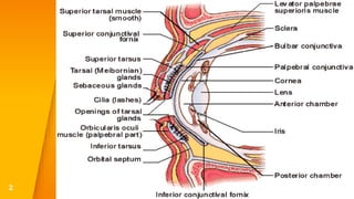
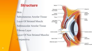
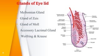
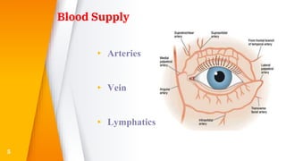
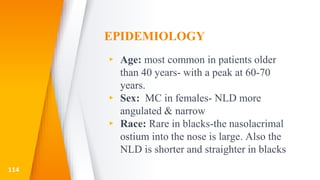
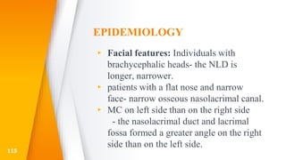
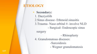
This document discusses the anatomy, structure, glands, blood supply, nerve supply, anomalies, diseases, and clinical features of the eyelid. It focuses on congenital and acquired ptosis, including the causes, clinical features, investigations, and treatment options. Specific conditions covered include congenital ptosis, neurogenic ptosis, myogenic ptosis, aponeurotic ptosis, mechanical ptosis, entropion, and ectropion. Surgical correction techniques are described for various types of ptosis and eyelid positioning anomalies.
Dr. Karan Bhatia provides an overview of keratoplasty (corneal transplantation). There are different types including penetrating keratoplasty (replacing the full thickness of the diseased cornea), partial thickness lamellar keratoplasty, and rotational keratoplasty. Penetrating keratoplasty has been the standard procedure but carries risks. The history and techniques of penetrating keratoplasty are described in detail including pre-operative evaluation, donor cornea excision, recipient cornea trephination, suturing, and post-operative management. Potential complications are also reviewed. Lamellar keratoplasty is introduced as an alternative to penetrating keratoplasty to reduce risks
Meibomian gland dysfunction (MGD) is a chronic abnormality of the meibomian glands characterized by terminal duct obstruction and changes in glandular secretion that can alter the tear film. It is a common cause of dry eye and estimated to affect 70% of Americans over age 60. Diagnosis involves examining the glands and assessing ocular surface damage. Treatment focuses on eyelid hygiene, warm compresses, and lubricating eye drops, with options for antibiotics, steroids, or procedures like probing, LipiFlow, or intense pulsed light. MGD is a chronic condition with periods of exacerbation and remission.
This document summarizes information about entropion, including eyelid anatomy, types of entropion, assessment, differential diagnosis, classification, and surgical procedures. It describes the anterior and posterior lamellae of the eyelid and the upper and lower lid retractors. Entropion is defined as inward turning of the eyelid margin against the eye. Types include congenital, involutional, and cicatricial. Surgical procedures to correct entropion are discussed such as suture techniques, tarsal strip procedures, plication of the inferior retractors, and posterior lamella grafts. Potential complications are also listed.
The document discusses the anatomy and structure of the normal crystalline lens. It describes the lens as being positioned behind the iris and composed of transparent, biconvex fibers that refract and focus light rays onto the retina. The lens maintains its own clarity, provides refractive power, and allows for accommodation through changes in curvature. With age, the lens becomes more curved and its refractive index decreases, resulting in hyperopic or myopic changes to the eye's refractive power. The lens is enclosed by an elastic capsule and supported by zonular fibers that attach along its equator.
Central serous retinopathy (CSR) is characterized by a serous retinal detachment in the macula due to leakage of fluid from the choroidal vasculature through the retinal pigment epithelium. It typically affects men in their 40s and 50s and can be associated with stress, corticosteroid use, hypertension, and type A personality. While often self-limiting, CSR can become chronic and cause long-lasting visual impairment without treatment. Management options include observation, stress reduction, oral medications like acetazolamide or ketoconazole to lower cortisol levels, and photodynamic therapy for severe or chronic cases.
The conjunctiva is a mucous membrane that lines the inner surface of the eyelids and covers the anterior sclera of the eye. It has several functions including lubricating the eye and contributing to immune surveillance. The conjunctiva consists of the palpebral, bulbar, and forniceal conjunctiva. The palpebral conjunctiva lines the inner surface of the eyelids and is subdivided into the marginal, tarsal, and orbital conjunctiva. The bulbar conjunctiva covers the anterior sclera and is separated from it by the episcleral tissue. It consists of the limbal and scleral conjunctiva. The conjunctival fornices are cul
The cornea is the transparent front part of the eye that allows light to enter. It has 6 layers - an epithelial layer, Bowman's membrane, a thick stromal layer, Duas layer, Descemet's membrane, and an endothelial layer. The stroma makes up around 90% of the cornea's thickness and contains collagen fibrils that give it strength and transparency. The cornea has no blood vessels and receives nutrients from vessels in the surrounding tissues. It has a rich nerve supply that provides its high sensitivity. The cornea refracts and helps focus light entering the eye and is essential for vision.
The document discusses the anatomy and physiology of the lens. It begins with the embryological development of the lens from surface ectoderm and describes the formation of the lens placode, pit, and vesicle. It details the anatomical structures of the lens including the capsule, epithelium, fibers, and zonules. The document also discusses the biochemical composition and metabolic processes that maintain lens transparency. Finally, it covers age-related changes to the lens and clinical significance including cataracts.
This presentation describes the background of the cornea and the corneal diseases in general, also it describes in detailed manner how to manage the corneal ulcer with its different causes
This document provides information on rhegmatogenous retinal detachment including:
- The pathogenesis which involves vitreoretinal traction from liquefaction of the vitreous gel creating a hole that allows fluid into the subretinal space.
- Symptoms include photopsia, floaters, and visual field defects.
- Signs include Marcus Gunn pupil, low intraocular pressure, retinal breaks, and mild iritis.
- Treatment options depend on factors like location and size of retinal break, state of vitreous gel, and include laser photocoagulation, cryotherapy, scleral buckling, pneumatic retinopexy, and drainage of subretinal fluid in
Uveitis classification & clinical features 1lijulk
Uveitis is inflammation of the uvea, which includes the iris, ciliary body, and choroid. It is classified based on anatomy, clinical course, etiology, and histology. The Standardization of Uveitis Nomenclature (SUN) Working Group developed standardized classifications including anatomical (anterior, intermediate, posterior, panuveitis), descriptors (onset, duration, course), and grading systems. Clinical features depend on the involved area and can include symptoms like pain, photophobia, blurred vision, and floaters. Signs on examination may reveal cells and flare in the anterior chamber, keratic precipitates, hypopyon, synechiae, and lesions of the iris, retina, or
Uveitis is an interesting disease of the with such a varied and diverse pathogenesis, various systemic causes and Dangerous complications in relation to the eye which makes it difficult and challenging to treat in a proper way. I hope this share will help.
Sympathetic ophthalmia and Vogt-Koyanagi-Harada disease are both rare granulomatous uveitides that can cause bilateral eye inflammation. Sympathetic ophthalmia occurs after trauma or surgery to one eye, while VKH disease is thought to be an autoimmune response against ocular and skin melanocytes. Both can lead to vision loss if not treated promptly with high-dose corticosteroids and immunosuppressants. Imaging like fluorescein angiography and optical coherence tomography are useful for monitoring disease activity and response to treatment.
The eyelids develop from the frontonasal and maxillary processes during embryogenesis. They contain skin, muscle, fibrous tissue and glands that work together to protect the eye. The orbicularis oculi muscle helps with blinking while the levator palpebrae superioris muscle elevates the upper eyelid. The tarsal plates provide structure and the meibomian glands secrete oils to form the tear film. Blood vessels from the internal and external carotid arteries provide a rich blood supply to support the eyelids' functions.
Cystoid macular edema is a pathological accumulation of fluid in the macula that can be caused by conditions like diabetic retinopathy, retinal vein occlusions, or following cataract surgery. It is diagnosed using optical coherence tomography or fluorescein angiography and treated initially with anti-inflammatory eye drops, corticosteroid injections, or anti-VEGF drugs to reduce fluid accumulation. Laser therapy or vitrectomy may also be used in some cases to treat underlying causes like vitreomacular traction.
This document discusses ophthalmic viscosurgical devices (OVDs), including their history, properties, composition, classification, and uses. It begins by describing the introduction of sodium hyaluronate as the first OVD used in ophthalmic surgery in 1972. It then covers the ideal properties of an OVD and the rheological properties of viscosity, elasticity, coatability, and others. OVDs are classified as cohesive, dispersive, or viscoadaptive based on their molecular structure and behavior. The document discusses the advantages and uses of OVDs in cataract surgery, glaucoma surgery, keratoplasty, and other ophthalmic procedures. It concludes by outlining complications like
This document discusses the treatment of non-healing corneal ulcers. It begins by describing the signs and symptoms of a non-healing corneal ulcer. Risk factors include compromised ocular surfaces, dry eye, previous trauma or surgery, and systemic conditions like diabetes or malnutrition. Treatment involves topical antibiotics, debridement of necrotic tissue, bandage contact lenses, and in severe cases, therapeutic keratoplasty.
This document discusses watering eye (epiphora) and its causes including hyperlacrimation and obstruction of tear outflow. It describes various mechanical obstructions that can cause epiphora such as punctal, canalicular, lacrimal sac or nasolacrimal duct obstructions. Clinical evaluation methods are outlined. Dacryocystitis, an infection of the lacrimal sac, is discussed including its congenital and acquired forms. Congenital dacryocystitis presents in newborns with epiphora, positive regurgitation test and swelling. Acquired dacryocystitis can be chronic or acute, with chronic forms including catarrhal dacryocystitis, lacrimal
Entropion is the in-turning of the eyelid margin. It can be congenital or acquired, with the most common type being involutional/senile entropion caused by laxity of the eyelid tissues and weakness of the retractors. Examination involves assessing lid laxity, snap back test, and tendon laxity. Treatment depends on severity and includes sutures, transverse lid splits with everting sutures, horizontal lid shortening procedures, and lower lid retractor procedures. Ectropion is eyelid eversion away from the globe and can also be congenital or acquired, with involutional being most common. Examination tests for laxity and muscle weakness.
Ectropion
It is an outward turning of the eyelid margin . This more frequently affects the lower eyelid.Upper eyelid ectropion is uncommon.Classified in 5 types
1)Congenital 2) Involutional 3) Paralytic 4) Cicatricial 5) Mechanical
Involutional ectropion is more common.Congenital ectropion is very rare.
Symptoms Epiphora :- excessive tearing.Excessive dryness.
Foreign body sensation Irritation.Burning.Redness.Chronic conjunctivitis KeratinizationCorneal exposure
Grading
Lid margin is out rolled and depending on out rolling ectropion can be classified as under:
Grade I –only punctum is everted
Grade II –lid margin is everted and palpebral conjunctiva is visible
Grade III –fornix is also visible
Etiological factors
Horizontal lid laxity:-can be demonstrated by pulling the central part of the lid 8 mm or more from the globe, with a failure to snap back to its normal position on release without the patient first blinking.
Medial canthal tendon laxity
demonstrated by pulling the lower lid laterally and observing the position of the inferior punctum If the lid is normal the punctum should not be displaced more than 1–2 mm
Lateral canthal tendon laxity
characterized by a rounded appearance of the lateral canthus and the ability to pull the lower lid medially more than 2 mm.
>Normally, the displacement should only be 0-2 mm.
Treatment
1 medical therapy
2 surgical therapy
The uvea consists of the iris, ciliary body, and choroid. It develops from both neuroectoderm and vascular mesoderm. The iris develops fully by age 5, with pigmentation continuing after birth. The ciliary body appears by 9 weeks and is fully developed by 6 months gestation. The choroid layers are seen by 5 months gestation. The uvea regulates light entry and provides blood supply to the outer retina. Congenital anomalies include heterochromia, polycoria, persistent pupillary membranes, and colobomas. Uveitis is inflammation of the uveal tract.
This document provides information on anterior ischemic optic neuropathy (AION), which is the most common cause of acute optic neuropathy in older age groups. It can be divided into two types: arteritic AION, which is due to giant cell arteritis; and non-arteritic AION, which makes up most cases. Both types present with sudden painless vision loss and optic disc swelling. Arteritic AION carries a worse prognosis and requires high-dose steroid treatment to prevent loss of vision in the fellow eye. Non-arteritic AION has a variable course but generally a poor rate of recovery without any proven effective treatments.
The document summarizes the anatomy and embryology of the lens. It discusses the gross anatomy, functions, embryological development from optic vesicles and lens placode, and structure of the lens including the capsule, epithelium, fibers, nucleus and zonules. It describes the aging changes that occur in the lens and various diseases that can affect it, including congenital disorders, cataracts, posterior capsular opacification, ectopia lentis and others. The document provides a detailed overview of the key anatomical structures and developmental processes involved in lens formation and function.
Tarsorrhaphy is a surgical procedure that joins the upper and lower eyelids together to partially or completely cover the eye. It is indicated to protect the cornea and promote healing in cases of corneal exposure from conditions like lagophthalmos, proptosis, or eyelid malposition, as well as non-healing corneal defects from issues like limbal stem cell deficiency or neurotrophic ulcers. Tarsorrhaphy can be temporary or permanent, partial or complete, and performed laterally, medially, or centrally using different suturing techniques. The goal is to safely and effectively manage corneal diseases and promote healing.
Endophthalmitis is an inflammation of the inner coats of the eyeball that can be infective or non-infective. Infective endophthalmitis is classified as exogenous or endogenous. Acute bacterial endophthalmitis is a complication of intraocular surgery where the infective source is usually the patient's own flora. Signs include lid edema, chemosis, hypopyon, and retinal periphlebitis. Treatment involves intravitreal and systemic antibiotics, intravitreal and topical steroids, and sometimes pars plana vitrectomy to treat severe or non-improving cases. Panophthalmitis is a more severe inflammation of the whole eyeball that
The document summarizes key anatomical structures and outflow pathways involved in aqueous humor drainage from the eye. It describes the transition from the trabecular meshwork to Schlemm's canal, noting structures like the scleral spur, Schwalbe's line, and trabecular endothelial cells. The trabecular meshwork consists of three layers - uveal meshwork, corneoscleral meshwork, and juxtacanalicular tissue. Aqueous humor drains through the trabecular meshwork into Schlemm's canal and collector channels before exiting into episcleral and conjunctival veins. The main outflow pathways are trabecular outflow, accounting for 85-95%
Vernal keratoconjunctivitis ophthalmology TONY SCARIA
This document discusses vernal keratoconjunctivitis (VKC), a recurrent, seasonal eye condition seen primarily in children and young adults. VKC involves inflammation of the conjunctiva and cornea and is caused by an allergic reaction, often to pollen allergens. Symptoms include itching, tearing, and thick discharge. Signs include raised papillae on the conjunctiva that resemble cobblestones. Treatment focuses on reducing inflammation and symptoms using topical steroids, mast cell stabilizers, and antihistamines. The condition typically resolves spontaneously by adulthood.
This document discusses ptosis, which is a drooping of the upper eyelid. It describes the functional anatomy of the levator palpebrae superioris muscle, which is responsible for eyelid elevation, as well as Muller's muscle and other muscles that can modify eyelid position. The document then covers evaluation of ptosis, including measurements of eyelid position, levator function testing, and classification of ptosis as congenital, acquired neurogenic, myogenic, aponeurotic, mechanical or pseudoptosis. Surgical management options are then briefly mentioned depending on the type and severity of ptosis.
This document discusses ptosis, or low-lying eyelids, including its definition, types, examination, and treatment. There are two main types of ptosis: congenital and acquired. Congenital ptosis can be simple or associated with other conditions. Acquired ptosis has neurological, muscular, aponeurotic, or mechanical causes. A full examination involves measuring lid position, levator function, ocular motility, and associated signs. Treatment options include observation, glasses, or surgical procedures like mullar resection, levator resection/advancement, or brow suspension depending on the type and severity of ptosis. Careful examination and documentation is important for successful ptosis management.
The document discusses ptosis, defined as an abnormally low resting position of the upper eyelid. It describes the anatomy and functions of the levator palpebrae superioris muscle and other structures involved in eyelid elevation. It then classifies and describes various types of ptosis including congenital, neurogenic, myogenic, aponeurotic, mechanical, and neurotoxic ptosis. Evaluation of ptosis involves assessing history, measurements like margin reflex distance, and ruling out pseudoptosis or ptosis mimics.
1) Cranial nerves III, IV, and VI are involved in eye movement control. A lesion in these cranial nerves can cause abnormalities like ophthalmoplegia or diplopia.
2) Examination of eye movements includes testing in 9 cardinal positions as well as tests like cover-uncover, forced duction, and versions to localize the lesion.
3) Common causes of eye movement abnormalities include thyroid eye disease, orbital pseudotumor, orbital tumors, and cranial nerve palsies. Accurate diagnosis relies on clinical history and examination findings.
This document discusses various topics related to accommodation and its anomalies. It begins by defining accommodation as the ability of the crystalline lens to change its diopter power to allow clear vision at different distances. It then provides tables showing how the amplitude of accommodation decreases with age. The rest of the document discusses various physiological and pathological conditions that can affect accommodation, including presbyopia, insufficiency of accommodation, spasm of accommodation, and paralysis of accommodation. For each condition, it provides details on definition, causes, signs and symptoms, and treatment approaches.
This document discusses hypospadias, a congenital anomaly where the urethra opens on the underside of the penis instead of at the tip. It covers the embryology, risk factors, classifications, clinical features, and various surgical treatment options for hypospadias repair. The goals of treatment are to position the urethral meatus at the tip of the penis, correct any penile curvature, and provide adequate skin coverage. Complications can include meatal stenosis, fistula formation, infection, and procedure failure. Careful patient selection and appropriate surgical technique are emphasized to achieve the best outcomes.
This document summarizes different types and causes of ptosis presented by Nazneen Nasir. It discusses acquired ptosis which can be neurogenic, myogenic, aponeurotic, or mechanical. Congenital ptosis and pseudo ptosis are also mentioned. Examination techniques for ptosis including margin-reflex distance, palpebral fissure height, and levator function are outlined. Surgical correction methods such as Müllerectomy, levator resection, and brow suspension are briefly described for different types and severities of ptosis.
1. Ptosis, or drooping of the upper eyelid, can be congenital or acquired through various mechanisms including myogenic, neurogenic, mechanical, or aponeurotic causes.
2. Clinical evaluation of ptosis involves measuring the margin-reflex distance, palpebral fissure height, and levator function to determine severity.
3. Simple congenital ptosis is usually treated through levator resection surgery during preschool years to prevent amblyopia, while Marcus Gunn jaw-winking syndrome may require levator disinsertion and brow suspension surgery.
This document discusses hypospadias, a congenital abnormality where the opening of the urethra is on the underside of the penis instead of at the tip. It begins with definitions and classifications of hypospadias. It then discusses the incidence, associated anomalies like cryptorchidism and inguinal hernia, role of imaging, timing of repair, anesthesia techniques, hemostasis methods, suture techniques, and dressings used postoperatively. It outlines indications for repair and the use of preoperative hormonal stimulation. It provides an intraoperative algorithm and details techniques for repair of distal, middle, and proximal hypospadias. Finally, it discusses potential complications like bleeding, meatal stenosis
The document describes a 50-year-old male who presented with a tender lump on his left shin and symptoms of fever and malaise for 5 days. The likely diagnosis is an abscess. Important investigations would include a blood sugar test. Management principles are adequate analgesia, antibiotics, adequate incision and drainage of the abscess under general anesthesia, cleaning and dressing of the wound.
Hypospadias is a congenital anomaly where the urethral opening is on the underside of the penis. It occurs in about 1 in 300 male births and has some genetic factors. Treatment is through surgical urethroplasty to reconstruct the urethra and correct any curvature. Outcomes include some urinary and sexual dysfunction compared to controls, with more issues for proximal versus distal hypospadias. The goal of surgery is a functional penis with normal appearance.
The document discusses the lacrimal system, which consists of the secretory system and excretory system. The secretory system includes the lacrimal gland and accessory glands that secrete the components of the tear film. The excretory system includes the puncta, canaliculi, lacrimal sac, and nasolacrimal duct that drain tears from the eye surface into the nose. Dacryocystitis is an infection of the lacrimal sac that can be acute or chronic. Evaluation and management of dacryocystitis as well as congenital nasolacrimal duct obstruction are discussed.
you will get knowledge about the ptosis, its different types, its examination, its measurement, its treatment in detail.
different eyelid muscles such as LPS, Orbicularis oculi and frontalis are also explained.
The document discusses various topics related to eyelid and facial reconstruction:
- It describes the different layers of the eyelid and techniques for repairing full-thickness eyelid lacerations.
- Entropion, ectropion, canalicular injuries, and dacryocystorhinostomy procedures are summarized.
- Facial nerve anatomy and various modalities for facial reanimation including nerve grafts, muscle transfers, and static procedures are outlined.
- Techniques for managing eyelid dysfunction in facial paralysis cases such as tarsorrhaphy and gold weights are also mentioned.
Ptosis, or drooping of the upper eyelid, can be congenital or acquired. It is caused by weakness or damage of the levator palpebrae superioris muscle or aponeurosis. Clinical evaluation involves measuring eyelid position and function of the levator muscle. Ptosis is classified based on severity and etiology. Surgical treatment options aim to strengthen the eyelid lift through techniques like levator resection or frontalis sling suspension. Proper diagnosis and management require a thorough history and examination.
Eyelid Surgery for Ophthalmic Clinical Course UPPGHAlex Tan
This document provides an overview of surgical techniques for managing various eyelid conditions including entropion, ectropion, and ptosis. It discusses the underlying causes and pathologies of each condition and describes various surgical procedures for correction. For entropion, procedures described include Quickert sutures, lateral tarsal strips, and reattachment of retractors for involutional cases, and tarsotomy with or without grafts for cicatricial cases. For ectropion, techniques include tendon tightening and lazy-T procedures. For ptosis, factors in evaluation are outlined and techniques covered include levator resection, frontalis suspension, and aponeurotic surgery.
Chemical Injury & Foreign Body Ocular Injury MEDICS india
This document discusses ocular injuries from open wounds and chemical exposures. It covers various types of eye trauma including lid lacerations, orbital fractures, globe injuries, and chemical burns. Specific injuries from penetrating wounds, foreign bodies, and different chemical agents like acids and alkalis are described. Diagnosis involves detailed history, exam of the anterior segment, imaging, and sometimes surgery to remove foreign materials. Treatment focuses on prevention of further damage, irrigation, antibiotic drops, steroids, and managing complications like infection, scarring, or glaucoma.
The abducens nerve is the sixth cranial nerve. It arises from the brainstem and exits through the subarachnoid space, piercing the dura mater. It travels through Dorello's canal and the cavernous sinus before innervating the lateral rectus muscle. Abducens nerve palsy presents with diplopia, the affected eye resting in adduction, and inability to abduct the eye. Common causes include space-occupying lesions, inflammation, diabetes, and trauma. Neuroimaging may be required to identify underlying causes if symptoms do not improve after 3 months.
The trochlear nerve is a motor nerve that arises from the midbrain and is the only cranial nerve that crosses to the other side of the brain. It innervates the superior oblique eye muscle and is involved in eye movement. Lesions or damage to the trochlear nerve can cause weakness of the superior oblique muscle resulting in symptoms like hyperdeviation of the eye, limited eye movement, and double vision when looking downward. The most common causes of trochlear nerve palsy are congenital, traumatic injury, and idiopathic.
The oculomotor nerve (3rd cranial nerve) is entirely motor and supplies several extraocular muscles and the levator palpebrae superioris muscle. It has nuclei located in the midbrain and courses through the fascicular, basilar, intracavernous, and intraorbital regions. Common causes of oculomotor nerve palsy include vascular issues like aneurysms and diabetes, neoplasms, trauma, and idiopathic cases. A complete oculomotor nerve palsy presents with ptosis, limitation of eye movements, and potentially a dilated pupil. Evaluation involves assessing pupil involvement and testing for underlying causes. Management consists of treating any identified cause as well as addressing diplopia.
CORNEAL ULCER ,DR M SAQUIB , OPHTHALMOLOGY MEDICS india
This document discusses corneal ulcers, including their classification, causes, signs and symptoms, and treatment. Corneal ulcers can be classified based on etiology (infective vs. non-infective), location (central, paracentral, peripheral), and depth (superficial, deep). Common causes are bacterial, viral, fungal or protozoal infections. Symptoms include pain, photophobia, and decreased vision. Treatment involves identifying and treating the underlying cause, using antibiotic or antifungal medications, cycloplegics, and sometimes therapeutic keratoplasty for severe cases. Complications can include hypopyon, glaucoma, perforation, and endophthalmitis if not properly
This document discusses optic nerve disease and optic neuropathy. It covers the clinical features, examination, investigations, and various causes of optic nerve disease including optic neuritis, nutritional optic neuropathy, anterior ischemic optic neuropathy, papilledema, and Leber's hereditary optic neuropathy. Examinations discussed include visual acuity testing, color vision testing, visual field testing, ophthalmoscopy, fluorescein angiography, and visual evoked potentials. Causes covered in more depth include optic neuritis, anterior ischemic optic neuropathy, nutritional optic neuropathy, papilledema, and Leber's hereditary optic neuropathy.
The document provides information about the optic nerve and visual pathway, including:
1. It describes the anatomy of the optic nerve from the retina through the optic chiasm and optic tracts to the visual cortex.
2. It discusses common lesions that can occur at different points along the visual pathway and their associated visual field defects.
3. It outlines the blood supply and venous drainage of the optic nerve and visual pathway structures.
1. Keratoplasty, or corneal transplantation, involves replacing part or all of a damaged cornea with donor corneal tissue.
2. There are several types of keratoplasty procedures including penetrating keratoplasty (full thickness), lamellar keratoplasty (partial thickness), DSAEK, and DMEK.
3. Keratoplasty is indicated for conditions such as corneal opacity, dystrophy, infection, trauma, ectasia, and refractive errors. Careful donor preparation, surgical technique, and postoperative management are required for successful outcomes. Complications can include rejection, infection, and recurrence of the underlying condition.
This document discusses retinopathy of prematurity (ROP), which occurs in premature infants who are exposed to high oxygen levels. Key points:
- ROP occurs when retinal vessels do not fully develop due to preterm birth. High oxygen can damage capillaries.
- Risk factors include low birth weight (<1500g), young gestational age (<32 weeks), and high oxygen exposure.
- ROP is diagnosed using an eye exam and classified by location, severity, and vascular characteristics. Treatment may include laser therapy or anti-VEGF injections depending on the stage and severity. Regular screening of high-risk infants allows for early detection and treatment.
This document discusses retinoblastoma, a rare eye cancer that affects children. It is the most common intraocular malignancy in childhood. If untreated, children typically die within 2-4 years of symptoms starting. The document covers epidemiology, genetics, clinical presentation, diagnosis, and management options like chemotherapy, radiation therapy, cryotherapy, laser therapy, and enucleation. Early detection and a multidisciplinary approach can help save the child's life and possibly preserve vision. Prognosis depends on factors like tumor size and extent of spread.
This document discusses retinal detachment, including the different types (rhegmatogenous, tractional, exudative), symptoms, risk factors, examination findings, diagnosis, treatment options including pneumatic retinopexy, scleral buckling, vitrectomy, and prevention techniques. It also covers traction retinal detachment and exudative retinal detachment, their causes, presentations, signs, and management approaches.
This document discusses retinal artery occlusion, specifically central retinal artery occlusion (CRAO). It provides details on:
- CRAO presentation as acute, painless monocular visual loss in older patients, often with severe final vision loss.
- Anatomy of the retinal blood supply from the ophthalmic artery.
- Risk factors including age, gender, cardiovascular/vascular conditions.
- Examination findings like RAPD and retinal signs of ischemia.
- Differential diagnosis, investigations like fluorescein angiography and treatment involving corticosteroids for CRAO related to temporal arteritis.
1. Retinal vein occlusion is a common retinal vascular disease caused by occlusion of retinal veins. Central retinal vein occlusion blocks the central retinal vein while branch retinal vein occlusion blocks smaller retinal veins.
2. Presentation includes sudden blurred vision, retinal hemorrhages, edema, and tortuous veins. Central retinal vein occlusion can be ischemic or non-ischemic depending on extent of occlusion.
3. Management depends on severity and includes treating any underlying conditions, observation, anti-VEGF injections, laser therapy, and sometimes surgery for complications like tractional retinal detachment.
This document discusses hypertensive retinopathy, including its clinical presentation, pathogenesis, staging, and fundus changes. It describes how long-standing hypertension can damage the retina, choroid, and optic nerve through vasoconstriction and increased vascular permeability. The staging systems of Keith Wagner and Wong-Mclntosh are outlined. Features of acute hypertensive retinopathy include cotton wool spots, flame-shaped hemorrhages, and exudates. Chronic changes include arteriolar narrowing, nicking, and sclerosis. Malignant hypertension can cause necrotizing retinitis and serous retinal detachment. Pregnancy-induced hypertension also causes a reversible retinopathy.
This document discusses different types of staphylomas, which are abnormal protrusions of the eyeball that can occur in various locations. Anterior staphylomas involve bulging of the cornea and sclera near the front of the eye. Posterior staphylomas occur behind the equator and involve bulging of the sclera lined by the choroid. Staphylomas can be caused by conditions like severe myopia, trauma, infection, inflammation, and certain collagen disorders. Complications may include glaucoma or reduced vision. Treatment depends on the type and severity but may involve surgery or steroids.
The document discusses diabetic retinopathy (DR) and its prevalence, risk factors, pathogenesis, stages of progression from non-proliferative DR to proliferative DR, complications like macular edema, and treatment approaches including anti-VEGF drugs, laser therapy, and panretinal photocoagulation. Key points covered include the global prevalence of DR being 27% on average from 2015-2019, microvascular damage and leakage due to hyperglycemia leading to complications, and treatment guided by disease severity and presence of macular edema or neovascularization.
The retina is a thin, light-sensitive tissue that lines the back of the eye. It contains 10 layers including photoreceptors that convert light into neural signals. The macula contains the highest concentration of cones and allows for sharp, central vision. The optic disc is where retinal blood vessels and optic nerve fibers exit the eye. The retina receives dual blood supply from the central retinal artery and choroidal vessels. It contains over 120 million light-sensitive rods and cones and transmits visual information through the optic nerve to the brain.
This document discusses scleritis, which is inflammation of the sclera. It describes the different types of scleritis including anterior diffuse, nodular, and necrotizing scleritis as well as posterior scleritis. Potential causes are noted such as rheumatoid arthritis, Wegener's granulomatosis, and connective tissue disorders. Signs, symptoms, and treatments are provided for each type of scleritis. Imaging techniques like ultrasound and CT scans are also mentioned for evaluating posterior scleritis.
1. The document describes various diseases and conditions that can affect the eyelids including inflammatory conditions like blepharitis and hordeolum, infections like molluscum contagiosum, benign growths like chalazions, and positional abnormalities of the eyelids.
2. The anatomy of the eyelid is also summarized including its layers and muscle components.
3. Specific conditions are further described like types of ectropion and entropion and their clinical features, testing, and treatment options.
Rasamanikya is a excellent preparation in the field of Rasashastra, it is used in various Kushtha Roga, Shwasa, Vicharchika, Bhagandara, Vatarakta, and Phiranga Roga. In this article Preparation& Comparative analytical profile for both Formulationon i.e Rasamanikya prepared by Kushmanda swarasa & Churnodhaka Shodita Haratala. The study aims to provide insights into the comparative efficacy and analytical aspects of these formulations for enhanced therapeutic outcomes.
NVBDCP.pptx Nation vector borne disease control programSapna Thakur
NVBDCP was launched in 2003-2004 . Vector-Borne Disease: Disease that results from an infection transmitted to humans and other animals by blood-feeding arthropods, such as mosquitoes, ticks, and fleas. Examples of vector-borne diseases include Dengue fever, West Nile Virus, Lyme disease, and malaria.
Adhd Medication Shortage Uk - trinexpharmacy.comreignlana06
The UK is currently facing a Adhd Medication Shortage Uk, which has left many patients and their families grappling with uncertainty and frustration. ADHD, or Attention Deficit Hyperactivity Disorder, is a chronic condition that requires consistent medication to manage effectively. This shortage has highlighted the critical role these medications play in the daily lives of those affected by ADHD. Contact : +1 (747) 209 – 3649 E-mail : sales@trinexpharmacy.com
Basavarajeeyam is an important text for ayurvedic physician belonging to andhra pradehs. It is a popular compendium in various parts of our country as well as in andhra pradesh. The content of the text was presented in sanskrit and telugu language (Bilingual). One of the most famous book in ayurvedic pharmaceutics and therapeutics. This book contains 25 chapters called as prakaranas. Many rasaoushadis were explained, pioneer of dhatu druti, nadi pareeksha, mutra pareeksha etc. Belongs to the period of 15-16 century. New diseases like upadamsha, phiranga rogas are explained.
Here is the updated list of Top Best Ayurvedic medicine for Gas and Indigestion and those are Gas-O-Go Syp for Dyspepsia | Lavizyme Syrup for Acidity | Yumzyme Hepatoprotective Capsules etc
These lecture slides, by Dr Sidra Arshad, offer a quick overview of the physiological basis of a normal electrocardiogram.
Learning objectives:
1. Define an electrocardiogram (ECG) and electrocardiography
2. Describe how dipoles generated by the heart produce the waveforms of the ECG
3. Describe the components of a normal electrocardiogram of a typical bipolar lead (limb II)
4. Differentiate between intervals and segments
5. Enlist some common indications for obtaining an ECG
6. Describe the flow of current around the heart during the cardiac cycle
7. Discuss the placement and polarity of the leads of electrocardiograph
8. Describe the normal electrocardiograms recorded from the limb leads and explain the physiological basis of the different records that are obtained
9. Define mean electrical vector (axis) of the heart and give the normal range
10. Define the mean QRS vector
11. Describe the axes of leads (hexagonal reference system)
12. Comprehend the vectorial analysis of the normal ECG
13. Determine the mean electrical axis of the ventricular QRS and appreciate the mean axis deviation
14. Explain the concepts of current of injury, J point, and their significance
Study Resources:
1. Chapter 11, Guyton and Hall Textbook of Medical Physiology, 14th edition
2. Chapter 9, Human Physiology - From Cells to Systems, Lauralee Sherwood, 9th edition
3. Chapter 29, Ganong’s Review of Medical Physiology, 26th edition
4. Electrocardiogram, StatPearls - https://www.ncbi.nlm.nih.gov/books/NBK549803/
5. ECG in Medical Practice by ABM Abdullah, 4th edition
6. Chapter 3, Cardiology Explained, https://www.ncbi.nlm.nih.gov/books/NBK2214/
7. ECG Basics, http://www.nataliescasebook.com/tag/e-c-g-basics
Promoting Wellbeing - Applied Social Psychology - Psychology SuperNotesPsychoTech Services
A proprietary approach developed by bringing together the best of learning theories from Psychology, design principles from the world of visualization, and pedagogical methods from over a decade of training experience, that enables you to: Learn better, faster!
Histololgy of Female Reproductive System.pptxAyeshaZaid1
Dive into an in-depth exploration of the histological structure of female reproductive system with this comprehensive lecture. Presented by Dr. Ayesha Irfan, Assistant Professor of Anatomy, this presentation covers the Gross anatomy and functional histology of the female reproductive organs. Ideal for students, educators, and anyone interested in medical science, this lecture provides clear explanations, detailed diagrams, and valuable insights into female reproductive system. Enhance your knowledge and understanding of this essential aspect of human biology.
REGULATION FOR COMBINATION PRODUCTS AND MEDICAL DEVICES.pptx
Eye Lid Disorders ,Ophthalmology
1. EYE LID
06/10/2020
DR M SAQUIB
Vice Principal , G.S.Medical College & Hospital
MBBS,MS , FSCEH DELHI,FHVDESAI PUNE,
EX REGISTRARA JNMCH,AMU
CONSULTANT OPHTHALMOLOGIST
HOD D/O OPHTHALMOLOGY
G.S .MEDICAL COLLEGE
Founder sec: MEDICS India ,
Mail-dms2k5@gmail.com , 9634123800
12. Congenital Ptosis
▸ A droopy eyelid, also called ptosis occurs when the muscle that
elevates the eyelid (the levator palpebrae superioris muscle) is
weak from various reasons.
▸ Congenital weakness of LPS due to maldevelopment .
12
14. Clinical Features:
▸ Drooping of Eye Lid – U/L , B/L
▸ Lid Crease –Diminished or Absent
▸ Lid Lag on Downgaze – Ptotis lid level
higher than other in downgaze
▸ LPS- Function –Poor, Fair ,Good
14
15. The Droopy Eyelid May Cause:
▸ Astigmatism- If the pressure on the front of the eye causes distortion and
refractive error it may need close observation, treatment with glasses or even
surgery.
▸ A chin up position- If the ptosis is severe enough it may cause the child to
adopt a chin up position in order to be able to see beneath the droopy eyelids
and use the eyes together. Contraction of the frontalis muscle (in the forehead)
to further elevate the upper eyelid is a very common compensatory
mechanism. This is also an indication for surgical correction.
▸ Amblyopia (abnormal visual development)- This results from astigmatism (a
misshaping of the cornea) or other refractive errors (refractive amblyopia).
(Stimulus deprivation amblyopia).
15
16. Associated Features with Congenital Ptosis
▸ Simple Congenital Ptosis
▸ Congenital Ptosis with Superior Rectus Muscle
weakness – Double elevator palsy
▸ Blephrophimosis Syndrome
▸ Congenital Synkinetic Ptosis : Ptotic lid retraction
with Jaw movement
16
22. Aponeurosis Ptosis
▸ Defect in LPS Aponeurosis.
▸ LPS muscle is normal
▸ Involutional Ptosis
▸ Postoperative Ptosis (Cataract,RD)
▸ Blephrochalasis weakness of aponeurosis
▸ Traumatic Dehiscence or Disintertion
▸ Characteristic features of this type of Ptosis are that patients have a
good levator function with a high lid crease, affected eyelid appears
lower on down gaze and a thin upper eyelid with redundant skin.
22
23. Mechanical Ptosis
▸ Lid Tumours
▸ Multiple Chalazion
▸ Lid Oedema
▸ Cicatricial Ptosis (Pemphigoid,Trauma )
23
24. History
▸ Age ‘Family History
▸ Trauma
▸ Surgery
▸ Systemic disorder
▸ A history of dry eyes, intermittent epiphora, or
chronic conjunctivitis can indicate a dry eye
disorder .
24
26. Clinical History
▸ Age
▸ Progression
▸ Diurnal variation
▸ Change with Jaw Movement
▸ History of Trauma
▸ History of Previous Ocular Surgery
▸ History of Muscle Fatigue
26
27. Clinical Examination▸ Vision
▸ Pupil Reaction
▸ Head Posture
▸ Frontalis Overaction /Brow Fold/Level
▸ Lid Crease
▸ MRD –
▸ Palpebral Fissure ( Height & Width)
▸ LPS Function –
▸ Vertical Palpebral Height
▸ Lid Lag on Downgaze
▸ Marcus Gunn Jaw Winking Phenomenon
▸ Bell’s Phenomenon
▸ Extraocular Movement
▸ Corneal Sensation
▸ Tear Film Evaluation
▸ Old Photograph27
28. ▸ Measure amount of Ptosis – Mild/Moderate /Severe
– 2/3/4 mm
▸ Marginal Reflex distance – MRD- Normal – 4-5 mm
▸
▸ LPS Function – Normal- 15 mm
▸ Good- 8mm ,fair- 5-7 mm ,Poor -4mm less
28
31. Specific Investigation
▸ Tensilon test – I . V Endrophonium ,cause
improvement in Ptosis ( myasthenia)
▸ Phenylephrine Test – Horner’s syndrome
Neurological investigation –MRI
Photographic Records
31
32. Treatment of Congenital Ptosis
▸ Severe Ptosis – Urgent to prevent Amblyopia
▸ Mild –Moderate – Till anatomical structure form
▸ 1.TARS-OCONJUCTIVAL –MULLERECTOMY
▸ 2. LEVATOR RESECTION –
▸ 3.FRONTALIS SLING OPEARTION
▸
32
44. A. Normal Lid Position
B. Spastic Entropion
C. Cicatricial Entropion
D. Senile Ectropion
44
45. Congenital Ectropion
▸ Vertical insufficiency of the anterior Lamella of the
Eye lid
▸ Associated with – Blephrophimosis Syndrome
Down Syndrome , Ichthyosis , Eublephron .
Treatment – Full Thickness Skin graft to lengthen the
anterior lamella
45
46. Congenital Entropion
▸ Congenital Inversion of Eye lid
▸ Developmental Factor associated – Lower Lid
Retractor Dysgenesis,Tarsus kink ,Relative
shortening of Posterior lamella
46
47. Senile (Involutional ) Entropion
▸ Very common
▸ Only lower lid
▸ Factors: Attenuation and disinternation of Eye lid
retractors
▸ Tissue atrophy with Horizontal laxity of the Eye lid
▸ Overriding of Pre tarsal fiber of orbicularis by
Preseptal Fibres of Orbicularis
47
50. Clinical Features -
▸ Foreign Body sensation
▸ Irritaion
▸ Lacrimation
▸ Photophobia
▸ Scarring of Palpebral Conjuctiva
▸ Recurrent corneal abrasion
▸ Superficial corneal Opacity
50
51. ▸ Snapback Test : Horizontal laxity detected by
poor eye lid tone ,
▸ Distraction Test – Ability to pull Eye lid more
than 6 mm from the Globe
▸ Grading of Entropion –
▸ Grade 1 –Posterior Lid Border is inrolled
▸ Grade 2- Inturing up to intermarginal strip
▸ Grade 3 –Whole lid margin inturned
51
52. Treatment
Reattachment of Lid Retractors
Shortening of Horizontal width of the Tarsal
plate
Forming cicatrix between pretarsal and preseptal
part of Orbicularis
52
53. Surgical Correction of Cicatrial
Entropion
▸ Mild Entropion -Anterior lamellar Resection
▸ Moderate Entropion with Atrophic Tarsus – Tarsal
Wedge Resection
▸ Mild to Moderate Cicatrial Entropion –
▸ Transposition of Tarsoconjuctival Wedge ) – Tarsus
Fracture and Eversion of Distal Tarsal
▸ Posterior Lamellar Graft
53
54. Surgical Technique for Senile
Entropion
1.- Very Old Patient – Temporary Transverse Everting
Sutures
▸ 2.Little Horizintal Laxity – Long Term – Wies Procedure
–Transverse Lid Split and Everting Sutures
▸ 3.Severe /Recurrence case- Plication of Lower Lid
Retractor – Jones Procedure
▸ 4.Severe Horizontal Lid Laxity – Quickert Procedure
54
59. Cicatrial Entropion
▸ Cicatrical contraction of Palpebral Conjuctiva
▸ Relative shortening of the inner tarsoconjuctival
lamina of the lid
▸ Inversion of Lid Margin
▸ Causes :
▸ Trachoma
▸ Trauma
▸ Chemical Burn
▸ Steven Jhonson Syndrome
▸ Cicatrical pemphigoid
59
60. Principle-
Lengthening of the posterior lid
lamina
Tarsal Plate Rotation
▸ Mild to Moderate Entropion – with Thick Tarsus-
Wedge Resection of Tarsus
▸ Severe Entropion / Entropion without Thick
Tarsus – Tarsal Fracture
60
61. Mechanical Entropion
▸ Due to lack of Support
▸ Pthisis Bulbi
▸ Enophthalmos
▸ After Enucleation or Evisceration
61
62. Ectropion
▸ Outward turning of the Eye lid margin.
Clinical Feature –
▸ Epiphora
▸ Irritation ,Discomfort ,
▸ Mild Photophobia
62
63. Senile( Involutional ) Ectropion
▸ Most common type of Ectropion
▸ Causes-
▸ Horizontal Laxity of Eyelid
▸ Medial Canthal Tendon laxity
▸ Lateral Canthal tendon laxity
▸ Disintertion of lower lid Retractors
63
64. Cicatrial Ectropion
▸ Due to scarring of lid skin
▸ Can involve both lids
Causes-
▸ Thermal burn
▸ Chemical burn
▸ Laceration, Ulcer
64
65. ▸ The skin, orbicularis muscle, and orbital septum are
normally flexible enough that spontaneous movement of the
eyelids can occur.
▸ Anterior lamella (skin and muscle) and posterior lamella
(tarsus and conjunctiva). Scarring of the anterior
lamella can cause a cicatricial ectropion .
▸ Lengthening of the shortened lamella by adding a full-
thickness skin graft is the treatment for cicatricial
ectropion.
▸ Scarring of the posterior lamella also causes a cicatricial
entropion.
65
67. Paralytic Ectropion
▸ Facial Nerve Palsy
▸ Lower Lid
▸ Causes-
▸ Facial Nerve Palsy
▸ Bell’s Palsy
▸ Head Injury
▸ Infection of middle Ear
▸ Operation of Parotid Gland
67
68. Signs
▸ Out rolling of Lid Margin – Grading
▸ Grade I - Puntum is everted
▸ Grade II -Lid Margin is everted ,Palpebral Conjuctiva is visible
▸ Grade III -Fornix is also visible .
68
69. If the ectropion is not cicatricial and not paralytic, it must be
involutional.
Involutional ectropion occurs in older patients with eyelid
laxity . The eyelid laxity can be demonstrated by the eyelid
distraction test and the eyelid snap test
▸ Involutional Ectropion – Positive snap Test –
Horizontal lid laxity ,lid can be easily pulled
away from the globe but fails to snape back to
the normal position on release .
▸ Medial Canthal Tendon laxity
▸ Lateral canthal tendon Laxity69
71. 71
The eyelid distraction test is
performed by manually pulling
the eyelid away from the eyeball.
The lower lid should not move
more than 6 mm off the eyeball.
72. The Eyelid Distraction
Test
The eyelid distraction
test is performed by
manually pulling the
eyelid away from the
eyeball. The lower lid
should not move more
than 6 mm off the
eyeball.
72
78. Involutional Ectropion
Age Related
Medial Conjuctivoplasty : Mild cases of ectropion involving punctal area
.Excising a spindle –shaped piece of conjuctiva and subconjuctival
tissue
Horizontal Lid Shortening – Full thickness pentagonal excision in
moderate ectropion .
Lateral Tarsal Strip Procedure – Generalised ectropion with horizontal
lid laxity
Byron Smith’s modified Kuhnt- Szymanowski78
99. SECRETORY SYSTEM-Development
Lacrimal gland- multiple solid ectodermal
buds- anterior supero-lateral orbit- 6-8 weeks
age of gestation
Buds branch and canalize- ducts and alveoli
Do not function fully- 6 weeks after birth
Newborn infants- do not produce tear- crying
Congenital NLDO seen following defective
canalization of the Caudal End .
50% of the Newborns will have obstruction of
the distal end (Valve of Hasner )
99
100. SECRETORY SYSTEM
▸ Lacrimal gland- Exocrine gland, almond size,
situated at lacrimal gland fossa- Superior
temporal orbit in frontal bone.
▸ LPS divides gland into orbital & palpebral lobe
anteriorly.
▸ Ducts from orbital lobe passes thru palpepral
lobe- empty into upper conj fornix temporally.
▸ Biopsy is avoided from Palpebral Lobe
100
102. ACCESSORY GLANDS
▸ Gland of Krause- Accessory exocrine gland
present deep in superior fornix
▸ Gland of Wolfring -situated near superior
border of tarsal plate.
▸ Previously- Main lacrimal gland- reflex
secretion
- Accessory glands- basal secretion
Recent evidence- All tearing –Reflex- single unit
102
103. TEAR FILM
▸ 1.Lipid layer- secreted by Meibomian glands
Increase surface tension of tear film &
decreases its rate of evaporation
▸ 2. Aqueous layer- secreted by main lacrimal
glands & gland of Krause & Wolfring.
Provides oxygen to cornea & antibacterial
function.
▸ 3.Mucinous layer- secreted by goblets cells of
conjunctiva-allows even distribution of tear
film over ocular surface
103
106. EXCRETORY SYSTEM
▸ The lacrimal drainage system includes the
- Upper & lower puncta
- Lacrimal canaliculi
- Upper canaliculi
- Common canaliculi
- Lower canaliculi
- Lacrimal Sac
- Nasolacrimal duct
106
107. Development : 5 weeks age of gestation
The lacrimal drainage system- ectodermal cord-
b/w the lateral nasal process and the maxillary
process.
Cord canalize- form NLD caudally and lacrimal sac
and canaliculi cranially
Caudally NLD extends intranasally- exiting-inferior
meatus
Canalization- complete around birth
NLD Obstruction- 50% of infants at time of birth
107
109. EXCRETORY SYSTEM
Lacrimal puncta- 0.3 mm in diameter located medially on upper
& lower lid margin, 6 mm & 6.5 mm from inner canthus
respectively- directed posteriorly towards tear lake
vertical canaliculus(ampulla)- 2 mm-turns 90 deg
horizontal canaliculus- 8 mm opens into sac by common
canaliculus(90%)
Common canaliculus- Mucosal fold- Valve of Rosenmuller- 1
way valve prevents tear reflux from lacrimal sac back into the
canaliculi
109
110. EXCRETORY SYSTEM
Lacrimal sac- lies b/w ant & post head MCT
in lacrimal fossa bordered by anterior &
posterior lacrimal crest
Size of sac 12-15x5 mm when distended,
Fundus - part of sac above common
canaliculus
Body of sac
Medial wall- lacrimal fossa- lacrimal bone posteriorly
& frontal process of maxilla anteriorly
Medial to lacrimal fossa- Middle meatus of nose
110
111. EXCRETORY -Nasolacrimal duct
▸ -12- 18 mm in length
- Travels thru bony nasolacrimal canal-
directed Inferiorly, Laterally And
Posteriorly
-opens thru an ostium into inferior nasal
meatus ,lateral & below inferior turbinate
- ostium- covered by partial mucosal fold
( Valve of Hasner)
111
112. DACRYOCYSTITIS
▸ Infection of Lacrimal sac- b,coz of stasis
of fluid(tears & mucus secretions) due to
NLD blockage
112
114. EPIDEMIOLOGY
▸ Age: most common in patients older
than 40 years- with a peak at 60-70
years.
▸ Sex: MC in females- NLD more
angulated & narrow
▸ Race: Rare in blacks-the nasolacrimal
ostium into the nose is large. Also the
NLD is shorter and straighter in blacks
114
115. EPIDEMIOLOGY
▸ Facial features: Individuals with
brachycephalic heads- the NLD is
longer, narrower.
▸ patients with a flat nose and narrow
face- narrow osseous nasolacrimal canal.
▸ MC on left side than on the right side
- the nasolacrimal duct and lacrimal
fossa formed a greater angle on the right
side than on the left side.
115
116. ETIOLOGY
▸ PrimaryAcquired Nasolacrimal Duct
Obstruction(PANDO): Idiopathic
▸ Involutional stenosis-older persons- due
to compression of NLD lumen b.coz of
fibrous obstruction by chronic
inflammation
116
118. PATHOGENESIS
▸ There are two main factors resulting in
vicious cycle-
1. Stasis of sac contents due to NLD
blockage
2. Infection may- ascend from nose
- descend from
conjunctiva
Both aerobic and anaerobic bacteria-
Mainly gram positive & gram negative.
118
119. ACUTE DACRYOCYSTITIS
▸ Acute dacryocystitis is characterized by
the sudden onset of
- severe pain
- redness &
- oedema in the medial canthal region
- Epiphora
119
120. ACUTE DACRYOCYSTITIS
▸ SIGNS
1. A Tender & Hyperemic Palpable Mass
Is Noted Inferior To The Medial Canthal
Tendon- Non Reducible
2. Purulent Discharge Is Noted From The
Puncta.
3.Lacimal Abscess- Rupture- Lacrimal
Fistula Through The Skin.
4.Conjunctival Injection And Preseptal
Cellulitis
120
122. COMPLICATIONS
▸ Orbital Cellulitis- bact overgrowth
which rupture thru lacrimal sac wall into
surrounding orbital soft tissue.
▸ Cavenous sinus thrombosis
▸ Blindness
▸ Osteomyelitis of lacrimal bone
122
123. MANAGMENT
▸ Syringing/Probing-
CONTRAINDICATED- extremely
painful- cellulitis
▸ Oral Broad Spectrum Antibiotics-
Amoxicillin ( 250-500 mg) Cloxacillin
(250-500 mg) TDS
▸ Anti inflammatory/ Analgesics drugs
▸ Hot compress
▸ Parentral antibiotics- severe cases-
orbital cellulitis
123
124. MANAGMENT
▸ Aspiration of lacrimal sac- If lacrimal
abcess is localised & approaching the
skin.
▸ Incision & drainage- in severe cases not
responding to conservative mgt
▸ abscess involving lacrimal sac &
adjacent soft tissue- vertical incision is
given- wound is packed- open- healing
by secondary intention- lacrimal fistula
formation
124
127. SYMPTOMS
1. Epiphora with or without mucopurulent
discharge
2. Swelling over sac area- present or
absent
3. Matting of eyelashes
4. Recurrent conjunctivitis
127
128. Sign
Swelling in lacrimal sac area (below MCT)
which is reducible- regurgitation test +ve
Fullness in medial canthal area with no
obvious swelling
Enlarging mucocele- due to chronic low grade
infection of sac-Increased glandular secretions
into the sac.
Pooling of tears at medial canthus.
Chronic discharging fistula of sac
128
131. DIFFERENTIAL DIAGNOSIS
▸ Dermoid Cyst
▸ Cavernous haemangioma
▸ Neurofibromas
▸ Lacrimal sac tumours
Above lesions are not reducible
NLD may be blocked on syringing but
DCG is diagnostic.
131
133. EXTERNAL DCR
▸ Standard gold treatment, success rate
>90%
▸ Indications:
Chronic dacryocystitis with NLD
blockage
Mucocele of lacrimal sac
Congenital dacryocystitis-failed
conservative managment
133
134. EXTERNAL DCR
▸ It is a lacrimal drainage surgery in which
a fistula is created b/w the lacrimal sac
& middle meatus of the nasal cavity in
order to bypass a obstruction in NLD
134
137. ENDONASAL DCR
Done with the help of a nasal endoscope.
Indication : Blockage at NLD
Advantage :
1.No Cutaneous Scar
2. Short Operative Time
3. Minimum disruption of adjacent
structures.
Disadvantage : Low success rate (80%-
85%)
137
140. EXTERNAL DCR WITH SILICONE
INTUBATION
▸ It is the procedure of DCR in which
there is insertion of a silicone stent. The
function of stent is to maintain patency
of fistula created.
Indications:
1. Failed DCR
2. Traumatic Dacryocystitis
3. Common canalicular blockage
4. Canalicular Blockage(<8mm)
140
145. PHYSIOLOGY OF TEAR DRAINAGE
Mechanism( Rosengren-Doane) of tear drainage
- Blinking- contraction of Orbicularis oculi- ampulla is
compressed- horizontal canaliculi is shortened- puncta
move medially
- Simultaneously lacrimal sac expands- negative
pressure- sucking tears from canaliculi
- Eye opens-muscle relaxes-sac collapses-positive
pressure-forces the tears down the NLD into nose
- Puncta move laterally- canaliculi lenghtens-fill with
tears145
146. CONGENITAL NLD OBSTRUCTION(NLDO)
▸ Caused by membranous block of valve
of Hasner
▸ Present in 50% of new borns
▸ Opens spontaneously in 4-6 weeks
▸ Becomes clinically evident in 2-6% at 4-
6 weeks of age. 1/3rd is B/L
▸ 90% of these symptomatic cases resolve
in 1st year of life
146
147. Evaluation
Congenital Tearing Evaluation-Straight forward
H/o tearing and/or mucopurulent discharge shortly
after birth
Constant tearing with minimal mucopurulence-
upper system block-punctal or canalicular stenosis/
dysgenesis
Constant tearing with frequent mucopurulence-
complete obstruction of NLD
Intermittent tearing with mucopurulence-
intermittent obstruction of NLD- impaction of
swollen inferior turbinate associated with URTI147
149. SYMPTOMS
1. Epiphora- usually few weeks of birth,
commonly unilateral, may be bilateral.
(normally tears are secreted after 4-6
weeks after birth).
2. Mucopurulent discharge.
Infective discharge at birth always due to
conjunctivitis.
149
150. SIGNS
1. Epiphora
2. Discharge- mucopurulent
3. Regurgitation test positive- Gentle
pressure over the lacrimal sac causes
reflex of mucopurulent material from
puncta- complete obstruction at the level
of NLD
4. Rarely Acute dacryocystitis
150
152. CONSERVATIVE MANAGMENT
Crigler massage over lacrimal sac area-Bilateral/
unilateral massage.
10 strokes four times a day.
Place index finger at common canaliculus & massage
firmly downwards
Mechanism: Massage increases hydrostatic pressure
in the sac & helps to open membranous occlusion
Broad spectrum antibiotic drops- instilled after
massage if discharge is present
90% cure rate
152
154. SURGICAL MANAGMENT
Probing & Irrigation
Done at 1 yr of age under GA
Punctal dilatation is required
Bowman’s probe is introduced first vertically then
horizontially towards medial canthus then rotate
superiorly- inferiorly, laterally & posteriorly.
Visualize with nasal endoscope
Syringing with saline mixed with fluorescein followed
by suction
Repeat probing- 6 weeks if no improvement154
156. Nasolacrimal Silicone Intubation
Indications
- Failed probing
- Older children with scarring or stenosis
- punctal stenosis or canalicular stenosis
Silicone stent having two lacrimal probes at both ends
passed through both canaliculi, common
canliculus, L. sac, NLD, pulled out through NLD
openings in inferior meatus where both ends are
tied with simple square knots
Removal of silicon tube after 3-6 months
156
157. Congenital Dacryocystocele/Mucocele
Also k/a Amniontocele
Collection of amniotic fluid or mucus in the
lacrimal sac- imperforate Hasner valve.
Presentation: Perinatal with a bluish cystic
swelling- at or below the medial canthal area
- accompanied epiphora
Sign: A tense lacrimal sac- mucus- secondarily
infected157
158. Differential Diagnosis
Encephalocele- pulsatile swelling above
the medial canthal tendon
Treatment
- Conservative initially
- If fails- probing should not be delayed
158