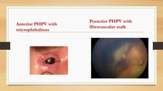
PERSISTENT
HYPERPLASTIC PRIMARY
VITREOUS
Dr Anupama Manoharan
1st year Diploma in Ophthalmology
Stanley Medical College and Hospital
DEFINITION
PHPV is a congenital anomaly of the eye that results following failure of
embryological primary vitreous and hyaloid vasculature to regress.
INTERNATIONAL CLASSIFICATION OF DISEASE
• ICD 9 743.51
• ICD 10 Q14.0
Synopses : Persistent tunica vasculosa lentis (PTVL)
Persistent posterior fetal fibrovascular sheath of lens .
Congenital retinal septum
Ablatio falciform congenita (AFC)
HISTORY
• PHPV in 1955 introduced by Mortan . F . Goldherg as a congenital
malformation of the anterior portion of the primary vitreous .
• Persistent fetal vasculature is now termed by Edward Jackson
memorial lecture A.B Reese in 1997 .
Classification
• Anterior PHPV- remnant vascular stalk is seen attached to back of lens causing traction in peripheral
retina , shallow AC, Poor pupil dilation and microphthalmos
• not extend to optic nerve .
( tunica vasculosa changes)
• Persistent posterior fetal fibrovascular sheath of lens .
• associated with cataract , glaucoma ,retrolenticular membrane .
• Posterior PHPV- remnant vascular stalk is seen arising the optic nerve from vitreous membrane
• not extend to lens. ( hyaloid artery changes)
• associated with abnormal development of retina( retinal fold , dysplasia , tractional retinal detachment ,
optic.N dysplasia , vitreal stalk , vitreal membrane, Congenital retinal septum ,total retinal detachment and
retrolental fibrous tissues suggestive of posterior PFV syndrome
• Vascularity through the patent blood vessel is sometimes detectable during the kinetic
examination .
• Anterior is less destructive than Posterior resulting better than surgical .
Anterior PHPV with
microphthalmos
Posterior PHPV with
fibrovascular stalk
Pathophysiology
• During embryonic development of eye the compartment between the retina
& crystalline lens contain vascular system (hyaloid artery ) that provides
nutrient are supposed to regress in the 3rd trimester .
• Failure of fetal vascular to regress over due to somatic mutation
• ATOH7 (autosomal recessive PHPV), and NDP (autosomal dominant
PHPV)
associated with pediatric retinal vitreopathies .
• OMIM 611308 (autosomal dominant PHPV) or OMIM 611311
(autosomal recessive PHPV).
• For normal vessel regression to occur, genes and growth factors
must be expressed at the precise timing and at the precise level,
balancing vaso-inhibitory and vaso-stimulatory factors, to allow
for apoptosis and macrophage activation.
• Some of these genes and factors that have been proposed in the
mechanism of PFV are the loss of Wnt7b signals in hyalocytes,
factors such as Angiopoietin 2 or p53 that may affect endothelial
cell apoptosis, deregulated vascular endothelial growth factor
(VEGF), and the absence of the Arf tumor suppressor
Leucocoria
Clinical feature
• Leucocoria ( white pupillary reflex due to dense retrolenticular membrane
or cataractous lens ).
• Vascularised plaque on the back of the lens & iris normal or small notches
at the pupillary margin
• Microphthalmos (small cornea & prominent ciliary process on the anterior
iris surface , pupillary margin ) .
• Occasionally intra vitreal haemorrhage , retinal detachment
• Visual acuity near normal
• Strabismus present at birth or develop shortly .
• Nystagmus
• PHPV unilateral
Syndrome associated
• Trisomy 13
• Norrie disease – express the most severe phenotype
• Walker Warburg syndrome
• Incontinentia pigmenti
• Cerebro-ocular-dysplasia –macular dystrophy
• Fetal alcohol syndrome
• Neurofibromatosis 2
• Axenfeld –Riager syndrome
• X linked familial exudative vitreoretinopathy , coats & ROP ( less phenotype )
Diagnosis
• USG , CT , RB B –Scan MRI , a cone shaped retrolental density is a
characteristic finding of PHPV on imaging studies
• Fluorescein angiography with slit lamp shows location of abnormal
vasculature the brittle star configuration .
• Peripheral retinal capillary non perfusion may also be noted in ROP
• Bcsan.
Differential diagnosis
• Leucocoria, the differential diagnosis of PFV, especially anterior
PFV,
• Retinoblastoma, the most common intraocular tumor of childhood.
• PFV occurs in a microphthalmia eye, while retinoblastoma occurs in
a normal-sized eye.
• Other causes of leukocoria are most commonly congenital cataract
or Coats’ Disease, astrocytic hamartomas (related to tuberous
sclerosis), uveitis, toxocariasis, and retinopathy of prematurity.
• PHPV is usually unilateral and less in full term infant
MEDICAL MANAGEMENT
• Visual rehabiltation ( aphakia contact lens & amblyopia therapy )
• With modern vitreoretinal techniques, aphakic rehabilitation, and
aggressive amblyopic therapy, useful vision can be obtained in
the majority of patients with both anterior and posterior
persistent fetal vasculature.
• Effective management of the anisometropic amblyopia requires
motivated patents to ensure compliance with occlusive therapy, as
well as parents willing to attend numerous visits to eye specialists.
Complication
• Untreated PFV leads to recurrent intraocular hemorrhage ,secondary
glaucoma, and phthisibulbi .
• Intractable glaucoma and the eventual phthisis bulbi have resulted in
enucleation of eyes with PFV.
• First, the media opacity in the affected eye should be cleared to allow visual
development and to save the eye from complications of untreated PFV, such
as glaucoma, intraocular hemorrhage, and phthisis.
• PFV eyes with myopia have less media opacity, normal corneal diameters,
are detected late and have good visual prognosis.
Surgical management
• Lensectomy with or without anterior or total vitrectomy
trabeculotomy .
• Strabismus surgery
Thank you

PERSISTENT HYPERPLASTIC PRIMARY VITREOUS

  • 1.
    PERSISTENT HYPERPLASTIC PRIMARY VITREOUS Dr AnupamaManoharan 1st year Diploma in Ophthalmology Stanley Medical College and Hospital
  • 2.
    DEFINITION PHPV is acongenital anomaly of the eye that results following failure of embryological primary vitreous and hyaloid vasculature to regress. INTERNATIONAL CLASSIFICATION OF DISEASE • ICD 9 743.51 • ICD 10 Q14.0 Synopses : Persistent tunica vasculosa lentis (PTVL) Persistent posterior fetal fibrovascular sheath of lens . Congenital retinal septum Ablatio falciform congenita (AFC)
  • 3.
    HISTORY • PHPV in1955 introduced by Mortan . F . Goldherg as a congenital malformation of the anterior portion of the primary vitreous . • Persistent fetal vasculature is now termed by Edward Jackson memorial lecture A.B Reese in 1997 .
  • 4.
    Classification • Anterior PHPV-remnant vascular stalk is seen attached to back of lens causing traction in peripheral retina , shallow AC, Poor pupil dilation and microphthalmos • not extend to optic nerve . ( tunica vasculosa changes) • Persistent posterior fetal fibrovascular sheath of lens . • associated with cataract , glaucoma ,retrolenticular membrane . • Posterior PHPV- remnant vascular stalk is seen arising the optic nerve from vitreous membrane • not extend to lens. ( hyaloid artery changes) • associated with abnormal development of retina( retinal fold , dysplasia , tractional retinal detachment , optic.N dysplasia , vitreal stalk , vitreal membrane, Congenital retinal septum ,total retinal detachment and retrolental fibrous tissues suggestive of posterior PFV syndrome • Vascularity through the patent blood vessel is sometimes detectable during the kinetic examination . • Anterior is less destructive than Posterior resulting better than surgical .
  • 5.
    Anterior PHPV with microphthalmos PosteriorPHPV with fibrovascular stalk
  • 6.
    Pathophysiology • During embryonicdevelopment of eye the compartment between the retina & crystalline lens contain vascular system (hyaloid artery ) that provides nutrient are supposed to regress in the 3rd trimester . • Failure of fetal vascular to regress over due to somatic mutation • ATOH7 (autosomal recessive PHPV), and NDP (autosomal dominant PHPV) associated with pediatric retinal vitreopathies .
  • 7.
    • OMIM 611308(autosomal dominant PHPV) or OMIM 611311 (autosomal recessive PHPV). • For normal vessel regression to occur, genes and growth factors must be expressed at the precise timing and at the precise level, balancing vaso-inhibitory and vaso-stimulatory factors, to allow for apoptosis and macrophage activation. • Some of these genes and factors that have been proposed in the mechanism of PFV are the loss of Wnt7b signals in hyalocytes, factors such as Angiopoietin 2 or p53 that may affect endothelial cell apoptosis, deregulated vascular endothelial growth factor (VEGF), and the absence of the Arf tumor suppressor
  • 8.
  • 9.
    Clinical feature • Leucocoria( white pupillary reflex due to dense retrolenticular membrane or cataractous lens ). • Vascularised plaque on the back of the lens & iris normal or small notches at the pupillary margin • Microphthalmos (small cornea & prominent ciliary process on the anterior iris surface , pupillary margin ) . • Occasionally intra vitreal haemorrhage , retinal detachment • Visual acuity near normal • Strabismus present at birth or develop shortly . • Nystagmus • PHPV unilateral
  • 10.
    Syndrome associated • Trisomy13 • Norrie disease – express the most severe phenotype • Walker Warburg syndrome • Incontinentia pigmenti • Cerebro-ocular-dysplasia –macular dystrophy • Fetal alcohol syndrome • Neurofibromatosis 2 • Axenfeld –Riager syndrome • X linked familial exudative vitreoretinopathy , coats & ROP ( less phenotype )
  • 11.
    Diagnosis • USG ,CT , RB B –Scan MRI , a cone shaped retrolental density is a characteristic finding of PHPV on imaging studies • Fluorescein angiography with slit lamp shows location of abnormal vasculature the brittle star configuration . • Peripheral retinal capillary non perfusion may also be noted in ROP • Bcsan.
  • 12.
    Differential diagnosis • Leucocoria,the differential diagnosis of PFV, especially anterior PFV, • Retinoblastoma, the most common intraocular tumor of childhood. • PFV occurs in a microphthalmia eye, while retinoblastoma occurs in a normal-sized eye. • Other causes of leukocoria are most commonly congenital cataract or Coats’ Disease, astrocytic hamartomas (related to tuberous sclerosis), uveitis, toxocariasis, and retinopathy of prematurity. • PHPV is usually unilateral and less in full term infant
  • 13.
    MEDICAL MANAGEMENT • Visualrehabiltation ( aphakia contact lens & amblyopia therapy ) • With modern vitreoretinal techniques, aphakic rehabilitation, and aggressive amblyopic therapy, useful vision can be obtained in the majority of patients with both anterior and posterior persistent fetal vasculature. • Effective management of the anisometropic amblyopia requires motivated patents to ensure compliance with occlusive therapy, as well as parents willing to attend numerous visits to eye specialists.
  • 14.
    Complication • Untreated PFVleads to recurrent intraocular hemorrhage ,secondary glaucoma, and phthisibulbi . • Intractable glaucoma and the eventual phthisis bulbi have resulted in enucleation of eyes with PFV. • First, the media opacity in the affected eye should be cleared to allow visual development and to save the eye from complications of untreated PFV, such as glaucoma, intraocular hemorrhage, and phthisis. • PFV eyes with myopia have less media opacity, normal corneal diameters, are detected late and have good visual prognosis.
  • 15.
    Surgical management • Lensectomywith or without anterior or total vitrectomy trabeculotomy . • Strabismus surgery
  • 16.