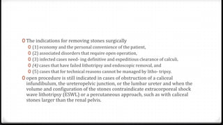
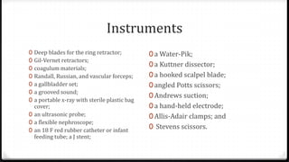
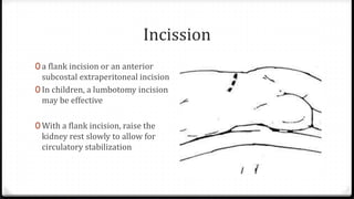
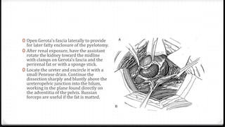
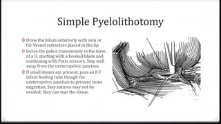
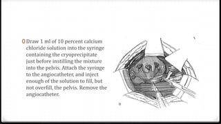
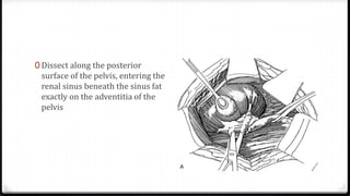
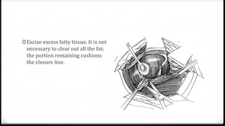
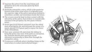
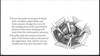
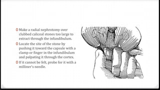
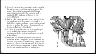
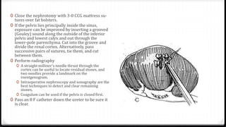
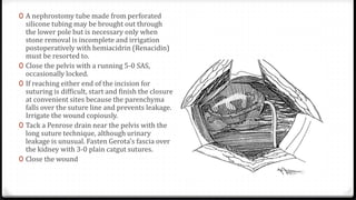
The document describes surgical procedures for removing kidney stones. It indicates that open surgery is still needed in cases of obstruction, infection, failed lithotripsy, or stones too large for other procedures. It then provides details on instruments, incisions, and techniques for simple pyelolithotomy, coagulum technique, extended pyelolithotomy, and managing stones extending into the ureteropelvic junction. The goal is to remove all stones and debris while minimizing damage to the kidney and ensuring the pelvis can be closed watertight.
Dr Ho Siew Hong shared his experience on how to perform the ideal puncture for PCNL in a lecture to Asian urologists during the Advanced Urology Course 2008 in Singapore
TURP step by step operative urology series
for more resources:
www.uronotes2012.blogspot.com
enter your mail & press follow us by mail to receive our daily feeds
Whipple's procedure - Indications, Steps, ComplicationsVikas V
Whipple's Procedure - Explaining the History of Whipple's Procedure, Indications, Contraindications, Step wise detailed procedure, Complications, Perioperative Management.
The Presentation Includes Detailed Step wise approach to the procedure assisted with Pictorial Representation of The steps
OPEN INGUINAL HERNIA REPAIR- OPERATIVE SURGERY
#surgicaleducator #operativesurgery #openinguinalherniarepair #usmle #babysurgeon #surgicaltutor
Dear viewers,
• Greetings from “Surgical Educator”
• Because of the popular demand by viewers of the YouTube channel “Surgical Educator”, I have decided to create and upload videos on common surgeries.
• I have already uploaded videos on open and Laparoscopic Appendicectomy, Thyroidectomy, Modified Radical Mastectomy and open and
Laparoscopic Cholecystectomy
• In this video today, I have discussed Open Inguinal Hernia Repair.
• However, these videos are not real surgeries but the theoretical aspect of operative surgery like going through an atlas of operative surgery.
• Along with these videos, I recommend you to watch real operative surgery videos as well and I will give a link for each surgery in the end of the video as end-cards, which I think will be very useful.
• This will give a very good opportunity for the surgical trainees to mentally rehearse various surgical steps in a sequential manner prior to actual surgery. You can watch the video in the following links:
• surgicaleducator.blogspot.com
• youtube.com/c/surgicaleducator
• Thank you for watching the videos.
HEMORRHOIDECTOMY- OPERATIVE SURGERY
#surgicaleducator #operativesurgery #hemorrhoidectomy #usmle #babysurgeon #surgicaltutor
Subscription Link: http://youtube.com/c/surgicaleducator...
Surgical Educator Android App link: https://play.google.com/store/apps/de...
Dear viewers,
• Greetings from “Surgical Educator”
• Because of the popular demand by viewers of the YouTube channel “Surgical Educator”, I have decided to create and upload videos on common surgeries.
• In this video today, I have discussed Hemorrhoidectomy- Barron’s banding, open and closed hemorrhoidectomy, Stapler hemorrhoidectomy and THD- Transanal Hemorroidal Dearterialisation. So, it is a 4in1 video.
• However, these videos are not real surgeries but the theoretical aspect of operative surgery like going through an atlas of operative surgery.
• Along with these videos, I recommend you watch real operative surgery videos as well and I will give a link for each surgery in the end of the video as end-cards, which I think will be very useful.
• This will give a very good opportunity for the surgical trainees to mentally rehearse various surgical steps in a sequential manner prior to actual surgery. You can watch the video in the following links:
• surgicaleducator.blogspot.com
• youtube.com/c/surgicaleducator
• Thank you for watching the videos.
LAPAROSCOPIC CHOLECYSTECTOMY- OPERATIVE SURGERY
#surgicaleducator #operativesurgery #laparoscopiccholecystectomy #usmle #babysurgeon #surgicaltutor
Dear viewers,
• Greetings from “Surgical Educator”
• Because of the popular demand by viewers of the YouTube channel “Surgical Educator”, I have decided to create and upload videos on common surgeries.
• I have already uploaded videos on open and Laparoscopic Appendicectomy, Thyroidectomy, Modified Radical Mastectomy and Open Cholecystectomy.
• In this video today, I have discussed Laparoscopic Cholecystectomy- the flagship procedure for laparoscopic surgeries.
• However, these videos are not real surgeries but the theoretical aspect of operative surgery like going through an atlas of operative surgery.
• Along with these videos, I recommend you to watch real operative surgery videos as well and the links are:
• https://www.youtube.com/watch?v=VStEzI1jL8Y
• https://www.youtube.com/watch?v=O8j4kwpzd24
• This will give a very good opportunity for the surgical trainees to mentally rehearse various surgical steps in a sequential manner prior to actual surgery. You can watch the video in the following links:
• surgicaleducator.blogspot.com
• youtube.com/c/surgicaleducator
The surgical treatment of an injury or defect within the urethra's walls is known as urethroplasty. The three most frequent factors leading to urethral damage that needs to be repaired are trauma, iatrogenic injury, and infections. The gold standard treatment for urethral strictures is urethroplasty, which has a lower recurrence rate than dilatations and urethrotomies. Although recurrence rates are higher for this challenging treatment group, it is likely the only effective treatment option for chronic and severe strictures.
Urethroplasty is not regarded as a small procedure, taking three to eight hours on average in the operating room. Between 20% and 30% of urethroplasty patients may benefit from the ease of going under the knife for a shorter period of time and going home the same day. On average, hospital stays last two to three days. Seven to ten days may be needed for hospitalization for more complicated surgeries.
Fewer than ten percent of patients experience significant complications after urethroplasty, while complications, particularly recurrences, are more frequent in long and complex strictures.
Retrograde Intrarenal Ureteroscopic Surgery (RIRS)Urovideo.org
Gerhard J. Fuchs, M.D., Dr. med., F.A.C.S.
Professor of Urology, UCLA School of Medicine
Vice Chair, Cedars Sinai Department of Surgery
Medallion Chair in Minimally Invasive Urology
Cedars-Sinai Medical Center
Los Angeles, USA
brief description of renal/ urinary system surgery
including: pyelolithotomy, nephrectomy, nephron-sparing surgery, Ureterotomy, Cystotomy, Nephrolithotomy
including surgical requirement for renal system surgery
In this presentation, I have shown how to do open anterior resection both high and low varieties in a step-by-step manner with clear pictures as if reading an atlas of operative surgery.
MANAGEMENT OF ATRIOVENTRICULAR CONDUCTION BLOCK.pdfJim Jacob Roy
Cardiac conduction defects can occur due to various causes.
Atrioventricular conduction blocks ( AV blocks ) are classified into 3 types.
This document describes the acute management of AV block.
TEST BANK for Operations Management, 14th Edition by William J. Stevenson, Ve...kevinkariuki227
TEST BANK for Operations Management, 14th Edition by William J. Stevenson, Verified Chapters 1 - 19, Complete Newest Version.pdf
Lung Cancer: Artificial Intelligence, Synergetics, Complex System Analysis, S...Oleg Kshivets
RESULTS: Overall life span (LS) was 2252.1±1742.5 days and cumulative 5-year survival (5YS) reached 73.2%, 10 years – 64.8%, 20 years – 42.5%. 513 LCP lived more than 5 years (LS=3124.6±1525.6 days), 148 LCP – more than 10 years (LS=5054.4±1504.1 days).199 LCP died because of LC (LS=562.7±374.5 days). 5YS of LCP after bi/lobectomies was significantly superior in comparison with LCP after pneumonectomies (78.1% vs.63.7%, P=0.00001 by log-rank test). AT significantly improved 5YS (66.3% vs. 34.8%) (P=0.00000 by log-rank test) only for LCP with N1-2. Cox modeling displayed that 5YS of LCP significantly depended on: phase transition (PT) early-invasive LC in terms of synergetics, PT N0—N12, cell ratio factors (ratio between cancer cells- CC and blood cells subpopulations), G1-3, histology, glucose, AT, blood cell circuit, prothrombin index, heparin tolerance, recalcification time (P=0.000-0.038). Neural networks, genetic algorithm selection and bootstrap simulation revealed relationships between 5YS and PT early-invasive LC (rank=1), PT N0—N12 (rank=2), thrombocytes/CC (3), erythrocytes/CC (4), eosinophils/CC (5), healthy cells/CC (6), lymphocytes/CC (7), segmented neutrophils/CC (8), stick neutrophils/CC (9), monocytes/CC (10); leucocytes/CC (11). Correct prediction of 5YS was 100% by neural networks computing (area under ROC curve=1.0; error=0.0).
CONCLUSIONS: 5YS of LCP after radical procedures significantly depended on: 1) PT early-invasive cancer; 2) PT N0--N12; 3) cell ratio factors; 4) blood cell circuit; 5) biochemical factors; 6) hemostasis system; 7) AT; 8) LC characteristics; 9) LC cell dynamics; 10) surgery type: lobectomy/pneumonectomy; 11) anthropometric data. Optimal diagnosis and treatment strategies for LC are: 1) screening and early detection of LC; 2) availability of experienced thoracic surgeons because of complexity of radical procedures; 3) aggressive en block surgery and adequate lymph node dissection for completeness; 4) precise prediction; 5) adjuvant chemoimmunoradiotherapy for LCP with unfavorable prognosis.
Anti ulcer drugs and their Advance pharmacology ||
Anti-ulcer drugs are medications used to prevent and treat ulcers in the stomach and upper part of the small intestine (duodenal ulcers). These ulcers are often caused by an imbalance between stomach acid and the mucosal lining, which protects the stomach lining.
||Scope: Overview of various classes of anti-ulcer drugs, their mechanisms of action, indications, side effects, and clinical considerations.
ARTIFICIAL INTELLIGENCE IN HEALTHCARE.pdfAnujkumaranit
Artificial intelligence (AI) refers to the simulation of human intelligence processes by machines, especially computer systems. It encompasses tasks such as learning, reasoning, problem-solving, perception, and language understanding. AI technologies are revolutionizing various fields, from healthcare to finance, by enabling machines to perform tasks that typically require human intelligence.
Tom Selleck Health: A Comprehensive Look at the Iconic Actor’s Wellness Journeygreendigital
Tom Selleck, an enduring figure in Hollywood. has captivated audiences for decades with his rugged charm, iconic moustache. and memorable roles in television and film. From his breakout role as Thomas Magnum in Magnum P.I. to his current portrayal of Frank Reagan in Blue Bloods. Selleck's career has spanned over 50 years. But beyond his professional achievements. fans have often been curious about Tom Selleck Health. especially as he has aged in the public eye.
Follow us on: Pinterest
Introduction
Many have been interested in Tom Selleck health. not only because of his enduring presence on screen but also because of the challenges. and lifestyle choices he has faced and made over the years. This article delves into the various aspects of Tom Selleck health. exploring his fitness regimen, diet, mental health. and the challenges he has encountered as he ages. We'll look at how he maintains his well-being. the health issues he has faced, and his approach to ageing .
Early Life and Career
Childhood and Athletic Beginnings
Tom Selleck was born on January 29, 1945, in Detroit, Michigan, and grew up in Sherman Oaks, California. From an early age, he was involved in sports, particularly basketball. which played a significant role in his physical development. His athletic pursuits continued into college. where he attended the University of Southern California (USC) on a basketball scholarship. This early involvement in sports laid a strong foundation for his physical health and disciplined lifestyle.
Transition to Acting
Selleck's transition from an athlete to an actor came with its physical demands. His first significant role in "Magnum P.I." required him to perform various stunts and maintain a fit appearance. This role, which he played from 1980 to 1988. necessitated a rigorous fitness routine to meet the show's demands. setting the stage for his long-term commitment to health and wellness.
Fitness Regimen
Workout Routine
Tom Selleck health and fitness regimen has evolved. adapting to his changing roles and age. During his "Magnum, P.I." days. Selleck's workouts were intense and focused on building and maintaining muscle mass. His routine included weightlifting, cardiovascular exercises. and specific training for the stunts he performed on the show.
Selleck adjusted his fitness routine as he aged to suit his body's needs. Today, his workouts focus on maintaining flexibility, strength, and cardiovascular health. He incorporates low-impact exercises such as swimming, walking, and light weightlifting. This balanced approach helps him stay fit without putting undue strain on his joints and muscles.
Importance of Flexibility and Mobility
In recent years, Selleck has emphasized the importance of flexibility and mobility in his fitness regimen. Understanding the natural decline in muscle mass and joint flexibility with age. he includes stretching and yoga in his routine. These practices help prevent injuries, improve posture, and maintain mobilit
The prostate is an exocrine gland of the male mammalian reproductive system
It is a walnut-sized gland that forms part of the male reproductive system and is located in front of the rectum and just below the urinary bladder
Function is to store and secrete a clear, slightly alkaline fluid that constitutes 10-30% of the volume of the seminal fluid that along with the spermatozoa, constitutes semen
A healthy human prostate measures (4cm-vertical, by 3cm-horizontal, 2cm ant-post ).
It surrounds the urethra just below the urinary bladder. It has anterior, median, posterior and two lateral lobes
It’s work is regulated by androgens which are responsible for male sex characteristics
Generalised disease of the prostate due to hormonal derangement which leads to non malignant enlargement of the gland (increase in the number of epithelial cells and stromal tissue)to cause compression of the urethra leading to symptoms (LUTS
New Directions in Targeted Therapeutic Approaches for Older Adults With Mantl...i3 Health
i3 Health is pleased to make the speaker slides from this activity available for use as a non-accredited self-study or teaching resource.
This slide deck presented by Dr. Kami Maddocks, Professor-Clinical in the Division of Hematology and
Associate Division Director for Ambulatory Operations
The Ohio State University Comprehensive Cancer Center, will provide insight into new directions in targeted therapeutic approaches for older adults with mantle cell lymphoma.
STATEMENT OF NEED
Mantle cell lymphoma (MCL) is a rare, aggressive B-cell non-Hodgkin lymphoma (NHL) accounting for 5% to 7% of all lymphomas. Its prognosis ranges from indolent disease that does not require treatment for years to very aggressive disease, which is associated with poor survival (Silkenstedt et al, 2021). Typically, MCL is diagnosed at advanced stage and in older patients who cannot tolerate intensive therapy (NCCN, 2022). Although recent advances have slightly increased remission rates, recurrence and relapse remain very common, leading to a median overall survival between 3 and 6 years (LLS, 2021). Though there are several effective options, progress is still needed towards establishing an accepted frontline approach for MCL (Castellino et al, 2022). Treatment selection and management of MCL are complicated by the heterogeneity of prognosis, advanced age and comorbidities of patients, and lack of an established standard approach for treatment, making it vital that clinicians be familiar with the latest research and advances in this area. In this activity chaired by Michael Wang, MD, Professor in the Department of Lymphoma & Myeloma at MD Anderson Cancer Center, expert faculty will discuss prognostic factors informing treatment, the promising results of recent trials in new therapeutic approaches, and the implications of treatment resistance in therapeutic selection for MCL.
Target Audience
Hematology/oncology fellows, attending faculty, and other health care professionals involved in the treatment of patients with mantle cell lymphoma (MCL).
Learning Objectives
1.) Identify clinical and biological prognostic factors that can guide treatment decision making for older adults with MCL
2.) Evaluate emerging data on targeted therapeutic approaches for treatment-naive and relapsed/refractory MCL and their applicability to older adults
3.) Assess mechanisms of resistance to targeted therapies for MCL and their implications for treatment selection
- Video recording of this lecture in English language: https://youtu.be/lK81BzxMqdo
- Video recording of this lecture in Arabic language: https://youtu.be/Ve4P0COk9OI
- Link to download the book free: https://nephrotube.blogspot.com/p/nephrotube-nephrology-books.html
- Link to NephroTube website: www.NephroTube.com
- Link to NephroTube social media accounts: https://nephrotube.blogspot.com/p/join-nephrotube-on-social-media.html
2. 0 The indications for removing stones surgically
0 (1) economy and the personal convenience of the patient,
0 (2) associated disorders that require open operation,
0 (3) infected cases need- ing definitive and expeditious clearance of calculi,
0 (4) cases that have failed lithotripsy and endoscopic removal, and
0 (5) cases that for technical reasons cannot be managed by litho- tripsy.
0 open procedure is still indicated in cases of obstruction of a caliceal
infundibulum, the ureteropelvic junction, or the lumbar ureter and when the
volume and configuration of the stones contraindicate extracorporeal shock
wave lithotripsy (ESWL) or a percutaneous approach, such as with caliceal
stones larger than the renal pelvis.
3. Instruments
0 Deep blades for the ring retractor;
0 Gil-Vernet retractors;
0 coagulum materials;
0 Randall, Russian, and vascular forceps;
0 a gallbladder set;
0 a grooved sound;
0 a portable x-ray with sterile plastic bag
cover;
0 an ultrasonic probe;
0 a flexible nephroscope;
0 an 18 F red rubber catheter or infant
feeding tube; a J stent;
0 a Water-Pik;
0 a Kuttner dissector;
0 a hooked scalpel blade;
0 angled Potts scissors;
0 Andrews suction;
0 a hand-held electrode;
0 Allis-Adair clamps; and
0 Stevens scissors.
4. Incission
0 a flank incision or an anterior
subcostal extraperitoneal incision
0 In children, a lumbotomy incision
may be effective
0 With a flank incision, raise the
kidney rest slowly to allow for
circulatory stabilization
sharply and bluntly above the ureteropelvic junction i
the hilum, working in the plane found directly on
adventitia of the pelvis. Russian forceps are useful if
fat is matted.
5. 0 Open Gerota's fascia laterally to provide
for later fatty enclosure of the pyelotomy.
0 After renal exposure, have the assistant
rotate the kidney toward the midline
with clamps on Gerota's fascia and the
perirenal fat or with a sponge stick.
0 Locate the ureter and encircle it with a
small Penrose drain. Continue the
dissection sharply and bluntly above the
ureteropelvic junction into the hilum,
working in the plane found directly on
the adventitia of the pelvis. Russian
forceps are useful if the fat is matted.
6. Simple Pyelolithotomy
0 Draw the hilum anteriorly with vein or
Gil-Vernet retractors placed in the lip
0 Incise the pelvis transversely in the form
of a U, starting with a hooked blade and
continuing with Potts scissors. Stay well
away from the ureteropelvic junction.
0 If small stones are present, pass an 8 F
infant feeding tube though the
ureteropelvic junction to prevent stone
migration. Stay sutures may not be
needed; they can tear the tissue.
slowly to allow for circulatory stabilization.
n Gerota's fascia laterally to provide for later
sure of the pyelotomy. After renal exposure,
ssistant rotate the kidney toward the midline
ps on Gerota's fascia and the perirenal fat or
onge stick. Locate the ureter and encircle it
mall Penrose drain. Continue the dissection
d bluntly above the ureteropelvic junction into
working in the plane found directly on the
of the pelvis. Russian forceps are useful if the
ed.
pelvis transversely in the form of a U, starting with a
hooked blade and continuing with Potts scissors. Stay
well away from the ureteropelvic junction. If small stones
are present, pass an 8 F infant feeding tube though the
ureteropelvic junction to prevent stone migration. Stay
sutures may not be needed; they can tear the tissue.
7. 0 Withdraw the stones with forceps or
a Mixter clamp. If a large stone
adheres to the pelvic wall, free it by
passing a probe around it.
0 Irrigate the interior with water
through a cut-off Robinson catheter.
Use a Water-Pik.
0 Insert a flexible nephroscope if
concern remains about residual
adherent stones. Alternatively, close
the pelvis and inject coagulum (Step
4).
8. Coagulum Technique
0 Obtain two bags of thawed
cryoprecipitate (about 15 ml each),
and keep them at room temperature.
Add a few drops of methylene blue to
them in a pan. Draw the
cryoprecipitate into the 35-ml
syringe.
0 Obstruct the ureter by placing
traction on the encircling Penrose
drain. Insert an angiocatheter into
the renal pelvis, withdraw the stylet,
and drain the urine, estimating its
volume.
9. 0 Draw 1 ml of 10 percent calcium
chloride solution into the syringe
containing the cryoprecipitate
just before instilling the mixture
into the pelvis. Attach the syringe
to the angiocatheter, and inject
enough of the solution to fill, but
not overfill, the pelvis. Remove the
angiocatheter.
10. 0 Wait 5 minutes; then open the
pelvis with a U- shaped incision,
and gingerly extract the coagulum
with the stone. Sometimes
pressure on the kidney
parenchyma helps extraction.
After removing the clot, flush the
ureter with saline through the 8 F
infant feeding tube.
11. 0 Inspect the coagulum to be
certain it is intact. Thoroughly
irrigate the pelvis and ureter.
12. 0 It may be worth-while before closure to
pass a ureteral catheter or infant feeding
tube to the bladder to be sure that no
fragments are caught in the ureter, which
would promote prolonged postoperative
drainage.
0 Make a watertight closure of the pelvis
with a running 4-0 or 5-0 SAS with an
occasional lock stitch.
0 Suture a Penrose drain by the long suture
technique near the closure, being sure its
end does not touch the anastomosis.
0 Tack the edges of Gerota's fascia
together, and close the wound.
13. Extended Pyelolithotomy (Gil-Vernet)
0 Alternatives are
0 anatrophic nephrolithotomy
0 partial nephrectomy
0 Contraindications to this
intrasinusal approach
0 previous extended pyelolithotomy,
0 extremely intrarenal pelvis,
0 staghorn calculi in clubbed
calyces.
0 Expose the kidney as for simple
pyelolithotomy.
0 Proceed with complete mobilization
of the kidney to allow control of the
renal artery and to facilitate
roentgenography.
0 Have the assistant rotate the kidney
toward the midline.
0 Feel for the arterial pulsation, and
expose the renal artery. Draw a sling
around it with a right-angle clamp.
0 Try applying a bulldog clamp on it for
size and clearance.
14. 0 Dissect along the posterior
surface of the pelvis, entering the
renal sinus beneath the sinus fat
exactly on the adventitia of the
pelvis
15. 0 Excise excess fatty tissue. It is not
necessary to clear out all the fat;
the portion remaining cushions
the closure line.
16. 0 Separate the pelvis from the renal hilum and
peripelvic fat in the avascular plane by blunt
dissection.
0 Avoid the retropelvic artery, which is the posterior
branch of the main renal artery. It originates near
the superior edge of the pelvis and passes behind it,
sometimes outside and sometimes inside the hilum.
0 The scissors must be kept in close contact with the
adventitia of the pelvis. Even if there is considerable
reaction in the peripelvic fat, this plane remains
intact.
0 Insert special Gil-Vernet retractors over the whole
mass of peripelvic fat, and insinuate the corner of a
moist, opened 4 X 8 gauze pad to expose the bases
of the infundibula.
0 Have your assistant lift and rotate the kidney to
bring the pelvis into view. If the pelvis is extrarenal,
the assistant should relax pressure on the
retractors occasionally to allow flow through the
retropelvic artery.
0 If exposure is difficult, place a bulldog clamp on the
renal artery to reduce parenchymal turgor.
1 0 4 4 KIDNEY: EXCISION
Separate the pelvis from the renal hilum and peripel-
vic fat in the avascular plane by blunt dissection.
Avoid the retropelvic artery, which is the posterior
branch of the main renal artery. It originates near the
superior edge of the pelvis and passes behind it, some-
times outside and sometimes inside the hilum. The scis-
sors must be kept in close contact with the adventitia of
the pelvis. Even if there is considerable reaction in the
peripelvic fat, this plane remains intact. Insert special Gil-
Vernet retractors over the whole mass of peripelvic fat,
and insinuate the corner of a moist, opened 4 X 8 gauze
pad to expose the bases of the infundibula. Have your
assistant lift and rotate the kidney to bring the pelvis into
view. If the pelvis is extrarenal, the assistant should relax
pressure on the retractors occasionally to allow flow
through the retropelvic artery. If exposure is difficult,
place a bulldog clamp on the renal artery to reduce
parenchymal turgor.
7
17. 0 Incise the pelvis in an open U shape
with a hooked scalpel blade and
Potts scissors. Design the cut to fit
the configuration of the periureteral
portion of the stone, keeping well
away from the ureteropelvic junction.
0 Usually make the incision from the
base of the lowest calyx to the base of
the uppermost. Stay sutures are not
needed and may tear the pelvic wall.
place a bulldog clamp on the renal artery to reduce
parenchymal turgor.
Incise the pelvis in an open U shape with a hooked
scalpel blade and Potts scissors. Design the cut to
fit the configuration of the periureteral portion of the
stone, keeping well away from the ureteropelvic junction.
Usually make the incision from the base of the lowest
calyx to the base of the uppermost. Stay sutures are not
needed and may tear the pelvic wall.
A, First wipe around the extension of the stone in
the ureteropelvic junction with a blunt probe to
free it from the pelvic epithelium.
B, Lever the periureteral extension out first, thereby
8
9
18. 0 First wipe around the extension of
the stone in the ureteropelvic
junction with a blunt probe to free
it from the pelvic epithelium.
A, First wipe around the extension of the stone in
the ureteropelvic junction with a blunt probe to
free it from the pelvic epithelium.
B, Lever the periureteral extension out first, thereby
exposing as much as 70 percent of the stone.
9
19. 0 Lever the periureteral extension
out first, thereby exposing as
much as 70 percent of the stone.
A, First wipe around the extension of the stone in
the ureteropelvic junction with a blunt probe to
free it from the pelvic epithelium.
B, Lever the periureteral extension out first, thereby
exposing as much as 70 percent of the stone.
9
20. 0 Grasp the stone with Randall forceps. Gently
rock and rotate it to extract its caliceal
extensions.
0 Extricate the shortest branch first.
0 If absolutely necessary, fracture the neck of one
or more of the branches and remove the
clubbed ends via transverse nephrotomies.
0 Often an infundibulum can be dilated with
forceps sufficiently to allow an extension of the
main stone to be extracted.
0 If the renal hilum is large enough, a vertical
incision along the involved infundibulum (cali-
cotomy) may assist in the removal of large
caliceal stones.
0 Remove the stone and fit the pieces together to
be sure all were retrieved.
0 Send the stone for culture and analysis.
21. 0 Inspect the interior of the calyces,
using a flexible nephroscope if
necessary, and remove any remaining
calculi, usually with stone forceps or
Mixter clamp.
0 If the stones are too large to pass
through an infundibulum, gently
dilate the opening with a clamp.
0 Try not to use a finger or high
pressure. Irrigate each calyx in turn,
using a large syringe and a cut-off 18
F red rubber catheter.
22. 0 Make a radial nephrotomy over
clubbed caliceal stones too large to
extract through the infundibulum.
0 Locate the site of the stone by
pushing it toward the capsule with a
clamp or finger in the infundibulum
and palpating it through the cortex.
0 If it cannot be felt, probe for it with a
milliner's needle.
23. 0 Sharply incise the capsule circumferentially
for a distance equal to the diameter of the
stone; then bluntly separate the kidney
parenchyma down to the stone, which is
supported by a clamp or finger in the
infundibulum.
0 Extract the stone with forceps inserted into
the nephrotomy. If the cortex is thick, it is
helpful to place a bulldog clamp on the renal
artery to soften the kidney long enough to
locate and remove the stone.
0 If these manipulations are to be prolonged,
cool the kidney and give mannitol
intravenously. Irrigate the calyx thoroughly
with saline.
0 Avulsion of the ureteropelvic junction is
possible. With a segment made ischemic by
chronic impaction of a relatively large stone,
avulsion can occur during dissection. Repair
and intubation are necessary), even though
the tissue has the quality of wet paper.
24. 0 Close the nephrotomy with 3-0 CCG mattress su-
tures over fat bolsters.
0 If the pelvis lies principally inside the sinus,
exposure can be improved by inserting a grooved
(Gouley) sound along the outside of the inferior
pelvis and lowest calyx and out through the
lower-pole parenchyma. Cut into the groove and
divide the renal cortex. Alternatively, pass
successive pairs of sutures, tie them, and cut
between them.
0 Perform radiography
0 A straight milliner's needle thrust through the
cortex can be useful to locate residual stones, and
two needles provide a landmark on the
roentgenogram.
0 Intraoperative nephroscopy and sonography are the
best techniques to detect and clear remaining
stones.
0 Coagulum can be used if the pelvis is closed first.
0 Pass an 8 F catheter down the ureter to be sure it
is clear.
25. 0 A nephrostomy tube made from perforated
silicone tubing may be brought out through
the lower pole but is necessary only when
stone removal is incomplete and irrigation
postoperatively with hemiacidrin (Renacidin)
must be resorted to.
0 Close the pelvis with a running 5-0 SAS,
occasionally locked.
0 If reaching either end of the incision for
suturing is difficult, start and finish the closure
at convenient sites because the parenchyma
falls over the suture line and prevents leakage.
Irrigate the wound copiously.
0 Tack a Penrose drain near the pelvis with the
long suture technique, although urinary
leakage is unusual. Fasten Gerota's fascia over
the kidney with 3-0 plain catgut sutures.
0 Close the wound
At a secondary operation, the kidney is found firmly attached
to the transversalis fascia and is readily entered inadvertently.
Identify the capsule early, and dissect it carefully from the
fibrous bed. Enter the sinus anteriorly and inferiorly, at a site-
distant from that for the initial pyelolithotomy.
A nephrostomy tube made from perforated sili-
cone tubing may be brought out through the
lower pole but is necessary only when stone removal is
incomplete and irrigation postoperatively with hemiaci-
drin (Renacidin) must be resorted to.
Close the pelvis with a running 5-0 SAS, occasionally
locked. If reaching either end of the incision for suturing
is difficult, start and finish the closure at convenient sites
because the parenchyma falls over the suture line and
prevents leakage. Irrigate the wound copiously. Tack a
Penrose drain near the pelvis with the long suture tech-
nique (see page 917), although urinary leakage is un-
usual. Fasten Gerota's fascia over the kidney with 3-0
plain catgut sutures. Close the wound.
13
26. 0 At a secondary operation, the
kidney is found firmly attached to
the transversalis fascia and is
readily entered inadvertently.
Identify the capsule early, and
dissect it carefully from the
fibrous bed. Enter the sinus
anteriorly and inferiorly, at a site-
distant from that for the initial
pyelolithotomy.