Download to read offline
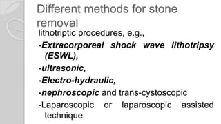
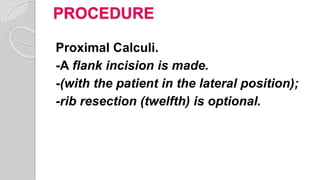


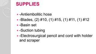
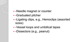
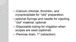
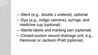


This document provides information on ureterolithotomy, a surgical procedure to remove stones from the ureter or kidney. It defines the procedure, describes different methods for stone removal including shock wave lithotripsy and various surgical techniques. It outlines the preparation of the patient, positioning, skin preparation, draping, instrumentation, supplies, and special notes for the procedure.


![Urolithiasis presented by Dr Muhammad nouman([Autosaved]-1.pptx](https://cdn.slidesharecdn.com/ss_thumbnails/urolithiasisautosaved-1-250603143258-c924c676-thumbnail.jpg?width=640&height=640&fit=bounds)


