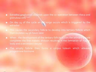
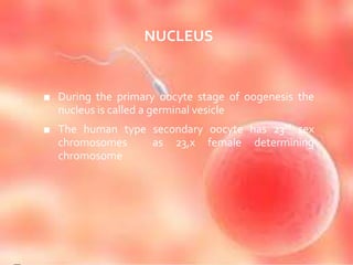
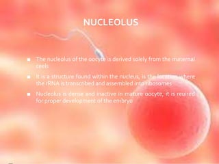
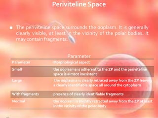
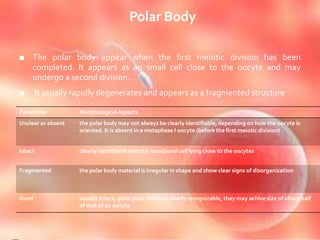
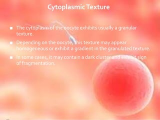
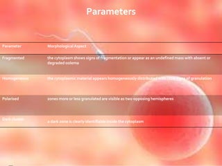
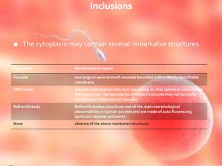
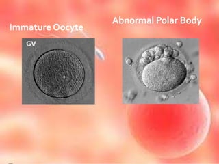
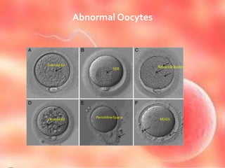
OOCYTE
LIPIKA PATTANAIK
EMBRYOLOGIST
FEMELIFE FERTILITY
www.femelife.com
www.wikiHealthNews.com
■ An oocyte is a female gametocyte or germ cell involved in
reproduction
■ It is an immature ovum or an egg
■ An oocyte is produced in the ovary during female
gametogenesis
■ The female germ cells produce a primordial germ cell pgc
which undergoes mitosis to form an oogonium
■ During oogenesis the oogonium becomes a primary
oocyte
■ It is the creation of an ovum
■ It is the female form of gametogenesis and involves the development of
various stages of the immature ovum
■ In mammals the first part of oogensis starts in germinal epithelium,
which gives rise to the development of ovarian follicles, the functional
unit of the ovary
■ Oogenesis consists of several sub processes – oocytogenesis,
ootidogenesis and the maturation to form an ovum
■ Folliculogenesis is a separate sub process and supports all three
oogenetic sub process
■ Oogenesis starts with the process of developing oogonia,which occurs
via the transformation of primordial follicles into primary oocytes, a
process called oocytogenesis
■ Oocytogenesis is complete either before or shortly after birth
NUMBER OF PRIMARY OOCYTES
■ Oocytogenesis is complete
■ Primary oocytes reach their max. development at 20
weeks of gestational age,when approx 7 million
primary oocytes have been created,this no. will be
approx. reduced to 1-2 million
OOTIDOGENESIS
■ The succeeding phase of ootidogenesis occurs when
the primary oocyte develops into an ootid.
■ This is achieved by the process of meiosis
■ Although this process begins at pre natal age, it
stops at prophase I.
■ All oocytes ,still primary oocytes, have halted at this
stage of development,called dictyate
■ After menarche these cells then continue to develop
MEIOSIS I
■ Meiosis I of ootidogenesis begins during embryonic
development ,but halts in the diplotene stage of
prophase I until puberty
■ For those primary oocytes that continue to develop
in each menstrual cycle synapsis occurs and tetrads
form enabling chromosomal cross over to occur
■ As a result of meiosis I the primary oocyte develops
into the secondary oocyte and the first polar body
MEIOSIS II
■ Immediately after meiosis I ,the haploid secondary oocyte
initiates meiosisII.
■ This process is also halted at the mataphase II stage until
fertilization
■ When meiosisII has completed ,an ootid and another polar
body have now been created
MATURATION INTO OVUM
■ Both polar bodies disintegrate at the end of meiosis
II,leaving only the ootid,which then undergoes maturation
into mature ovum
■ The function of forming polar bodies is to discard the extra
haploid sets of chromosomes
FOLLICULOGENESIS
■ It is the maturation of ovarian follicle,a densely packed
shell of somatic cells that contains an immature oocyte
■ This describes the progression of number of small
primordial follicles into large pre ovulatory follicles that
enter the menstrual cycle
■ Folliculogenesis ends when the remaining follicles in the
ovaries are incapable of responding to the hormones that
had recruited some of the follicles to mature
■ This depletion of follicle supply signals the beginning of
menopause
Female Gametogenesis
HUMAN OOGENESIS
■ At the start of the menstrual cycle some 12-20
primary follicles begin to develop under the
influence of elevated FSH to form secondary
follicles.
■ By around day 9 of the cycle only one healthy
secondary follicle remains with the rest having
undergone ovarian follicle atrasia
■ The remaining follicle is called the dominant follicle
and is responsible for producing large amount of
estradiol during the late follicular phase.
■ Estradiol production depends upon the co operation between theca and
granulosa cells
■ On day 14 of the cycle an LH surge occurs which is triggered by the
estradiol
■ This causes the secondary follicle to develop into tertiary follicle which
then ovulates 24-36 hours later
■ When the development of the tertiary follicle occurs the primary oocyte
completes the first meiotic division resulting in the formation of a polar
body and a secondary oocyte
■ The empty follicle then forms a corpus luteum which releases
progestrone hormone
CHARACTERISTICS
■ CYTOPLASM
■ Oocytes are rich in cytoplasm which contains yolk
granules to nourish the cell in early development
NUCLEUS
■ During the primary oocyte stage of oogenesis the
nucleus is called a germinal vesicle
■ The human type secondary oocyte has 23rd sex
chromosomes as 23,x female determining
chromosome
NEST
• The space where an ovum or immature ovum is
located is the cell nest
MITOCHONDRIA
■ The oocyte receives mitochondria from maternal cells,which
will go on to control embryonic metabolism
■ The partitioning of mitochondria is carried out by a system of
microtubules which will localize mitochondria throughout the
oocyte
NUCLEOLUS
■ The nucleolus of the oocyte is derived solely from the maternal
ceels
■ It is a structure found within the nucleus, is the location where
the rRNA is transcribed and assembled into ribosomes
■ Nucleolus is dense and inactive in mature oocyte, it is reuired
for proper development of the embryo
RIBOSOMES
■ Maternal cells also synthesise and contribute a store
of ribosomes that are reuired for the translation of
proteins before the zygotic genomes is activated
■ Maternally derived ribosomes and some mRNAs are
stored in a structure called cytoplasmic lattices
■ They are the network of fibrils, protein and RNA s
PATERNAL CONTRIBUTIONS
• The spermatozoon which fertilizes an oocyte will
contribute its pronucleus ,the other half of the
zygotic genome
• The sperm provides three essential parts
• . An activating factor which causes the dormant
oocyte to activate
• The haploid paternal genome
• The centrosome which is responsible for maintaining
the microtubule system
ABNORMALITIES
■ Nondisjunction
■ A failure of proper homolog separation in meiosisI or
sister chromatid separation in meiosis II can lead to
aneuploidy in which the oocyte has the wrong
number of chromosomes -22x,or 24x
■ Downs syndrome and edwards syndrome
■ Advanced maternal age
■ Some oocytes have multiple nuclei ,although it is
thought they never mature
Different Patterns of Cumulus cells
Oocytes under the Stereo-microscope
Grading of Oocyte
■ Zona Pellucida
■ Perivetiline Space
■ Polar Body
■ CytoplasmicTexture
■ Inclusions
Zona Pellucida
■ The zona pellucida (ZP) may vary in thickness and
rigidity.
■ Some patients have clearly thiner ZP than others.
■ It is usually a characteristics associated with the
patient more than with the oocyte.
■ Although thickness may vary slightly around the
oocyte, the following categories can be defined.
Parameters
Parameters Morphological aspects
Thin the ZP is less than 15 µm
Thick the ZP is greater or equal to 25 µm
Normal the ZP is around 20 µm (15-25)
Abnormal the ZP is quite irregular both in thickness and in shape
Periviteline Space
■ The periviteline space surrounds the ooplasm. It is generally
clearly visible, at least in the vicinity of the polar bodies. It
may contain fragments.
Parameter
Parameter Morphological aspect
Small the ooplasma is adherent to the ZP and the periviteline
space is almsot inexistent
Large the ooplasma is clearly retraced away from the ZP leaving
a clearly identifiable space all around the cytoplasm
With fragments presence of clearly identifiable fragments
Normal the ooplasm is slightly retracted away from the ZP at least
in the vicinity of the polar body
Polar Body
■ The polar body appear when the first meiotic division has been
completed. It appears as an small cell close to the oocyte and may
undergo a second division.
■ It usually rapidly degenerates and appears as a fragmented structure
Parameter MorphologicalAspects
Unclear or absent the polar body may not always be clearly identifiable, depending on how the oocyte is
oriented. It is absent in a metaphase I oocyte (before the first meiotic division)
Intact clearly identifiable smooth round/oval cell lying close to the oocytes
Fragmented the polar body material is irregular in shape and show clear signs of disorganization
Giant usually intact, giant polar bodies a clearly recognizable, they may achive size of about half
of that of an oocyte
CytoplasmicTexture
■ The cytoplasm of the oocyte exhibits usually a granular
texture.
■ Depending on the oocyte, this texture may appear
homogeneous or exhibit a gradient in the granulated texture.
■ In some cases, it may contain a dark cluster and exhibit sign
of fragmentation.
Parameters
Parameter MorphologicalAspect
Fragmented the cytoplasm shows signs of fragmentation or appear as an undefined mass with absent or
degraded oolema
Homogeneous the cytoplasmic material appears homogeneously distributed with little signs of granulation
Polarised zones more or less granulated are visible as two opposing hemispheres
Dark cluster
a dark zone is clearly identifiable inside the cytoplasm
Inclusions
■ The cytoplasm may contain several remarkable structures.
Parameter Morphological aspect
Vacuole one large or several small vacuoles bounded with a clearly identifiable
membrane
SER cluster smooth endoplasmic reticulum appearing as clear spherical zones inside
the cytoplasm.The boundaries of these structures may not be easily
identified as in the case of vacuoles
Refractile body Refractile bodies constitute one of the main morphological
abnormalities in human oocytes and are made of auto fluorescing
lipofucsin (appear yellowish)
None absence of the above mentioned structures
Abnormal Polar Body
Immature Oocyte
Abnormal Oocytes
Granulated
SER
Refractile Bodies
Vacuolated
Perivitiline Space
MUGS
THANK
YOU

Oocyte

  • 1.
  • 2.
    ■ An oocyteis a female gametocyte or germ cell involved in reproduction ■ It is an immature ovum or an egg ■ An oocyte is produced in the ovary during female gametogenesis ■ The female germ cells produce a primordial germ cell pgc which undergoes mitosis to form an oogonium ■ During oogenesis the oogonium becomes a primary oocyte
  • 3.
    ■ It isthe creation of an ovum ■ It is the female form of gametogenesis and involves the development of various stages of the immature ovum ■ In mammals the first part of oogensis starts in germinal epithelium, which gives rise to the development of ovarian follicles, the functional unit of the ovary ■ Oogenesis consists of several sub processes – oocytogenesis, ootidogenesis and the maturation to form an ovum ■ Folliculogenesis is a separate sub process and supports all three oogenetic sub process
  • 5.
    ■ Oogenesis startswith the process of developing oogonia,which occurs via the transformation of primordial follicles into primary oocytes, a process called oocytogenesis ■ Oocytogenesis is complete either before or shortly after birth
  • 6.
    NUMBER OF PRIMARYOOCYTES ■ Oocytogenesis is complete ■ Primary oocytes reach their max. development at 20 weeks of gestational age,when approx 7 million primary oocytes have been created,this no. will be approx. reduced to 1-2 million
  • 7.
    OOTIDOGENESIS ■ The succeedingphase of ootidogenesis occurs when the primary oocyte develops into an ootid. ■ This is achieved by the process of meiosis ■ Although this process begins at pre natal age, it stops at prophase I. ■ All oocytes ,still primary oocytes, have halted at this stage of development,called dictyate ■ After menarche these cells then continue to develop
  • 8.
    MEIOSIS I ■ MeiosisI of ootidogenesis begins during embryonic development ,but halts in the diplotene stage of prophase I until puberty ■ For those primary oocytes that continue to develop in each menstrual cycle synapsis occurs and tetrads form enabling chromosomal cross over to occur ■ As a result of meiosis I the primary oocyte develops into the secondary oocyte and the first polar body
  • 9.
    MEIOSIS II ■ Immediatelyafter meiosis I ,the haploid secondary oocyte initiates meiosisII. ■ This process is also halted at the mataphase II stage until fertilization ■ When meiosisII has completed ,an ootid and another polar body have now been created
  • 10.
    MATURATION INTO OVUM ■Both polar bodies disintegrate at the end of meiosis II,leaving only the ootid,which then undergoes maturation into mature ovum ■ The function of forming polar bodies is to discard the extra haploid sets of chromosomes
  • 11.
    FOLLICULOGENESIS ■ It isthe maturation of ovarian follicle,a densely packed shell of somatic cells that contains an immature oocyte ■ This describes the progression of number of small primordial follicles into large pre ovulatory follicles that enter the menstrual cycle ■ Folliculogenesis ends when the remaining follicles in the ovaries are incapable of responding to the hormones that had recruited some of the follicles to mature ■ This depletion of follicle supply signals the beginning of menopause
  • 12.
  • 13.
    HUMAN OOGENESIS ■ Atthe start of the menstrual cycle some 12-20 primary follicles begin to develop under the influence of elevated FSH to form secondary follicles. ■ By around day 9 of the cycle only one healthy secondary follicle remains with the rest having undergone ovarian follicle atrasia ■ The remaining follicle is called the dominant follicle and is responsible for producing large amount of estradiol during the late follicular phase.
  • 14.
    ■ Estradiol productiondepends upon the co operation between theca and granulosa cells ■ On day 14 of the cycle an LH surge occurs which is triggered by the estradiol ■ This causes the secondary follicle to develop into tertiary follicle which then ovulates 24-36 hours later ■ When the development of the tertiary follicle occurs the primary oocyte completes the first meiotic division resulting in the formation of a polar body and a secondary oocyte ■ The empty follicle then forms a corpus luteum which releases progestrone hormone
  • 15.
    CHARACTERISTICS ■ CYTOPLASM ■ Oocytesare rich in cytoplasm which contains yolk granules to nourish the cell in early development
  • 16.
    NUCLEUS ■ During theprimary oocyte stage of oogenesis the nucleus is called a germinal vesicle ■ The human type secondary oocyte has 23rd sex chromosomes as 23,x female determining chromosome
  • 17.
    NEST • The spacewhere an ovum or immature ovum is located is the cell nest
  • 18.
    MITOCHONDRIA ■ The oocytereceives mitochondria from maternal cells,which will go on to control embryonic metabolism ■ The partitioning of mitochondria is carried out by a system of microtubules which will localize mitochondria throughout the oocyte
  • 19.
    NUCLEOLUS ■ The nucleolusof the oocyte is derived solely from the maternal ceels ■ It is a structure found within the nucleus, is the location where the rRNA is transcribed and assembled into ribosomes ■ Nucleolus is dense and inactive in mature oocyte, it is reuired for proper development of the embryo
  • 20.
    RIBOSOMES ■ Maternal cellsalso synthesise and contribute a store of ribosomes that are reuired for the translation of proteins before the zygotic genomes is activated ■ Maternally derived ribosomes and some mRNAs are stored in a structure called cytoplasmic lattices ■ They are the network of fibrils, protein and RNA s
  • 21.
    PATERNAL CONTRIBUTIONS • Thespermatozoon which fertilizes an oocyte will contribute its pronucleus ,the other half of the zygotic genome • The sperm provides three essential parts • . An activating factor which causes the dormant oocyte to activate • The haploid paternal genome • The centrosome which is responsible for maintaining the microtubule system
  • 22.
    ABNORMALITIES ■ Nondisjunction ■ Afailure of proper homolog separation in meiosisI or sister chromatid separation in meiosis II can lead to aneuploidy in which the oocyte has the wrong number of chromosomes -22x,or 24x ■ Downs syndrome and edwards syndrome ■ Advanced maternal age ■ Some oocytes have multiple nuclei ,although it is thought they never mature
  • 23.
    Different Patterns ofCumulus cells
  • 24.
    Oocytes under theStereo-microscope
  • 25.
    Grading of Oocyte ■Zona Pellucida ■ Perivetiline Space ■ Polar Body ■ CytoplasmicTexture ■ Inclusions
  • 26.
    Zona Pellucida ■ Thezona pellucida (ZP) may vary in thickness and rigidity. ■ Some patients have clearly thiner ZP than others. ■ It is usually a characteristics associated with the patient more than with the oocyte. ■ Although thickness may vary slightly around the oocyte, the following categories can be defined.
  • 27.
    Parameters Parameters Morphological aspects Thinthe ZP is less than 15 µm Thick the ZP is greater or equal to 25 µm Normal the ZP is around 20 µm (15-25) Abnormal the ZP is quite irregular both in thickness and in shape
  • 28.
    Periviteline Space ■ Theperiviteline space surrounds the ooplasm. It is generally clearly visible, at least in the vicinity of the polar bodies. It may contain fragments. Parameter Parameter Morphological aspect Small the ooplasma is adherent to the ZP and the periviteline space is almsot inexistent Large the ooplasma is clearly retraced away from the ZP leaving a clearly identifiable space all around the cytoplasm With fragments presence of clearly identifiable fragments Normal the ooplasm is slightly retracted away from the ZP at least in the vicinity of the polar body
  • 29.
    Polar Body ■ Thepolar body appear when the first meiotic division has been completed. It appears as an small cell close to the oocyte and may undergo a second division. ■ It usually rapidly degenerates and appears as a fragmented structure Parameter MorphologicalAspects Unclear or absent the polar body may not always be clearly identifiable, depending on how the oocyte is oriented. It is absent in a metaphase I oocyte (before the first meiotic division) Intact clearly identifiable smooth round/oval cell lying close to the oocytes Fragmented the polar body material is irregular in shape and show clear signs of disorganization Giant usually intact, giant polar bodies a clearly recognizable, they may achive size of about half of that of an oocyte
  • 30.
    CytoplasmicTexture ■ The cytoplasmof the oocyte exhibits usually a granular texture. ■ Depending on the oocyte, this texture may appear homogeneous or exhibit a gradient in the granulated texture. ■ In some cases, it may contain a dark cluster and exhibit sign of fragmentation.
  • 31.
    Parameters Parameter MorphologicalAspect Fragmented thecytoplasm shows signs of fragmentation or appear as an undefined mass with absent or degraded oolema Homogeneous the cytoplasmic material appears homogeneously distributed with little signs of granulation Polarised zones more or less granulated are visible as two opposing hemispheres Dark cluster a dark zone is clearly identifiable inside the cytoplasm
  • 32.
    Inclusions ■ The cytoplasmmay contain several remarkable structures. Parameter Morphological aspect Vacuole one large or several small vacuoles bounded with a clearly identifiable membrane SER cluster smooth endoplasmic reticulum appearing as clear spherical zones inside the cytoplasm.The boundaries of these structures may not be easily identified as in the case of vacuoles Refractile body Refractile bodies constitute one of the main morphological abnormalities in human oocytes and are made of auto fluorescing lipofucsin (appear yellowish) None absence of the above mentioned structures
  • 33.
  • 34.
  • 35.