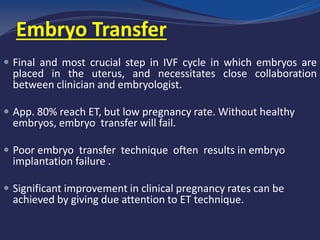
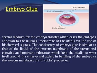
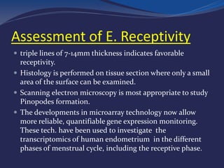
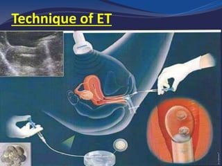
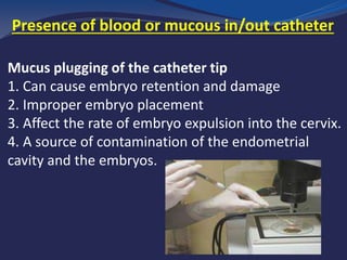
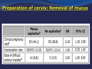
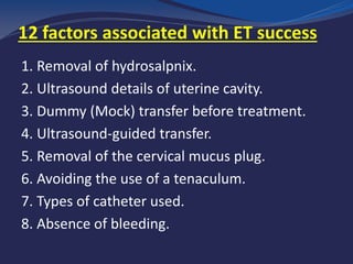
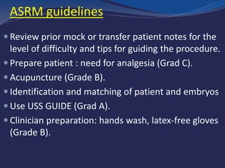
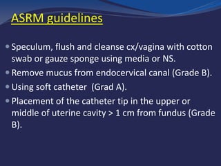
Aisha M. Elbareg, MD, PhD
Associate Clinical Professor & Senior Consultant
IVF Unit, Al-Amal Hospital, Misurata, Libya
The entire IVF cycle depends on a delicate placement of the
embryos at the proper location near the middle of the
endometrial cavity where implantation is maximized with
minimal trauma and manipulation.
Embryo Transfer
Embryo Transfer
 Final and most crucial step in IVF cycle in which embryos are
placed in the uterus, and necessitates close collaboration
between clinician and embryologist.
 App. 80% reach ET, but low pregnancy rate. Without healthy
embryos, embryo transfer will fail.
 Poor embryo transfer technique often results in embryo
implantation failure .
 Significant improvement in clinical pregnancy rates can be
achieved by giving due attention to ET technique.
• Select the best seed
• Prepare the soil
• Plant at the appropriate time with
proper technique
Factors affecting embryo implantation
 Implantation capacity of the embryo
 Endometrial receptivity
 Embryo transfer technique
Preimplantation embryo development
The embryo
 Failure of embryo to implant is the major limiting step in
determining ART success rate.
 A number of strategies aimed at increasing chances of
embryo to successfully implant:
Blastocyst culture - PGD
Assisted hatching
Co-culture with human endomet -epithelial cells
Embryo Glue
GlueEmbryo
•
special medium for the embryo transfer which eases the embryo's
adhesion to the mucous membrane of the uterus via the use of
biochemical signals. The consistency of embryo glue is similar to
that of the liquid of the mucous membrane of the uterus and
contains an important substance which help the media to wrap
itself around the embryo and assists in bonding of the embryo to
the mucous membrane via its ‘sticky' properties.
The endometrium
 Human endometrium is a dynamic tissue.
 In the phase of receptivity, the endometrium
undergoes morphological, cytoskeletal,
biochemical, and genetic changes.
 The period of E. receptivity is known as WOI.
 WOI opens on day 19 or 20 of the cycle, remains
for just 4-5 days when the P4 reaches peak.
Assessment of E. Receptivity
 USS measurement of end.:
Thickness, pattern, volume,
doppler of uterine and
subendometrial blood flow in
prediction of successful
implantation
 Histological assessment of
endometrial biopsy.
 Gene expression profiling.
(ERA)
Assessment of E. Receptivity
 triple lines of 7-14mm thickness indicates favorable
receptivity.
 Histology is performed on tissue section where only a small
area of the surface can be examined.
 Scanning electron microscopy is most appropriate to study
Pinopodes formation.
 The developments in microarray technology now allow
more reliable, quantifiable gene expression monitoring.
These tech. have been used to investigate the
transcriptomics of human endometrium in the different
phases of menstrual cycle, including the receptive phase.
Technique of ET
1. Disruption of endometrium
2. Induction of uterine contractions
3. Deposition of embryos in a suboptimal location
4. Damage of embryos during the process
What may go wrong during transfer ?
What is difficult Transfer ?
 Prolonged time to negotiate uterocervical angle
 Tenaculum application
 Blood in or on the catheter
 Stiff embryo transfer catheter
Why is it difficult to transfer ?
 Cervical stenosis
 Acute angle of uterocervical portion
 (anteversion/retroversion)
 Unexperienced operator
Laboratory Part
1. Embryo Selection (Grade, Stage, no. and timing)
2. Handling of catheter
3. Embryo loaded method of embryo in catheter
4. Embryo Loading medium
5. Duration of embryo loading
6. Check presence of blood or mucus in/out catheter
Variables Influencing the Success of
Embryo Transfer Technique
Clinician Part
1. The preparation before embryo transfer
2. Uterine Contractions
3. Way of Catheter Insertion
4. Placement of the Catheter Tip
5. Types of catheter!!
Variables Influencing the Success of
Embryo Transfer Technique
3 stages of preparation
First stage: the preparation before ET
1. Evaluating the cervico-uterine axis by:
 USS
 A dummy (Mock transfer) (Madani et al., 2009)
2. Appropriate evaluation of uterine cavity.
 USS, Hysteroscopy
 Many unexpected agents make entering the uterine cavity
difficult, such as cervical polyps or fibroids, a pin-point external
os, and cervical deformation due to congenital anomalies or
previous surgery, all of which can be discovered by a ‘dummy’ or
‘mock’ transfer. In the case of cervical stenosis, cervical
dilatation should be performed before ovarian stimulation.
Second stage: measures during ET
1. Cervical preparation, removing of
mucous or blood
2. Straightening of the utero-cervical angle
3. Use of US guidance
4. Types of catheter: Wallace, Labotect,
Cook and Frydman. Benefits of one over the
Other are controversial!!
5. Loading the embryo medium/embryo Glue
6. Embryo load method for transfer
3 stages of preparation
Presence of blood or mucous in/out catheter
The presence of blood on the outside of the
catheter tip :
1. May be a sign of difficult embryo transfer
2. Associated with lower pregnancy rates
3. A higher incidence of retained embryos
Mucus plugging of the catheter tip
1. Can cause embryo retention and damage
2. Improper embryo placement
3. Affect the rate of embryo expulsion into the cervix.
4. A source of contamination of the endometrial
cavity and the embryos.
Presence of blood or mucous in/out catheter
Preparation of cervix: Removal of mucus
6. Embryo load method for transfer
• Embryos could expel into vagina, if transfer
medium exceeds 60 μl.
• Implantation could be affected when medium
was less than 10 μl .
7. Time of ET with regard to uterine contraction
problem:
 Serum P at the time of ET
 Difficult ET with use of tenaculum
 Progesterone into the luteal phase- blastocyst
transfer.
8. Injection of air before ET catheter withdrawal with
Slow withdrawal of transfer catheter.
Third stage: measures after ET
Prevention of the expulsion of fluids & embryo
from cx
Closing the cx
Fibrin sealant
Bed rest!!!!!!!!!!!!
βhCG test 14 days after ET.
Adequate luteal support+ low dose Aspirin&
or LMWH wherever indicated.
Consider
Single embryo transfer (blastocyst)
Later frozen embryo transfer
Blastocyst ET
 Blastocyst culture
 Blastocyst development
 Compaction, cavitation
 Blastocyst scoring & grading system
 Blastocyst selection for transfer
Way of catheter insertion
Favorable prognosis for Elective
Single ET
 Expectation of one or more high-quality embryos
available for cryopreservation
 Eupolid embryos
 Previous live birth after an IVF cycle
 The availability of vitrified, high-quality, day-5, or
day-6 blastocysts for transfer
embryo transfer cycles-In frozen
 Favorable characteristics should be based on The age
of the woman when the embryos were frozen
 presence of high quality vitrified embryos.
 1st FET cycle
 previous live birth after an IVF cycle.
 Embryo transfer numbers should not exceed the
recommended limit on the number of fresh embryos
transferred for each age group.
factors associated with ET success12
1. Removal of hydrosalpnix.
2. Ultrasound details of uterine cavity.
3. Dummy (Mock) transfer before treatment.
4. Ultrasound-guided transfer.
5. Removal of the cervical mucus plug.
6. Avoiding the use of a tenaculum.
7. Types of catheter used.
8. Absence of bleeding.
9. Not touching the fundus.
10. Keeping catheter stationary for at least 1 min.
11. 30 minutes rest after transfer.
12. Giving antiprostaglandins to prevent contraction.
factors associated with ET success12
ASRM guidelines
 Review prior mock or transfer patient notes for the
level of difficulty and tips for guiding the procedure.
 Prepare patient : need for analgesia (Grad C).
 Acupuncture (Grade B).
 Identification and matching of patient and embryos
 Use USS GUIDE (Grad A).
 Clinician preparation: hands wash, latex-free gloves
(Grade B).
 Speculum, flush and cleanse cx/vagina with cotton
swab or gauze sponge using media or NS.
 Remove mucus from endocervical canal (Grade B).
 Using soft catheter (Grad A).
 Placement of the catheter tip in the upper or
middle of uterine cavity > 1 cm from fundus (Grade
B).
ASRM guidelines
Embryo transfer

Embryo transfer

  • 1.
    Aisha M. Elbareg,MD, PhD Associate Clinical Professor & Senior Consultant IVF Unit, Al-Amal Hospital, Misurata, Libya
  • 2.
    The entire IVFcycle depends on a delicate placement of the embryos at the proper location near the middle of the endometrial cavity where implantation is maximized with minimal trauma and manipulation. Embryo Transfer
  • 3.
    Embryo Transfer  Finaland most crucial step in IVF cycle in which embryos are placed in the uterus, and necessitates close collaboration between clinician and embryologist.  App. 80% reach ET, but low pregnancy rate. Without healthy embryos, embryo transfer will fail.  Poor embryo transfer technique often results in embryo implantation failure .  Significant improvement in clinical pregnancy rates can be achieved by giving due attention to ET technique.
  • 4.
    • Select thebest seed • Prepare the soil • Plant at the appropriate time with proper technique
  • 5.
    Factors affecting embryoimplantation  Implantation capacity of the embryo  Endometrial receptivity  Embryo transfer technique
  • 6.
  • 7.
    The embryo  Failureof embryo to implant is the major limiting step in determining ART success rate.  A number of strategies aimed at increasing chances of embryo to successfully implant: Blastocyst culture - PGD Assisted hatching Co-culture with human endomet -epithelial cells Embryo Glue
  • 8.
    GlueEmbryo • special medium forthe embryo transfer which eases the embryo's adhesion to the mucous membrane of the uterus via the use of biochemical signals. The consistency of embryo glue is similar to that of the liquid of the mucous membrane of the uterus and contains an important substance which help the media to wrap itself around the embryo and assists in bonding of the embryo to the mucous membrane via its ‘sticky' properties.
  • 9.
    The endometrium  Humanendometrium is a dynamic tissue.  In the phase of receptivity, the endometrium undergoes morphological, cytoskeletal, biochemical, and genetic changes.  The period of E. receptivity is known as WOI.  WOI opens on day 19 or 20 of the cycle, remains for just 4-5 days when the P4 reaches peak.
  • 10.
    Assessment of E.Receptivity  USS measurement of end.: Thickness, pattern, volume, doppler of uterine and subendometrial blood flow in prediction of successful implantation  Histological assessment of endometrial biopsy.  Gene expression profiling. (ERA)
  • 11.
    Assessment of E.Receptivity  triple lines of 7-14mm thickness indicates favorable receptivity.  Histology is performed on tissue section where only a small area of the surface can be examined.  Scanning electron microscopy is most appropriate to study Pinopodes formation.  The developments in microarray technology now allow more reliable, quantifiable gene expression monitoring. These tech. have been used to investigate the transcriptomics of human endometrium in the different phases of menstrual cycle, including the receptive phase.
  • 12.
  • 13.
    1. Disruption ofendometrium 2. Induction of uterine contractions 3. Deposition of embryos in a suboptimal location 4. Damage of embryos during the process What may go wrong during transfer ?
  • 14.
    What is difficultTransfer ?  Prolonged time to negotiate uterocervical angle  Tenaculum application  Blood in or on the catheter  Stiff embryo transfer catheter
  • 15.
    Why is itdifficult to transfer ?  Cervical stenosis  Acute angle of uterocervical portion  (anteversion/retroversion)  Unexperienced operator
  • 16.
    Laboratory Part 1. EmbryoSelection (Grade, Stage, no. and timing) 2. Handling of catheter 3. Embryo loaded method of embryo in catheter 4. Embryo Loading medium 5. Duration of embryo loading 6. Check presence of blood or mucus in/out catheter Variables Influencing the Success of Embryo Transfer Technique
  • 17.
    Clinician Part 1. Thepreparation before embryo transfer 2. Uterine Contractions 3. Way of Catheter Insertion 4. Placement of the Catheter Tip 5. Types of catheter!! Variables Influencing the Success of Embryo Transfer Technique
  • 18.
    3 stages ofpreparation First stage: the preparation before ET 1. Evaluating the cervico-uterine axis by:  USS  A dummy (Mock transfer) (Madani et al., 2009) 2. Appropriate evaluation of uterine cavity.  USS, Hysteroscopy  Many unexpected agents make entering the uterine cavity difficult, such as cervical polyps or fibroids, a pin-point external os, and cervical deformation due to congenital anomalies or previous surgery, all of which can be discovered by a ‘dummy’ or ‘mock’ transfer. In the case of cervical stenosis, cervical dilatation should be performed before ovarian stimulation.
  • 19.
    Second stage: measuresduring ET 1. Cervical preparation, removing of mucous or blood 2. Straightening of the utero-cervical angle 3. Use of US guidance 4. Types of catheter: Wallace, Labotect, Cook and Frydman. Benefits of one over the Other are controversial!! 5. Loading the embryo medium/embryo Glue 6. Embryo load method for transfer 3 stages of preparation
  • 20.
    Presence of bloodor mucous in/out catheter The presence of blood on the outside of the catheter tip : 1. May be a sign of difficult embryo transfer 2. Associated with lower pregnancy rates 3. A higher incidence of retained embryos
  • 21.
    Mucus plugging ofthe catheter tip 1. Can cause embryo retention and damage 2. Improper embryo placement 3. Affect the rate of embryo expulsion into the cervix. 4. A source of contamination of the endometrial cavity and the embryos. Presence of blood or mucous in/out catheter
  • 22.
    Preparation of cervix:Removal of mucus
  • 25.
    6. Embryo loadmethod for transfer • Embryos could expel into vagina, if transfer medium exceeds 60 μl. • Implantation could be affected when medium was less than 10 μl .
  • 26.
    7. Time ofET with regard to uterine contraction problem:  Serum P at the time of ET  Difficult ET with use of tenaculum  Progesterone into the luteal phase- blastocyst transfer. 8. Injection of air before ET catheter withdrawal with Slow withdrawal of transfer catheter.
  • 27.
    Third stage: measuresafter ET Prevention of the expulsion of fluids & embryo from cx Closing the cx Fibrin sealant Bed rest!!!!!!!!!!!! βhCG test 14 days after ET. Adequate luteal support+ low dose Aspirin& or LMWH wherever indicated.
  • 28.
    Consider Single embryo transfer(blastocyst) Later frozen embryo transfer
  • 29.
    Blastocyst ET  Blastocystculture  Blastocyst development  Compaction, cavitation  Blastocyst scoring & grading system  Blastocyst selection for transfer
  • 30.
    Way of catheterinsertion
  • 31.
    Favorable prognosis forElective Single ET  Expectation of one or more high-quality embryos available for cryopreservation  Eupolid embryos  Previous live birth after an IVF cycle  The availability of vitrified, high-quality, day-5, or day-6 blastocysts for transfer
  • 32.
    embryo transfer cycles-Infrozen  Favorable characteristics should be based on The age of the woman when the embryos were frozen  presence of high quality vitrified embryos.  1st FET cycle  previous live birth after an IVF cycle.  Embryo transfer numbers should not exceed the recommended limit on the number of fresh embryos transferred for each age group.
  • 33.
    factors associated withET success12 1. Removal of hydrosalpnix. 2. Ultrasound details of uterine cavity. 3. Dummy (Mock) transfer before treatment. 4. Ultrasound-guided transfer. 5. Removal of the cervical mucus plug. 6. Avoiding the use of a tenaculum. 7. Types of catheter used. 8. Absence of bleeding.
  • 34.
    9. Not touchingthe fundus. 10. Keeping catheter stationary for at least 1 min. 11. 30 minutes rest after transfer. 12. Giving antiprostaglandins to prevent contraction. factors associated with ET success12
  • 35.
    ASRM guidelines  Reviewprior mock or transfer patient notes for the level of difficulty and tips for guiding the procedure.  Prepare patient : need for analgesia (Grad C).  Acupuncture (Grade B).  Identification and matching of patient and embryos  Use USS GUIDE (Grad A).  Clinician preparation: hands wash, latex-free gloves (Grade B).
  • 36.
     Speculum, flushand cleanse cx/vagina with cotton swab or gauze sponge using media or NS.  Remove mucus from endocervical canal (Grade B).  Using soft catheter (Grad A).  Placement of the catheter tip in the upper or middle of uterine cavity > 1 cm from fundus (Grade B). ASRM guidelines