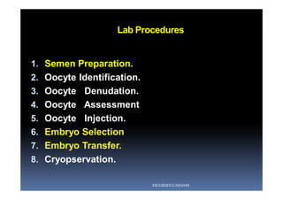
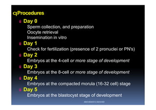
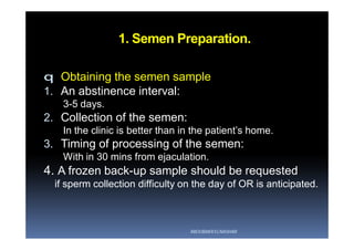
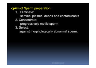
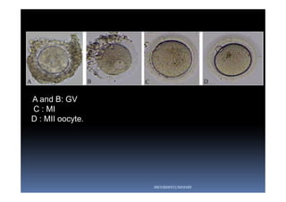
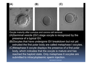
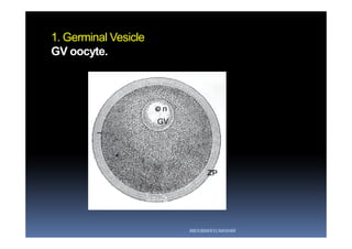
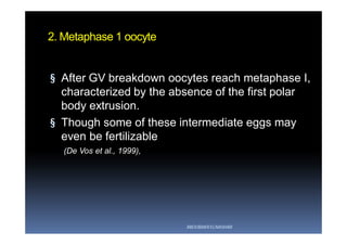
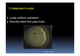
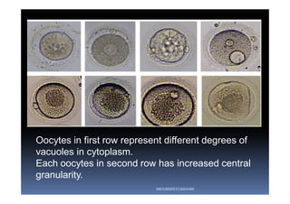
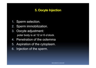
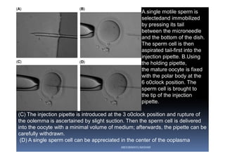
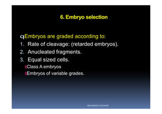
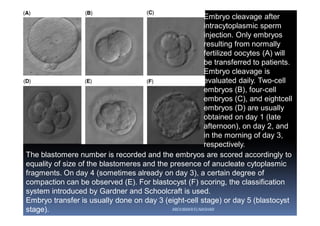
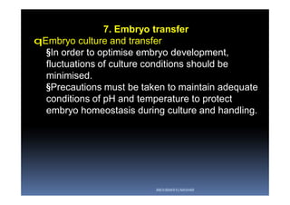
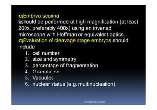
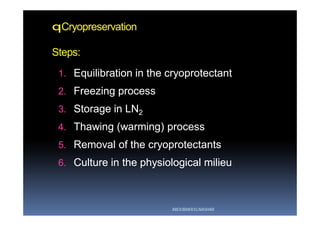
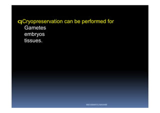
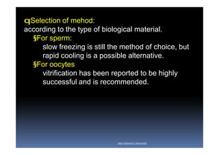
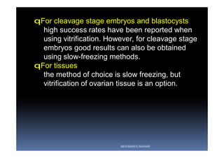
ICSI Lab
Procedures
For Gynecologist
Aboubakr Elnashar
Benha University, Egypt
ABOUBAKR ELNASHAR
Lab Procedures
1. Semen Preparation.
2. Oocyte Identification.
3. Oocyte Denudation.
4. Oocyte Assessment
5. Oocyte Injection.
6. Embryo Selection
7. Embryo Transfer.
8. Cryopservation.
ABOUBAKR ELNASHAR
qProcedures
ú Day 0
Sperm collection, and preparation
Oocyte retrieval
Insemination in vitro
ú Day 1
Check for fertilization (presence of 2 pronuclei or PN's)
ú Day 2
Embryos at the 4-cell or more stage of development
ú Day 3
Embryos at the 8-cell or more stage of development
ú Day 4
Embryos at the compacted morula (16-32 cell) stage
ú Day 5
Embryos at the blastocyst stage of development
ABOUBAKR ELNASHAR
1. Semen Preparation.
q Obtaining the semen sample
1. An abstinence interval:
3-5 days.
2. Collection of the semen:
In the clinic is better than in the patient’s home.
3. Timing of processing of the semen:
With in 30 mins from ejaculation.
4. A frozen back-up sample should be requested
if sperm collection difficulty on the day of OR is anticipated.
ABOUBAKR ELNASHAR
qAim of Sperm preparation:
1. Eliminate:
seminal plasma, debris and contaminants
2. Concentrate:
progressively motile sperm
3. Select:
against morphologically abnormal sperm.
ABOUBAKR ELNASHAR
q Methods of Sperm preparation:
1. Discontinuous density gradient.
2. Double wash.
3. Swim up.
The swim-up technique and discontinuous density-
gradient centrifugation are most frequently used
and widely accepted.
ABOUBAKR ELNASHAR
qSelection of Sperm preparation method:
•Characteristics
•Origin of individual samples.
qIn absence of motile sperm.
-Phosphodiesterase inhibitors (pentoxifylline,
theophylline) or
-Hypo-osmotic swelling (HOS) test
ABOUBAKR ELNASHAR
qAzoospermia on the day of OR and in the absence
of a back-up sample:
•Sperm retrieval procedures or
•Oocyte cryopreservation
qIf no sperm are observed.
Enzymatic digestion of testicular tissue by
collagenase
ABOUBAKR ELNASHAR
2. Oocyte identification
§ Aim:
Identify the oocytes from the aspirated follicular fluid.
oocytes are washed from the surrounding follicular
fluid (mimic physiology)
§ The time between OR and culture of washed
oocytes
should be minimal.
{Prolonged oocyte exposure to follicular fluid is not
recommended}
§ Exposure of oocytes to light
should be minimized.
ABOUBAKR ELNASHAR
3. Oocyte Denudation
§ Chemical:
repeated pipetting in hylaundase enzyme.
§ Mechanical:
using thin pipettes of varying diameters (175-125 µ)
ABOUBAKR ELNASHAR
A and B: GV
C : MI
D : MII oocyte.
ABOUBAKR ELNASHAR
4. Oocyte Assessment
qImportance of oocyte quality:
§ has a great influence on:
§ Fertilization
§ early development
§ implantation of embryos.
§ In order that the oocyte would be successfully
fertilized and cleaved, the nucleus and the
cytoplasm of the oocyte must be mature at the
same time.
ABOUBAKR ELNASHAR
qNuclear Maturation
1. Germinal Vesicle.
2. Metaphase 1 oocytes.
3. Metaphase 2 oocytes.
ABOUBAKR ELNASHAR
Oocyte maturity after cumulus and corona cell removal.
(A)Germinal vesicle (GV) stage oocyte is recognized by the
presence of a typical GV.
(B)Oocytes that have undergone GV breakdown but not yet
extruded the first polar body are called metaphase I oocytes.
(C)Metaphase II oocyte displays the presence of a first polar
body, which indicates that the oocyte is mature and has
reached the haploid state. Only metaphase II oocytes are
submitted to intracytoplasmic sperm injection.
ABOUBAKR ELNASHAR
1. Germinal Vesicle
GV oocyte.
ABOUBAKR ELNASHAR
2. Metaphase 1 oocyte
§ After GV breakdown oocytes reach metaphase I,
characterized by the absence of the first polar
body extrusion.
§ Though some of these intermediate eggs may
even be fertilizable
(De Vos et al., 1999),
ABOUBAKR ELNASHAR
qRetrieved immature oocytes after COH
ú Usually a variable percentage of the retrieved
oocytes from mature follicles after COH are GV or
M I.
ú These oocytes may change to M II oocytes in vitro,
and hence can be injected.
ú However, the reproductive outcome of these
oocytes is very poor.
ú This effect may not be evident until pre and early
postimplantation period.
ú This poor outcome can be explained by the
asynchronous nuclear and cytoplasmic maturity.
ABOUBAKR ELNASHAR
qFertilization and pregnancy potential of immature
oocytes from stimulated ICSI cycles.
(Shin et el , 2013)
§ The aim was to compare the outcome of retrieved
mature oocytes and retrieved immature oocytes that
were injected after maturation.
§ The fertilization rate of retrieved immature oocytes
was half that of retrieved mature oocytes (37% Vs
73%).
ABOUBAKR ELNASHAR
3. Metaphase II oocyte
§ Large uniform cytoplasm
§ Discrete intact first polar body
ABOUBAKR ELNASHAR
Oocytes in first row represent different degrees of
vacuoles in cytoplasm.
Each oocytes in second row has increased central
granularity.
ABOUBAKR ELNASHAR
qCytoplasmic maturation
§ The only method:
oocyte fertilization
embryo development in vitro.
§ If the cytoplasm of oocytes is immature:
embryo cannot develop normally after
fertilization.
ABOUBAKR ELNASHAR
qDefects in Cytoplasmic maturation lead to:
1. Retarded embryos
2. Cleavage arrest
3. Failure of implantation
ABOUBAKR ELNASHAR
5. Oocyte Injection
1. Sperm selection.
2. Sperm immobilization.
3. Oocyte adjustment:
polar body is at 12 or 6 o'clock.
4. Penetration of the oolemma
5. Aspiration of the cytoplasm.
6. Injection of the sperm.
ABOUBAKR ELNASHAR
(C) The injection pipette is introduced at the 3 o0clock position and rupture of
the oolemma is ascertained by slight suction. Then the sperm cell is delivered
into the oocyte with a minimal volume of medium; afterwards, the pipette can be
carefully withdrawn.
(D) A single sperm cell can be appreciated in the center of the ooplasma
A.single motile sperm is
selectedand immobilized
by pressing its tail
between the microneedle
and the bottom of the dish.
The sperm cell is then
aspirated tail-first into the
injection pipette. B.Using
the holding pipette,
the mature oocyte is fixed
with the polar body at the
6 o0clock position. The
sperm cell is brought to
the tip of the injection
pipette.
ABOUBAKR ELNASHAR
6. Embryo selection
qEmbryos are graded according to:
1. Rate of cleavage: (retarded embryos).
2. Anucleated fragments.
3. Equal sized cells.
§Class A embryos
§Embryos of variable grades.
ABOUBAKR ELNASHAR
qScoring for fertilization
§All inseminated or injected oocytes should be
examined for the presence of pronuclei (PN) and
polar bodies at 16 to 18 hours post insemination.
§A normally fertilized oocyte contains 2PN and 2
polar bodies.
§For conventional IVF, cumulus cells must be
removed and 2PN oocytes transferred into new
dishes containing pre-equilibrated culture
medium.
ABOUBAKR ELNASHAR
§Abnormal fertilizations.
A : egg with1 pronucleus (PN) B with 3 C with 4 PN.
§Normal fertilization
Oocyte in D, E or F, each has 2 PN.
ABOUBAKR ELNASHAR
Fertilization outcome after intracytoplasmic sperm injection.
(A) Oocytes are considered normally fertilized when two
individualized or fragmented polar bodies are present together
with two clearly visible pronuclei (2-PN) that contain nucleoli.
(B) Abnormal fertilization may occur as one pronuclear (1-PN)
oocyte, probably due to parthenogenic activation.
(C) The occasional finding of three pronuclear (3-PN) oocytes
after injection of a single spermatozoon into the ooplasm is
probably caused by non-extrusion of the second polar body at
the time of fertilization.
ABOUBAKR ELNASHAR
§Embryos derived from ≥ 3PN oocytes should never
be transferred or cryopreserved.
§Under exceptional circumstances, if no
transferable embryos derived from 2PN oocytes are
available, embryos derived from 1PN oocytes or
oocytes showing no PN but going through normal
cleavage may be used for transfer.
§Even if no transferable embryos derived from 2PN
oocytes are available, the use of embryos derived
from 1PN oocytes or oocytes showing no PN is not
recommended.
ABOUBAKR ELNASHAR
The blastomere number is recorded and the embryos are scored accordingly to
equality of size of the blastomeres and the presence of anucleate cytoplasmic
fragments. On day 4 (sometimes already on day 3), a certain degree of
compaction can be observed (E). For blastocyst (F) scoring, the classification
system introduced by Gardner and Schoolcraft is used.
Embryo transfer is usually done on day 3 (eight-cell stage) or day 5 (blastocyst
stage).
Embryo cleavage after
intracytoplasmic sperm
injection. Only embryos
resulting from normally
fertilized oocytes (A) will
be transferred to patients.
Embryo cleavage is
evaluated daily. Two-cell
embryos (B), four-cell
embryos (C), and eightcell
embryos (D) are usually
obtained on day 1 (late
afternoon), on day 2, and
in the morning of day 3,
respectively.
ABOUBAKR ELNASHAR
Timeline for optimal blastocyst
development.
ABOUBAKR ELNASHAR
7. Embryo transfer
qEmbryo culture and transfer
§In order to optimise embryo development,
fluctuations of culture conditions should be
minimised.
§Precautions must be taken to maintain adequate
conditions of pH and temperature to protect
embryo homeostasis during culture and handling.
ABOUBAKR ELNASHAR
qEmbryo scoring
§should be performed at high magnification (at least
200x, preferably 400x) using an inverted
microscope with Hoffman or equivalent optics.
qEvaluation of cleavage stage embryos should
include
1. cell number
2. size and symmetry
3. percentage of fragmentation
4. Granulation
5. Vacuoles
6. nuclear status (e.g. multinucleation).
ABOUBAKR ELNASHAR
qBlastocyst scoring should include
1. expansion grade,
2. blastocoel cavity size
3. morphology of the inner cell mass (ICM)
4. trophectoderm (TE).
qAssessment should be performed at standardised
times post-insemination.
ABOUBAKR ELNASHAR
Scoring system for human blastocysts. Initially blastocysts are given a
numerical score from 1 to 6 based upon their degree of expansion and hatching
status: (1) early blastocyst; the blastocoel being less than half the volume of the
embryo; (2) blastocyst; the blastocoel being greater than or equal to half of the
volume of the embryo; (3) full blastocyst; the blastocoel completely fills the
embryo; (4) expanded blastocyst; the blastocoel volume is now larger than that
of the early embryo and the zona is thinning; (5) hatching blastocyst; the
trophectoderm has started to herniate through the zona; (6) hatched blastocyst;
the blastocyst has completely escaped from the zona.ABOUBAKR ELNASHAR
qTime-lapse imaging
§Assess embryo development
§more precise evaluation of the timing of
consecutive events
§not interfering with the embryo culture
environment.
ABOUBAKR ELNASHAR
qEmbryo selection for transfer is primarily based on
1. developmental stage
2. morphological aspects.
3. Other selection parameters, such as time-
lapse kinetics, may be considered.
ABOUBAKR ELNASHAR
qNumber of embryos to be transferred:
§should be based on:
1. embryo quality
2. stage of development
3. female age
4. ovarian response
5. rank of treatment.
§Single embryo transfer is recommended to avoid
multiple pregnancies.
qIt is advisable not to transfer more than two
embryos.
qSupernumerary embryos may be cryopreserved,
donated to research or discarded, according to their
quality, patient wishes and national legislation.
ABOUBAKR ELNASHAR
qEmbryo loading:
Air bubbles to bracket the embryo-containing medium
Insufficient evidence to suggest that the fluid-only
method is superior to the use of air brackets during
embryo loading.
(Abou-Setta, 2007)
ABOUBAKR ELNASHAR
qTime interval between embryo catheter loading
and discharging the embryos in the uterine cavity
§ The longer the duration, the lower the pregnancy
and implantation rates.
§ The decrease in pregnancy and implantation
rates is gradual until the duration of 120 s, and
decreases sharply afterwards.
(Hum Reprod. 2004)
ABOUBAKR ELNASHAR
9. Cryopreservation
qMethods:
1. Slow freezing
2. Vitrification
ABOUBAKR ELNASHAR
qVitrification
§ solidification of a solution (water is rapidly cooled
and formed into a glassy, vitrified state from the
liquid phase) at low temperature
§ Not by ice crystallization but
by extreme elevation in viscosity during cooling.
§ During vitrification the
§ entire solution remains unchanged
§ water does not precipitate: no ice crystals are
formed.
ABOUBAKR ELNASHAR
ABOUBAKR ELNASHAR
qCryopreservation
Steps:
1.1. Equilibration in the cryoprotectantEquilibration in the cryoprotectant
2.2. Freezing processFreezing process
3.3. Storage in LNStorage in LN22
4.4. Thawing (warming) processThawing (warming) process
5.5. Removal of the cryoprotectantsRemoval of the cryoprotectants
6.6. Culture in the physiological milieuCulture in the physiological milieu
ABOUBAKR ELNASHAR
qAction of the cryoprotectant
Penetration of cryoprotectant in the cell andPenetration of cryoprotectant in the cell and
partially replacing thepartially replacing the
↓↓
intracellular waterintracellular water
↓↓
dehydration of the celldehydration of the cell
ABOUBAKR ELNASHAR
qCryopreservation can be performed for
Gametes
embryos
tissues.
ABOUBAKR ELNASHAR
qSelection of mehod:
according to the type of biological material.
§For sperm:
slow freezing is still the method of choice, but
rapid cooling is a possible alternative.
§For oocytes
vitrification has been reported to be highly
successful and is recommended.
ABOUBAKR ELNASHAR
qFor cleavage stage embryos and blastocysts
high success rates have been reported when
using vitrification. However, for cleavage stage
embryos good results can also be obtained
using slow-freezing methods.
qFor tissues
the method of choice is slow freezing, but
vitrification of ovarian tissue is an option.
ABOUBAKR ELNASHAR
ABOUBAKR ELNASHAR
You can get this lecture from:
1.My scientific page on Face book:
Aboubakr Elnashar Lectures.
https://www.facebook.com/groups/2277
44884091351/
2.Slide share web site
3.elnashar53@hotmail.com

ICSI Lab procedures for gynecologist

  • 1.
    ICSI Lab Procedures For Gynecologist AboubakrElnashar Benha University, Egypt ABOUBAKR ELNASHAR
  • 2.
    Lab Procedures 1. SemenPreparation. 2. Oocyte Identification. 3. Oocyte Denudation. 4. Oocyte Assessment 5. Oocyte Injection. 6. Embryo Selection 7. Embryo Transfer. 8. Cryopservation. ABOUBAKR ELNASHAR
  • 3.
    qProcedures ú Day 0 Spermcollection, and preparation Oocyte retrieval Insemination in vitro ú Day 1 Check for fertilization (presence of 2 pronuclei or PN's) ú Day 2 Embryos at the 4-cell or more stage of development ú Day 3 Embryos at the 8-cell or more stage of development ú Day 4 Embryos at the compacted morula (16-32 cell) stage ú Day 5 Embryos at the blastocyst stage of development ABOUBAKR ELNASHAR
  • 4.
    1. Semen Preparation. qObtaining the semen sample 1. An abstinence interval: 3-5 days. 2. Collection of the semen: In the clinic is better than in the patient’s home. 3. Timing of processing of the semen: With in 30 mins from ejaculation. 4. A frozen back-up sample should be requested if sperm collection difficulty on the day of OR is anticipated. ABOUBAKR ELNASHAR
  • 5.
    qAim of Spermpreparation: 1. Eliminate: seminal plasma, debris and contaminants 2. Concentrate: progressively motile sperm 3. Select: against morphologically abnormal sperm. ABOUBAKR ELNASHAR
  • 6.
    q Methods ofSperm preparation: 1. Discontinuous density gradient. 2. Double wash. 3. Swim up. The swim-up technique and discontinuous density- gradient centrifugation are most frequently used and widely accepted. ABOUBAKR ELNASHAR
  • 7.
    qSelection of Spermpreparation method: •Characteristics •Origin of individual samples. qIn absence of motile sperm. -Phosphodiesterase inhibitors (pentoxifylline, theophylline) or -Hypo-osmotic swelling (HOS) test ABOUBAKR ELNASHAR
  • 8.
    qAzoospermia on theday of OR and in the absence of a back-up sample: •Sperm retrieval procedures or •Oocyte cryopreservation qIf no sperm are observed. Enzymatic digestion of testicular tissue by collagenase ABOUBAKR ELNASHAR
  • 9.
    2. Oocyte identification §Aim: Identify the oocytes from the aspirated follicular fluid. oocytes are washed from the surrounding follicular fluid (mimic physiology) § The time between OR and culture of washed oocytes should be minimal. {Prolonged oocyte exposure to follicular fluid is not recommended} § Exposure of oocytes to light should be minimized. ABOUBAKR ELNASHAR
  • 10.
    3. Oocyte Denudation §Chemical: repeated pipetting in hylaundase enzyme. § Mechanical: using thin pipettes of varying diameters (175-125 µ) ABOUBAKR ELNASHAR
  • 11.
    A and B:GV C : MI D : MII oocyte. ABOUBAKR ELNASHAR
  • 12.
    4. Oocyte Assessment qImportanceof oocyte quality: § has a great influence on: § Fertilization § early development § implantation of embryos. § In order that the oocyte would be successfully fertilized and cleaved, the nucleus and the cytoplasm of the oocyte must be mature at the same time. ABOUBAKR ELNASHAR
  • 13.
    qNuclear Maturation 1. GerminalVesicle. 2. Metaphase 1 oocytes. 3. Metaphase 2 oocytes. ABOUBAKR ELNASHAR
  • 14.
    Oocyte maturity aftercumulus and corona cell removal. (A)Germinal vesicle (GV) stage oocyte is recognized by the presence of a typical GV. (B)Oocytes that have undergone GV breakdown but not yet extruded the first polar body are called metaphase I oocytes. (C)Metaphase II oocyte displays the presence of a first polar body, which indicates that the oocyte is mature and has reached the haploid state. Only metaphase II oocytes are submitted to intracytoplasmic sperm injection. ABOUBAKR ELNASHAR
  • 15.
    1. Germinal Vesicle GVoocyte. ABOUBAKR ELNASHAR
  • 16.
    2. Metaphase 1oocyte § After GV breakdown oocytes reach metaphase I, characterized by the absence of the first polar body extrusion. § Though some of these intermediate eggs may even be fertilizable (De Vos et al., 1999), ABOUBAKR ELNASHAR
  • 17.
    qRetrieved immature oocytesafter COH ú Usually a variable percentage of the retrieved oocytes from mature follicles after COH are GV or M I. ú These oocytes may change to M II oocytes in vitro, and hence can be injected. ú However, the reproductive outcome of these oocytes is very poor. ú This effect may not be evident until pre and early postimplantation period. ú This poor outcome can be explained by the asynchronous nuclear and cytoplasmic maturity. ABOUBAKR ELNASHAR
  • 18.
    qFertilization and pregnancypotential of immature oocytes from stimulated ICSI cycles. (Shin et el , 2013) § The aim was to compare the outcome of retrieved mature oocytes and retrieved immature oocytes that were injected after maturation. § The fertilization rate of retrieved immature oocytes was half that of retrieved mature oocytes (37% Vs 73%). ABOUBAKR ELNASHAR
  • 19.
    3. Metaphase IIoocyte § Large uniform cytoplasm § Discrete intact first polar body ABOUBAKR ELNASHAR
  • 20.
    Oocytes in firstrow represent different degrees of vacuoles in cytoplasm. Each oocytes in second row has increased central granularity. ABOUBAKR ELNASHAR
  • 21.
    qCytoplasmic maturation § Theonly method: oocyte fertilization embryo development in vitro. § If the cytoplasm of oocytes is immature: embryo cannot develop normally after fertilization. ABOUBAKR ELNASHAR
  • 22.
    qDefects in Cytoplasmicmaturation lead to: 1. Retarded embryos 2. Cleavage arrest 3. Failure of implantation ABOUBAKR ELNASHAR
  • 23.
    5. Oocyte Injection 1.Sperm selection. 2. Sperm immobilization. 3. Oocyte adjustment: polar body is at 12 or 6 o'clock. 4. Penetration of the oolemma 5. Aspiration of the cytoplasm. 6. Injection of the sperm. ABOUBAKR ELNASHAR
  • 24.
    (C) The injectionpipette is introduced at the 3 o0clock position and rupture of the oolemma is ascertained by slight suction. Then the sperm cell is delivered into the oocyte with a minimal volume of medium; afterwards, the pipette can be carefully withdrawn. (D) A single sperm cell can be appreciated in the center of the ooplasma A.single motile sperm is selectedand immobilized by pressing its tail between the microneedle and the bottom of the dish. The sperm cell is then aspirated tail-first into the injection pipette. B.Using the holding pipette, the mature oocyte is fixed with the polar body at the 6 o0clock position. The sperm cell is brought to the tip of the injection pipette. ABOUBAKR ELNASHAR
  • 25.
    6. Embryo selection qEmbryosare graded according to: 1. Rate of cleavage: (retarded embryos). 2. Anucleated fragments. 3. Equal sized cells. §Class A embryos §Embryos of variable grades. ABOUBAKR ELNASHAR
  • 26.
    qScoring for fertilization §Allinseminated or injected oocytes should be examined for the presence of pronuclei (PN) and polar bodies at 16 to 18 hours post insemination. §A normally fertilized oocyte contains 2PN and 2 polar bodies. §For conventional IVF, cumulus cells must be removed and 2PN oocytes transferred into new dishes containing pre-equilibrated culture medium. ABOUBAKR ELNASHAR
  • 27.
    §Abnormal fertilizations. A :egg with1 pronucleus (PN) B with 3 C with 4 PN. §Normal fertilization Oocyte in D, E or F, each has 2 PN. ABOUBAKR ELNASHAR
  • 28.
    Fertilization outcome afterintracytoplasmic sperm injection. (A) Oocytes are considered normally fertilized when two individualized or fragmented polar bodies are present together with two clearly visible pronuclei (2-PN) that contain nucleoli. (B) Abnormal fertilization may occur as one pronuclear (1-PN) oocyte, probably due to parthenogenic activation. (C) The occasional finding of three pronuclear (3-PN) oocytes after injection of a single spermatozoon into the ooplasm is probably caused by non-extrusion of the second polar body at the time of fertilization. ABOUBAKR ELNASHAR
  • 29.
    §Embryos derived from≥ 3PN oocytes should never be transferred or cryopreserved. §Under exceptional circumstances, if no transferable embryos derived from 2PN oocytes are available, embryos derived from 1PN oocytes or oocytes showing no PN but going through normal cleavage may be used for transfer. §Even if no transferable embryos derived from 2PN oocytes are available, the use of embryos derived from 1PN oocytes or oocytes showing no PN is not recommended. ABOUBAKR ELNASHAR
  • 30.
    The blastomere numberis recorded and the embryos are scored accordingly to equality of size of the blastomeres and the presence of anucleate cytoplasmic fragments. On day 4 (sometimes already on day 3), a certain degree of compaction can be observed (E). For blastocyst (F) scoring, the classification system introduced by Gardner and Schoolcraft is used. Embryo transfer is usually done on day 3 (eight-cell stage) or day 5 (blastocyst stage). Embryo cleavage after intracytoplasmic sperm injection. Only embryos resulting from normally fertilized oocytes (A) will be transferred to patients. Embryo cleavage is evaluated daily. Two-cell embryos (B), four-cell embryos (C), and eightcell embryos (D) are usually obtained on day 1 (late afternoon), on day 2, and in the morning of day 3, respectively. ABOUBAKR ELNASHAR
  • 31.
    Timeline for optimalblastocyst development. ABOUBAKR ELNASHAR
  • 32.
    7. Embryo transfer qEmbryoculture and transfer §In order to optimise embryo development, fluctuations of culture conditions should be minimised. §Precautions must be taken to maintain adequate conditions of pH and temperature to protect embryo homeostasis during culture and handling. ABOUBAKR ELNASHAR
  • 33.
    qEmbryo scoring §should beperformed at high magnification (at least 200x, preferably 400x) using an inverted microscope with Hoffman or equivalent optics. qEvaluation of cleavage stage embryos should include 1. cell number 2. size and symmetry 3. percentage of fragmentation 4. Granulation 5. Vacuoles 6. nuclear status (e.g. multinucleation). ABOUBAKR ELNASHAR
  • 34.
    qBlastocyst scoring shouldinclude 1. expansion grade, 2. blastocoel cavity size 3. morphology of the inner cell mass (ICM) 4. trophectoderm (TE). qAssessment should be performed at standardised times post-insemination. ABOUBAKR ELNASHAR
  • 35.
    Scoring system forhuman blastocysts. Initially blastocysts are given a numerical score from 1 to 6 based upon their degree of expansion and hatching status: (1) early blastocyst; the blastocoel being less than half the volume of the embryo; (2) blastocyst; the blastocoel being greater than or equal to half of the volume of the embryo; (3) full blastocyst; the blastocoel completely fills the embryo; (4) expanded blastocyst; the blastocoel volume is now larger than that of the early embryo and the zona is thinning; (5) hatching blastocyst; the trophectoderm has started to herniate through the zona; (6) hatched blastocyst; the blastocyst has completely escaped from the zona.ABOUBAKR ELNASHAR
  • 36.
    qTime-lapse imaging §Assess embryodevelopment §more precise evaluation of the timing of consecutive events §not interfering with the embryo culture environment. ABOUBAKR ELNASHAR
  • 37.
    qEmbryo selection fortransfer is primarily based on 1. developmental stage 2. morphological aspects. 3. Other selection parameters, such as time- lapse kinetics, may be considered. ABOUBAKR ELNASHAR
  • 38.
    qNumber of embryosto be transferred: §should be based on: 1. embryo quality 2. stage of development 3. female age 4. ovarian response 5. rank of treatment. §Single embryo transfer is recommended to avoid multiple pregnancies. qIt is advisable not to transfer more than two embryos. qSupernumerary embryos may be cryopreserved, donated to research or discarded, according to their quality, patient wishes and national legislation. ABOUBAKR ELNASHAR
  • 39.
    qEmbryo loading: Air bubblesto bracket the embryo-containing medium Insufficient evidence to suggest that the fluid-only method is superior to the use of air brackets during embryo loading. (Abou-Setta, 2007) ABOUBAKR ELNASHAR
  • 40.
    qTime interval betweenembryo catheter loading and discharging the embryos in the uterine cavity § The longer the duration, the lower the pregnancy and implantation rates. § The decrease in pregnancy and implantation rates is gradual until the duration of 120 s, and decreases sharply afterwards. (Hum Reprod. 2004) ABOUBAKR ELNASHAR
  • 41.
    9. Cryopreservation qMethods: 1. Slowfreezing 2. Vitrification ABOUBAKR ELNASHAR
  • 42.
    qVitrification § solidification ofa solution (water is rapidly cooled and formed into a glassy, vitrified state from the liquid phase) at low temperature § Not by ice crystallization but by extreme elevation in viscosity during cooling. § During vitrification the § entire solution remains unchanged § water does not precipitate: no ice crystals are formed. ABOUBAKR ELNASHAR
  • 43.
  • 44.
    qCryopreservation Steps: 1.1. Equilibration inthe cryoprotectantEquilibration in the cryoprotectant 2.2. Freezing processFreezing process 3.3. Storage in LNStorage in LN22 4.4. Thawing (warming) processThawing (warming) process 5.5. Removal of the cryoprotectantsRemoval of the cryoprotectants 6.6. Culture in the physiological milieuCulture in the physiological milieu ABOUBAKR ELNASHAR
  • 45.
    qAction of thecryoprotectant Penetration of cryoprotectant in the cell andPenetration of cryoprotectant in the cell and partially replacing thepartially replacing the ↓↓ intracellular waterintracellular water ↓↓ dehydration of the celldehydration of the cell ABOUBAKR ELNASHAR
  • 46.
    qCryopreservation can beperformed for Gametes embryos tissues. ABOUBAKR ELNASHAR
  • 47.
    qSelection of mehod: accordingto the type of biological material. §For sperm: slow freezing is still the method of choice, but rapid cooling is a possible alternative. §For oocytes vitrification has been reported to be highly successful and is recommended. ABOUBAKR ELNASHAR
  • 48.
    qFor cleavage stageembryos and blastocysts high success rates have been reported when using vitrification. However, for cleavage stage embryos good results can also be obtained using slow-freezing methods. qFor tissues the method of choice is slow freezing, but vitrification of ovarian tissue is an option. ABOUBAKR ELNASHAR
  • 49.
    ABOUBAKR ELNASHAR You canget this lecture from: 1.My scientific page on Face book: Aboubakr Elnashar Lectures. https://www.facebook.com/groups/2277 44884091351/ 2.Slide share web site 3.elnashar53@hotmail.com