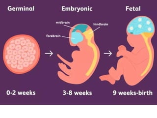
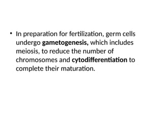
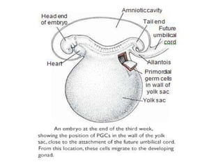
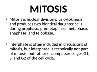
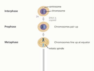
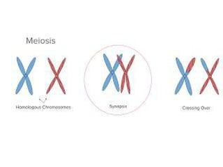
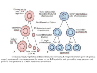
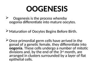
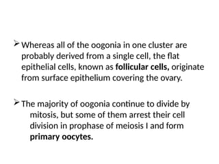
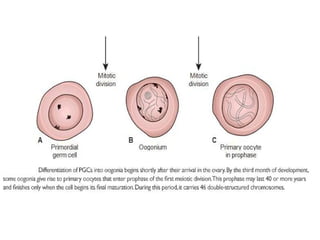
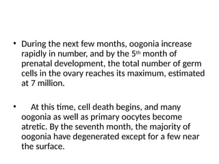
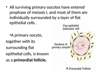
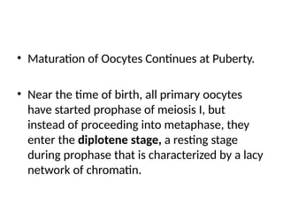
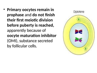
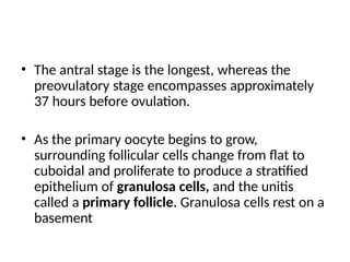
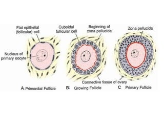
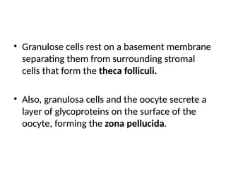
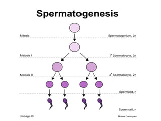
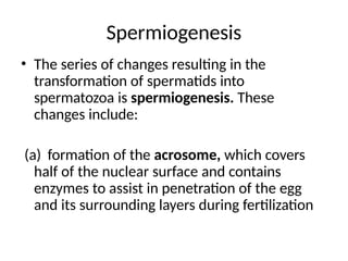
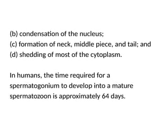
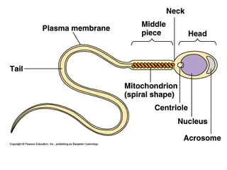
GAMETOGENESIS
Dr. Abshir Osman
MBBS, PGD in Medicine
Chapter 1
SCHOOL OF MEDICINE
GENERAL EMBRYOLOGY
1
INTRODUCTION
Embryology ( embryo + logos, study)
 general embryology (embryogenesis)
 Systemic embryology (organogenesis)
 Is the study of the prenatal development of
gamates (sex cells), fertilization, and development
of Embryos and Fetuses. Additionally, embryology
encompasses the study of congenital disorders that
occur before birth, known as teratology.
• Thus, our increasingly better understanding of
embryology has resulted in new techniques for
prenatal diagnoses and treatments, therapeutic
procedures to circumvent problems with
infertility, and mechanisms to prevent birth
defects, the leading cause of infant mortality.
• These improvements in prenatal and
reproductive health care are significant not only
for their contributions to improved birth
outcomes but also for their long-term effects
postnatally.
The process of prenatal development occurs in
three main stages.
1. The first 2 weeks after conception are known
as the germinal stage,
2. The time between 3rd- 8th weeks is known as
the embryonic period, &
3. The time from the 9th week until
birth is known as the fetal period.
• The study of the embryological origins and
causes for these birth defects was called
teratology.
Primordial Germ Cells
• Development begins with fertilization, the
process by which the male gamete, the
sperm, and the female gamete, the oocyte,
unite to give rise to a zygote.
• Gametes are derived from primordial germ
cells (PGCs) that are formed in the epiblast
during the second week and that move to the
wall of the yolk sac.
• During the fourth week these cells begin to
migrate from the yolk sac toward the
developing gonads, where they arrive by the
end of the fifth week.
• Mitotic divisions increase their number during
their migration and also when they arrive in
the gonad.
• In preparation for fertilization, germ cells
undergo gametogenesis, which includes
meiosis, to reduce the number of
chromosomes and cytodifferentiation to
complete their maturation.
MITOSIS
• Mitosis is nuclear division plus cytokinesis,
and produces two identical daughter cells
during prophase, prometaphase, metaphase,
anaphase, and telophase.
• Interphase is often included in discussions of
mitosis, but interphase is technically not part
of mitosis, but rather encompasses stages G1,
S, and G2 of the cell cycle.
MEIOSIS
• Meiosis is the cell division that takes place in
the germ cells to generate male and female
gametes, sperm and egg cells, respectively.
• Meiosis requires two cell divisions, meiosis I
and meiosis II, to reduce the number of
chromosomes to the haploid number of 23.
• (spermatocytes and primary oocytes) at the
beginning of meiosis I replicate their DNA so
that each of the 46 chromosomes is
duplicated into sister chromatids.
• In contrast to mitosis, however, homologous
chromosomes then align themselves in pairs,
a process called synapsis
• The pairing is exact and point for point except
for the XY combination.
• Homologous pairs then separate into two
daughter cells.
• Shortly thereafter meiosis II separates sister
chromatids. Each gamete then contains 23
chromosomes.
Crossover
• Crossovers, critical events in meiosis I, are the
interchange of chromatid segments between
paired homologous chromosomes .
• Segments of chromatids break and are exchanged
as homologous chromosomes separate.
• As separation occurs, points of interchange are
temporarily united and form an X-like structure, a
chiasma .
• Genetic variability is enhanced through
– crossover, which redistributes genetic material
– random distribution of homologous chromosomes
to the daughter cells
• Each germ cell contains a haploid number of
chromosomes, so that at fertilization the
diploid number of 46 is restored.
Morphological Changes
During Maturation of the
Gametes
OOGENESIS
 Oogenesis is the process whereby
oogonia differentiate into mature oocytes.
Maturation of Oocytes Begins Before Birth.
Once primordial germ cells have arrived in the
gonad of a genetic female, they differentiate into
oogonia. These cells undergo a number of mitotic
divisions and, by the end of the 3rd month, are
arranged in clusters surrounded by a layer of flat
epithelial cells.
Whereas all of the oogonia in one cluster are
probably derived from a single cell, the flat
epithelial cells, known as follicular cells, originate
from surface epithelium covering the ovary.
The majority of oogonia continue to divide by
mitosis, but some of them arrest their cell
division in prophase of meiosis I and form
primary oocytes.
• During the next few months, oogonia increase
rapidly in number, and by the 5th month of
prenatal development, the total number of germ
cells in the ovary reaches its maximum, estimated
at 7 million.
• At this time, cell death begins, and many
oogonia as well as primary oocytes become
atretic. By the seventh month, the majority of
oogonia have degenerated except for a few near
the surface.
• All surviving primary oocytes have entered
prophase of meiosis I, and most of them are
individually surrounded by a layer of flat
epithelial cells .
•A primary oocyte,
together with its
surrounding flat
epithelial cells, is known
as a primordial follicle.
• Maturation of Oocytes Continues at Puberty.
• Near the time of birth, all primary oocytes
have started prophase of meiosis I, but
instead of proceeding into metaphase, they
enter the diplotene stage, a resting stage
during prophase that is characterized by a lacy
network of chromatin.
• Primary oocytes remain in
prophase and do not finish
their first meiotic division
before puberty is reached,
apparently because of
oocyte maturation inhibitor
(OMI), substance secreted
by follicular cells.
• The total number of primary oocytes at birth is
estimated to vary from 700,000 to 2 million.
• During childhood most oocytes become atretic;
only approximately 400,000 are present by the
beginning of puberty, and fewer than 500 will be
ovulated. Some oocytes that reach maturity late
in life have been dormant in the diplotene stage
of the first meiotic division for 40 years or more
before ovulation.
• Whether the diplotene stage is the most
suitable phase to protect the oocyte against
environmental influences is unknown.
• The fact that the risk of having children with
chromosomal abnormalities increases with
maternal age indicates that primary oocytes
are vulnerable to damage as they age
• At puberty, a pool of growing follicles is established
and continuously maintained from the supply of
primordial follicles.
• Each month, 15 to 20 follicles selected from this pool
begin to mature, passing through three stages:
1) Primary or preantral;
2) Secondary or antral (also called
vesicular or Graafian); and
3) Preovulatory.
• The antral stage is the longest, whereas the
preovulatory stage encompasses approximately
37 hours before ovulation.
• As the primary oocyte begins to grow,
surrounding follicular cells change from flat to
cuboidal and proliferate to produce a stratified
epithelium of granulosa cells, and the unitis
called a primary follicle. Granulosa cells rest on a
basement
• Granulose cells rest on a basement membrane
separating them from surrounding stromal
cells that form the theca folliculi.
• Also, granulosa cells and the oocyte secrete a
layer of glycoproteins on the surface of the
oocyte, forming the zona pellucida.
• As follicles continue to grow, cells of the theca
folliculi organize into an inner layer of secretory
cells, the theca interna, and an outer fibrous
capsule, the theca externa
• As development continues, fluid-filled spaces
appear between granulosa cells. Coalescence
of these spaces forms the antrum, and the
follicle is termed a secondary (vesicular,
Graafian) follicle.
• Initially, the antrum is crescent shaped,
but with time, it enlarges.
• Granulosa cells surrounding the oocyte remain
intact and form the cumulus oophorus. At
maturity, the secondary follicle may be 25 mm or
more in diameter.
• It is surrounded by the theca interna, which is
composed of cells having characteristics of
steroid secretion, rich in blood vessels, and the
theca externa, which gradually merges with the
ovarian stroma.
• With each ovarian cycle, a number of follicles
begin to develop, but usually only one reaches
full maturity. The others degenerate and become
atretic.
• When the secondary follicle is mature, a surge in
luteinizing hormone (LH) induces the
preovulatory growth phase. Meiosis I is
completed, resulting in formation of two
daughter cells of unequal size, each with 23
double structured chromosomes
• One cell, the secondary oocyte, receives most of
the cytoplasm; the other, the first polar body,
receives practically none.
• The first polar body lies between the zona
pellucida and the cell membrane of the
secondary oocyte in the perivitelline space. The
cell then enters meiosis II but arrests in
metaphase approximately 3 hours before
ovulation.
• Meiosis II is completed only if the oocyte
is fertilized; otherwise, the cell degenerates
approximately 24 hours after ovulation. The
first polar body also undergoes a second
division.
SPERMATOGENESIS
 Maturation of Sperm Begins at Puberty
•Spermatogenesis, which begins at puberty,
includes all of the events by which spermatogonia
are transformed into spermatozoa.
•At birth, germ cells in the male can be
recognized in the sex cords of the testis as large,
pale cells surrounded by supporting cells.
Spermiogenesis
• The series of changes resulting in the
transformation of spermatids into
spermatozoa is spermiogenesis. These
changes include:
(a) formation of the acrosome, which covers
half of the nuclear surface and contains
enzymes to assist in penetration of the egg
and its surrounding layers during fertilization
(b) condensation of the nucleus;
(c) formation of neck, middle piece, and tail; and
(d) shedding of most of the cytoplasm.
In humans, the time required for a
spermatogonium to develop into a mature
spermatozoon is approximately 64 days.
• Spermatogenesis is regulated by luteinizing
hormone (LH) production by the pituitary. LH
binds to receptors on Leydig cells and stimulates
testosterone production, which in turn binds to
Sertoli cells to promote spermatogenesis.
• Follicle stimulating hormone (FSH) is also
essential because its binding to Sertoli cells
stimulates testicular fluid production and
synthesis of intracellular androgen receptor
proteins
Introduction of  Embryo and how it develop .pptx

Introduction of Embryo and how it develop .pptx

  • 1.
    GAMETOGENESIS Dr. Abshir Osman MBBS,PGD in Medicine Chapter 1 SCHOOL OF MEDICINE GENERAL EMBRYOLOGY 1
  • 2.
    INTRODUCTION Embryology ( embryo+ logos, study)  general embryology (embryogenesis)  Systemic embryology (organogenesis)  Is the study of the prenatal development of gamates (sex cells), fertilization, and development of Embryos and Fetuses. Additionally, embryology encompasses the study of congenital disorders that occur before birth, known as teratology.
  • 3.
    • Thus, ourincreasingly better understanding of embryology has resulted in new techniques for prenatal diagnoses and treatments, therapeutic procedures to circumvent problems with infertility, and mechanisms to prevent birth defects, the leading cause of infant mortality. • These improvements in prenatal and reproductive health care are significant not only for their contributions to improved birth outcomes but also for their long-term effects postnatally.
  • 4.
    The process ofprenatal development occurs in three main stages. 1. The first 2 weeks after conception are known as the germinal stage, 2. The time between 3rd- 8th weeks is known as the embryonic period, & 3. The time from the 9th week until birth is known as the fetal period.
  • 6.
    • The studyof the embryological origins and causes for these birth defects was called teratology.
  • 7.
    Primordial Germ Cells •Development begins with fertilization, the process by which the male gamete, the sperm, and the female gamete, the oocyte, unite to give rise to a zygote. • Gametes are derived from primordial germ cells (PGCs) that are formed in the epiblast during the second week and that move to the wall of the yolk sac.
  • 8.
    • During thefourth week these cells begin to migrate from the yolk sac toward the developing gonads, where they arrive by the end of the fifth week. • Mitotic divisions increase their number during their migration and also when they arrive in the gonad.
  • 9.
    • In preparationfor fertilization, germ cells undergo gametogenesis, which includes meiosis, to reduce the number of chromosomes and cytodifferentiation to complete their maturation.
  • 11.
    MITOSIS • Mitosis isnuclear division plus cytokinesis, and produces two identical daughter cells during prophase, prometaphase, metaphase, anaphase, and telophase. • Interphase is often included in discussions of mitosis, but interphase is technically not part of mitosis, but rather encompasses stages G1, S, and G2 of the cell cycle.
  • 17.
    MEIOSIS • Meiosis isthe cell division that takes place in the germ cells to generate male and female gametes, sperm and egg cells, respectively. • Meiosis requires two cell divisions, meiosis I and meiosis II, to reduce the number of chromosomes to the haploid number of 23.
  • 18.
    • (spermatocytes andprimary oocytes) at the beginning of meiosis I replicate their DNA so that each of the 46 chromosomes is duplicated into sister chromatids. • In contrast to mitosis, however, homologous chromosomes then align themselves in pairs, a process called synapsis
  • 19.
    • The pairingis exact and point for point except for the XY combination. • Homologous pairs then separate into two daughter cells. • Shortly thereafter meiosis II separates sister chromatids. Each gamete then contains 23 chromosomes.
  • 20.
    Crossover • Crossovers, criticalevents in meiosis I, are the interchange of chromatid segments between paired homologous chromosomes . • Segments of chromatids break and are exchanged as homologous chromosomes separate. • As separation occurs, points of interchange are temporarily united and form an X-like structure, a chiasma .
  • 22.
    • Genetic variabilityis enhanced through – crossover, which redistributes genetic material – random distribution of homologous chromosomes to the daughter cells • Each germ cell contains a haploid number of chromosomes, so that at fertilization the diploid number of 46 is restored.
  • 24.
  • 25.
    OOGENESIS  Oogenesis isthe process whereby oogonia differentiate into mature oocytes. Maturation of Oocytes Begins Before Birth. Once primordial germ cells have arrived in the gonad of a genetic female, they differentiate into oogonia. These cells undergo a number of mitotic divisions and, by the end of the 3rd month, are arranged in clusters surrounded by a layer of flat epithelial cells.
  • 26.
    Whereas all ofthe oogonia in one cluster are probably derived from a single cell, the flat epithelial cells, known as follicular cells, originate from surface epithelium covering the ovary. The majority of oogonia continue to divide by mitosis, but some of them arrest their cell division in prophase of meiosis I and form primary oocytes.
  • 28.
    • During thenext few months, oogonia increase rapidly in number, and by the 5th month of prenatal development, the total number of germ cells in the ovary reaches its maximum, estimated at 7 million. • At this time, cell death begins, and many oogonia as well as primary oocytes become atretic. By the seventh month, the majority of oogonia have degenerated except for a few near the surface.
  • 29.
    • All survivingprimary oocytes have entered prophase of meiosis I, and most of them are individually surrounded by a layer of flat epithelial cells . •A primary oocyte, together with its surrounding flat epithelial cells, is known as a primordial follicle.
  • 30.
    • Maturation ofOocytes Continues at Puberty. • Near the time of birth, all primary oocytes have started prophase of meiosis I, but instead of proceeding into metaphase, they enter the diplotene stage, a resting stage during prophase that is characterized by a lacy network of chromatin.
  • 31.
    • Primary oocytesremain in prophase and do not finish their first meiotic division before puberty is reached, apparently because of oocyte maturation inhibitor (OMI), substance secreted by follicular cells.
  • 32.
    • The totalnumber of primary oocytes at birth is estimated to vary from 700,000 to 2 million. • During childhood most oocytes become atretic; only approximately 400,000 are present by the beginning of puberty, and fewer than 500 will be ovulated. Some oocytes that reach maturity late in life have been dormant in the diplotene stage of the first meiotic division for 40 years or more before ovulation.
  • 33.
    • Whether thediplotene stage is the most suitable phase to protect the oocyte against environmental influences is unknown. • The fact that the risk of having children with chromosomal abnormalities increases with maternal age indicates that primary oocytes are vulnerable to damage as they age
  • 34.
    • At puberty,a pool of growing follicles is established and continuously maintained from the supply of primordial follicles. • Each month, 15 to 20 follicles selected from this pool begin to mature, passing through three stages: 1) Primary or preantral; 2) Secondary or antral (also called vesicular or Graafian); and 3) Preovulatory.
  • 35.
    • The antralstage is the longest, whereas the preovulatory stage encompasses approximately 37 hours before ovulation. • As the primary oocyte begins to grow, surrounding follicular cells change from flat to cuboidal and proliferate to produce a stratified epithelium of granulosa cells, and the unitis called a primary follicle. Granulosa cells rest on a basement
  • 37.
    • Granulose cellsrest on a basement membrane separating them from surrounding stromal cells that form the theca folliculi. • Also, granulosa cells and the oocyte secrete a layer of glycoproteins on the surface of the oocyte, forming the zona pellucida.
  • 38.
    • As folliclescontinue to grow, cells of the theca folliculi organize into an inner layer of secretory cells, the theca interna, and an outer fibrous capsule, the theca externa
  • 39.
    • As developmentcontinues, fluid-filled spaces appear between granulosa cells. Coalescence of these spaces forms the antrum, and the follicle is termed a secondary (vesicular, Graafian) follicle. • Initially, the antrum is crescent shaped, but with time, it enlarges.
  • 40.
    • Granulosa cellssurrounding the oocyte remain intact and form the cumulus oophorus. At maturity, the secondary follicle may be 25 mm or more in diameter. • It is surrounded by the theca interna, which is composed of cells having characteristics of steroid secretion, rich in blood vessels, and the theca externa, which gradually merges with the ovarian stroma.
  • 42.
    • With eachovarian cycle, a number of follicles begin to develop, but usually only one reaches full maturity. The others degenerate and become atretic. • When the secondary follicle is mature, a surge in luteinizing hormone (LH) induces the preovulatory growth phase. Meiosis I is completed, resulting in formation of two daughter cells of unequal size, each with 23 double structured chromosomes
  • 43.
    • One cell,the secondary oocyte, receives most of the cytoplasm; the other, the first polar body, receives practically none. • The first polar body lies between the zona pellucida and the cell membrane of the secondary oocyte in the perivitelline space. The cell then enters meiosis II but arrests in metaphase approximately 3 hours before ovulation.
  • 45.
    • Meiosis IIis completed only if the oocyte is fertilized; otherwise, the cell degenerates approximately 24 hours after ovulation. The first polar body also undergoes a second division.
  • 46.
    SPERMATOGENESIS  Maturation ofSperm Begins at Puberty •Spermatogenesis, which begins at puberty, includes all of the events by which spermatogonia are transformed into spermatozoa. •At birth, germ cells in the male can be recognized in the sex cords of the testis as large, pale cells surrounded by supporting cells.
  • 48.
    Spermiogenesis • The seriesof changes resulting in the transformation of spermatids into spermatozoa is spermiogenesis. These changes include: (a) formation of the acrosome, which covers half of the nuclear surface and contains enzymes to assist in penetration of the egg and its surrounding layers during fertilization
  • 49.
    (b) condensation ofthe nucleus; (c) formation of neck, middle piece, and tail; and (d) shedding of most of the cytoplasm. In humans, the time required for a spermatogonium to develop into a mature spermatozoon is approximately 64 days.
  • 50.
    • Spermatogenesis isregulated by luteinizing hormone (LH) production by the pituitary. LH binds to receptors on Leydig cells and stimulates testosterone production, which in turn binds to Sertoli cells to promote spermatogenesis. • Follicle stimulating hormone (FSH) is also essential because its binding to Sertoli cells stimulates testicular fluid production and synthesis of intracellular androgen receptor proteins