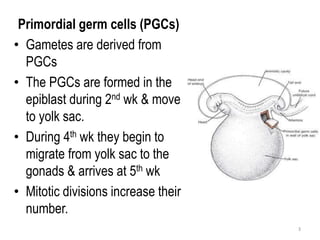
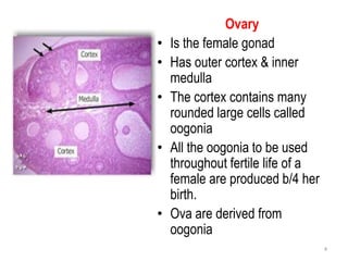
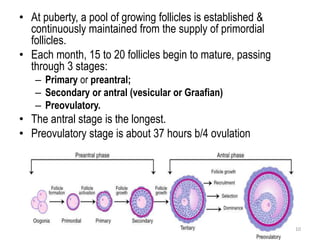
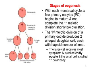
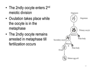
Ovary & Oogenesis
By
Dr. A.U. Agu
MBBS, MSc, PhD
1
Introduction
• Male gamete is called spermatozoa & the process
of it’s production is spermatogenesis
• Female gamete is called ova & the process of it’s
production is oogenesis
• Spermatogenesis & oogenesis are called
gametogenesis
2
Primordial germ cells (PGCs)
• Gametes are derived from
PGCs
• The PGCs are formed in the
epiblast during 2nd wk & move
to yolk sac.
• During 4th wk they begin to
migrate from yolk sac to the
gonads & arrives at 5th wk
• Mitotic divisions increase their
number.
3
Ovary
• Is the female gonad
• Has outer cortex & inner
medulla
• The cortex contains many
rounded large cells called
oogonia
• All the oogonia to be used
throughout fertile life of a
female are produced b/4 her
birth.
• Ova are derived from
oogonia
4
OOGENESIS
Maturation of Oocytes Begins Before Birth
• As the primordial germ cells arrives in the ovary,
they differentiate into oogonia
• The oogonia undergo mitotic divisions &, by the end
of the 3rd month, are arranged in clusters at the
ovarian cortex & surrounded flat epithelial cells.
• All the oogonia in one cluster are from a single
follicular cells.
5
• The oogonia continue to divide by mitosis
• Some of them are arrested at prophase of meiosis I
& differentiate into primary oocytes
• During the next few months, oogonia increase
rapidly in number, & by the 5th month, the total
number in the ovary reaches its maximum of 7
million.
• Thereafter, cell death begins, & many oogonia &
primary oocytes become atretic.
6
• By 7th month, the majority of oogonia have
degenerated except for a few near the surface.
• All surviving primary oocytes enters prophase of
meiosis I, & are individually surrounded by a layer
of follicular cell (flat epithelial cells)
• A primary oocyte, with its surrounding flat epithelial
cells, is called a primordial follicle
7
Maturation of Oocytes Continues at Puberty
• Close to the time of birth, all primary oocytes have
started prophase of meiosis I, but instead of proceeding
into metaphase, they enter the diplotene stage.
• Primary oocytes remain in prophase & do not finish
their 1st meiotic division b/4 puberty is reached
• This is due to oocyte maturation inhibitor (OMI),
secreted by follicular cells.
• Total number of primary oocytes at birth is about
700,000 to 2 million.
• During childhood most oocytes become atretic & about
400,000 are present by the beginning of puberty
• Less than 500 will be ovulated.
8
• Thus, some oocytes that reach maturity late in life
have been dormant in the diplotene stage of the 1st
meiotic division for 40 years or more b/4 ovulation.
• The fact that the risk of having children with
chromosomal abnormalities increases with maternal
age indicates that primary oocytes are vulnerable to
damage as they age.
9
• At puberty, a pool of growing follicles is established &
continuously maintained from the supply of primordial
follicles.
• Each month, 15 to 20 follicles begin to mature, passing
through 3 stages:
– Primary or preantral;
– Secondary or antral (vesicular or Graafian)
– Preovulatory.
• The antral stage is the longest.
• Preovulatory stage is about 37 hours b/4 ovulation
10
Stages of oogenesis
• With each menstrual cycle, a
few primary oocytes (PO)
begins to mature & one
complete the 1st meiotic
division shortly b/4 ovulation.
• The 1st meiotic division of a
primary oocyte produces 2
unequal daughter cell, each
with haploid number of xme .
– The large cell receives most
cytoplasm & is called 2ndly
oocyte & the small cell is called
1st polar body
11
• The 2ndly oocyte enters 2nd
meiotic division
• Ovulation takes place while
the oocyte is in the
metaphase
• The 2ndly oocyte remains
arrested in metaphase till
fertilization occurs
12
• The 2nd meiotic division is completed only if
fertilization occurs.
• This results in another 2 unequal daughter cell.
• The smaller one becomes 2nd polar body the larger
one is the ovum
• If fertilization does not occur, the 2ndly oocyte fails
to complete the 2nd meiotic division & degenerates
within 24 hours after ovulation
13
Formation of ovarian follicle
• Some cells of the stroma
become flattened &
surround the oocyte
• The flattened cells form the
ovarian follicle & are called
follicular cells
• The primary oocytes,
surrounded by the layers of
the flat epithelial (follicular)
cells & are primordial
follicles
14
• A layer of glycoproteins
appears b/w oocyte &
follicular cells & is called
the zona pellucida
• The follicular cell proliferate
& form several layers called
granulosa cells
(membrana granulosa)
15
• A cavity (antrum) appears
within the granulosa forming
a follicle
• The cavity of the follicle
increases in size & the wall of
the follicle becomes thin
• The oocyte now lies
eccentrically surrounded by
granulosa cells called
cumulus oophorus
• Cells that attach it to the wall
of the follicle is called discus
proligerus
16
• As the follicles expands, the
stromal cells surrounding the
granulosa organize into an
– Inner layer of secretory cells
(theca interna) that secretes
oestrogen
– Outer fibrous capsule (theca
externa).
17
Differences b/w spermatogenesis & oogenesis
• One primary spermatocyte gives rise to 4 spermatozoa
• One primary oocyte forms only one ovum
• When primary spermatocyte divides, it’s cytoplasm is
equally distributed b/w the 2 spermatocytes
• When primary oocyte divides, almost all it’s cytoplasm
goes to the daughter cell & the polar body gets ½ the
chromosome
• The polar body is used to get rid of the unwanted
chromosomes
18
19

Ovary & Oogenesis.pptx

  • 1.
    Ovary & Oogenesis By Dr.A.U. Agu MBBS, MSc, PhD 1
  • 2.
    Introduction • Male gameteis called spermatozoa & the process of it’s production is spermatogenesis • Female gamete is called ova & the process of it’s production is oogenesis • Spermatogenesis & oogenesis are called gametogenesis 2
  • 3.
    Primordial germ cells(PGCs) • Gametes are derived from PGCs • The PGCs are formed in the epiblast during 2nd wk & move to yolk sac. • During 4th wk they begin to migrate from yolk sac to the gonads & arrives at 5th wk • Mitotic divisions increase their number. 3
  • 4.
    Ovary • Is thefemale gonad • Has outer cortex & inner medulla • The cortex contains many rounded large cells called oogonia • All the oogonia to be used throughout fertile life of a female are produced b/4 her birth. • Ova are derived from oogonia 4
  • 5.
    OOGENESIS Maturation of OocytesBegins Before Birth • As the primordial germ cells arrives in the ovary, they differentiate into oogonia • The oogonia undergo mitotic divisions &, by the end of the 3rd month, are arranged in clusters at the ovarian cortex & surrounded flat epithelial cells. • All the oogonia in one cluster are from a single follicular cells. 5
  • 6.
    • The oogoniacontinue to divide by mitosis • Some of them are arrested at prophase of meiosis I & differentiate into primary oocytes • During the next few months, oogonia increase rapidly in number, & by the 5th month, the total number in the ovary reaches its maximum of 7 million. • Thereafter, cell death begins, & many oogonia & primary oocytes become atretic. 6
  • 7.
    • By 7thmonth, the majority of oogonia have degenerated except for a few near the surface. • All surviving primary oocytes enters prophase of meiosis I, & are individually surrounded by a layer of follicular cell (flat epithelial cells) • A primary oocyte, with its surrounding flat epithelial cells, is called a primordial follicle 7
  • 8.
    Maturation of OocytesContinues at Puberty • Close to the time of birth, all primary oocytes have started prophase of meiosis I, but instead of proceeding into metaphase, they enter the diplotene stage. • Primary oocytes remain in prophase & do not finish their 1st meiotic division b/4 puberty is reached • This is due to oocyte maturation inhibitor (OMI), secreted by follicular cells. • Total number of primary oocytes at birth is about 700,000 to 2 million. • During childhood most oocytes become atretic & about 400,000 are present by the beginning of puberty • Less than 500 will be ovulated. 8
  • 9.
    • Thus, someoocytes that reach maturity late in life have been dormant in the diplotene stage of the 1st meiotic division for 40 years or more b/4 ovulation. • The fact that the risk of having children with chromosomal abnormalities increases with maternal age indicates that primary oocytes are vulnerable to damage as they age. 9
  • 10.
    • At puberty,a pool of growing follicles is established & continuously maintained from the supply of primordial follicles. • Each month, 15 to 20 follicles begin to mature, passing through 3 stages: – Primary or preantral; – Secondary or antral (vesicular or Graafian) – Preovulatory. • The antral stage is the longest. • Preovulatory stage is about 37 hours b/4 ovulation 10
  • 11.
    Stages of oogenesis •With each menstrual cycle, a few primary oocytes (PO) begins to mature & one complete the 1st meiotic division shortly b/4 ovulation. • The 1st meiotic division of a primary oocyte produces 2 unequal daughter cell, each with haploid number of xme . – The large cell receives most cytoplasm & is called 2ndly oocyte & the small cell is called 1st polar body 11
  • 12.
    • The 2ndlyoocyte enters 2nd meiotic division • Ovulation takes place while the oocyte is in the metaphase • The 2ndly oocyte remains arrested in metaphase till fertilization occurs 12
  • 13.
    • The 2ndmeiotic division is completed only if fertilization occurs. • This results in another 2 unequal daughter cell. • The smaller one becomes 2nd polar body the larger one is the ovum • If fertilization does not occur, the 2ndly oocyte fails to complete the 2nd meiotic division & degenerates within 24 hours after ovulation 13
  • 14.
    Formation of ovarianfollicle • Some cells of the stroma become flattened & surround the oocyte • The flattened cells form the ovarian follicle & are called follicular cells • The primary oocytes, surrounded by the layers of the flat epithelial (follicular) cells & are primordial follicles 14
  • 15.
    • A layerof glycoproteins appears b/w oocyte & follicular cells & is called the zona pellucida • The follicular cell proliferate & form several layers called granulosa cells (membrana granulosa) 15
  • 16.
    • A cavity(antrum) appears within the granulosa forming a follicle • The cavity of the follicle increases in size & the wall of the follicle becomes thin • The oocyte now lies eccentrically surrounded by granulosa cells called cumulus oophorus • Cells that attach it to the wall of the follicle is called discus proligerus 16
  • 17.
    • As thefollicles expands, the stromal cells surrounding the granulosa organize into an – Inner layer of secretory cells (theca interna) that secretes oestrogen – Outer fibrous capsule (theca externa). 17
  • 18.
    Differences b/w spermatogenesis& oogenesis • One primary spermatocyte gives rise to 4 spermatozoa • One primary oocyte forms only one ovum • When primary spermatocyte divides, it’s cytoplasm is equally distributed b/w the 2 spermatocytes • When primary oocyte divides, almost all it’s cytoplasm goes to the daughter cell & the polar body gets ½ the chromosome • The polar body is used to get rid of the unwanted chromosomes 18
  • 19.