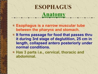
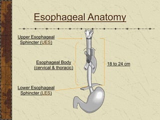
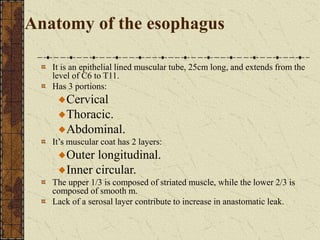
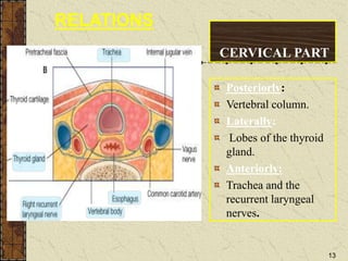
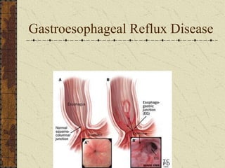
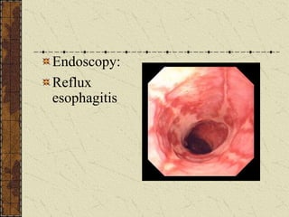
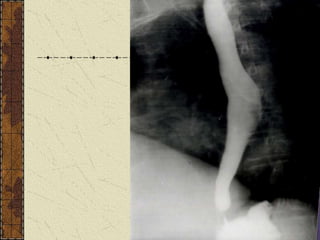
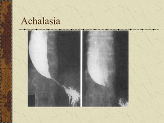
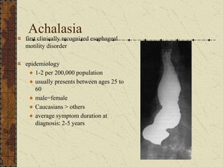
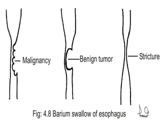
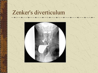
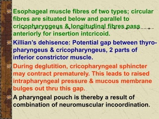
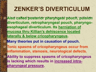
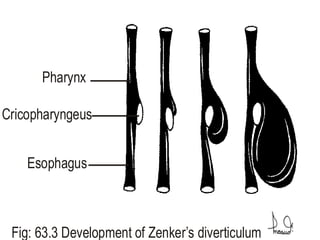
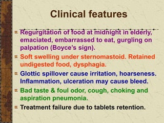
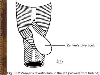
D Y S P H A G I A
Difficulty in swallowing, that is for liquids,
semisolids, solids, complete, incomplete.
ESOPHAGUS
Anatomy
Esophagus is a narrow muscular tube
between the pharynx and stomach.
It forms passage for food that passes thru
it during 3rd stage of deglutition, 25 cm in
length, collapsed antero posteriorly under
normal conditions.
Has 3 parts i.e., cervical, thoracic and
abdominal.
Esophageal Anatomy
Upper Esophageal
Sphincter (UES)
Lower Esophageal
Sphincter (LES)
Esophageal Body
(cervical & thoracic)
18 to 24 cm
Soft tissue neck
Positioned the same as an AP & Lateral
C-spine: ½ the mAs.
Trachea
Nasopharynx
Esophagus
Adnoids
Hyoid bone
Expose
during
inspiration
Oropharynx
Done to assess the patenc
of the airway
* masses
* foreign bodies
* enlarged adnoids (kids)
* epiglottitis (kids)
Anatomy of the esophagus
It is an epithelial lined muscular tube, 25cm long, and extends from the
level of C6 to T11.
Has 3 portions:
Cervical
Thoracic.
Abdominal.
It’s muscular coat has 2 layers:
Outer longitudinal.
Inner circular.
The upper 1/3 is composed of striated muscle, while the lower 2/3 is
composed of smooth m.
Lack of a serosal layer contribute to increase in anastomatic leak.
Oesophagus
A muscular tube; 25 cm in length
Collapsed at rest,
Flat in upper 2/3 & rounded in lower 1/3
Commences at the lower border of the cricoid cartilage.(C6).
Descends along the front of the spine, through the posterior
mediastinum, passes through the Diaphragm, and, entering the
abdomen, terminates at the cardiac orifice of the stomach, opposite
the eleventh dorsal vertebra.
In the newborn Upper limit at the level of 4th or 5th CerVertb and it
ends at 9th Dorsal
General direction of the oesophagus is vertical
Presents two or three slight
curvatures
At commencement, in the median
line
Inclines to the left side at the root
of the neck
Gradually passes to the middle
line
Again deviates to the left
Cricopharyngeus
Arch of aorta
Diaphragm
Fig: 8.15 Physiological narrowings in esophagus Fig: 8.8 Lateral view of upper aerodigestive tract
Speculum
Septum
Pouch
Esophagus
Extended
Fig: 63.6 Esophageal speculum
Oesophagus is the narrowest region of
alimentary tract except vermiform appendix.
During its course it has three indentations:
At 15 cm from incisor teeth
is crico-pharyngues
sphincter (normally closed)
(UES)
At 25 cm aortic arch and left
main bronchus
At 40 cms where it pierces
the diaphragm where a
physiological sphincter is
sited (LES)
Surgical Anatomy
The relations of the oesophagus are of considerable practical
interest to the surgeon, as he is frequently required, in cases of
stricture of this tube to dilate the canal by a bougie
In cases of malignant disease of the oesophagus,, the greatest
care is requisite in directing the bougie through the strictured
part, as a false passage may easily be made, and the instrument
may pass into the mediastinum, or into one or the other pleural
cavity, or even into the pericardium
CERVICAL PART
Posteriorly:
Vertebral column.
Laterally:
Lobes of the thyroid
gland.
Anteriorly:
Trachea and the
recurrent laryngeal
nerves.
13
RELATIONS
Physiology of swallowing
Process by which food passes from mouth,
thru pharynx & esophagus into stomach due
to a reflex phenomenon called deglutition.
Voluntary oral stage when bolus is ready for
swallowing after chewing, tongue exerts up
& back pressure, bolus is forced in pharynx.
Involuntary pharyngeal stage when bolus
stimulates swallow receptor area. Impulses
travel via trigeminal and glossopharyngeal
to swallowing centre in medulla.
Efferents pass to pharynx & esophagus,
cords appoximated & larynx pulled up.
Esophageal sphincter relaxes, superior
constrictor of pharynx initiates peristalsis.
Involuntary esophageal stage is continuation
of primary peristaltic wave from the pharynx
propels the bolus into stomach.
Secondary peristaltic waves originate in
esophagus thru myenteric & vagal fibres with
relaxation of gastroesophageal sphincter &
remaining food is passed into stomach.
Gravity has little effect on the passage of food
through esophagus.
DYSPHAGIA
Difficulty in swallowing, which may be for
liquids, semisolids, solids, complete and
is a distinct feature incomplete from;
Odynophagia which is painful swallowing,
Sensation of swallowing difficulty with no
real swallowing impairmen is globus
sensation.
◄ ORAL CAUSES: Mastication disturbance:
trauma, tumors, trismus. Oral cavity causes:
ulcers, stomatitis, Ludwig’s angina. Unable to
produce saliva: postradiotherapy, Sjogren’s
Tongue disorder: palsy, ulcers, abscess,
glossectomy. Hard palate: clefts, oroantral
fistula, tumors.
◄ PHARYNGEAL CAUSES: Palate palsy, CVA.
Hypertrophied tonsils, tumors. Acute
tonsillitis, peritonsillar abscess. Retro-
pharyngeal abscess. Infections: tetanus,
rabies. Nasopharyngeal diphtheria. Glottic /
supraglottic edema. Epiglottitis. Abscess of
PP space. Laryngopharyngeal tumors.
◄ ESOPHAGEAL CAUSES: Esophageal
trauma: chemical burns, foreign bodies,
surgical trauma.
Systemic disease (A to D): Alcohol neuropathy,
B2 vitamin deficiency, Collagen disease,
Diabetic neuropathy. Occult disease (tumors):
Carcinoma, carcinosarcoma. Pharmacy (4 Anti
drugs): Anti-biotics, Anti-arrythmics, Anti-
convulsants, Anti-rheumatics. Hereditary
(congenital: A to F): Agenesis, Atresia, Big
(mega) esophagus, Congenital stenoses,
Dysphagia lusoria, Esophageal diverticula,
Fistula (tracheoeso-phageal).
◄ Investigations
■ History: Age, sex, rate of progress, onset
sudden / gradual, tolerance to fruit juices,
for solids, semisolids, liquids, intermittent,
constant, hoarseness, pain, regurgitation.
■ Examination: Complete ENT exam, indirect
laryngoscopy, orodental, neck, general,
systemic and cranial nerves examination.
■ Lab tests: Blood complete, liver function
tests, blood sugar estimation.
■ Radiography: CXR & lateral view neck, barium
video fluoroscopy. Advanced radiogy CT/MR
for malignancies & ultrasound abdomen.
■ Manometric studies: Measure intraesophageal
& sphincteric pressures thru pressure
transducer with the PH measuring electrode to
measure acid reflux, study motility disorders,
sphinc-teric abilities and esophageal spasms.
■ Esophagoscopy: Fibreoptic & rigid scopes
provide opportunity to examine esophagus &
take biopsy. May be combined with broncho-
scopy or direct laryngoscopy.
Gastroesophageal Reflux Disease
Endoscopy:
Reflux
esophagitis
Esophageal achalasia
usually at age 30 to
50 years
Absence of
peristalsis of body
of esophagus
Failure of the LES
to relax with
swallowing
Smooth,tapered or
Achalasia
Achalasia
first clinically recognized esophageal
motility disorder
epidemiology
1-2 per 200,000 population
usually presents between ages 25 to
60
male=female
Caucasians > others
average symptom duration at
diagnosis: 2-5 years
Malignancy Benign tumor Stricture
Fig: 4.8 Barium swallow of esophagus
Endotracheal tube
Esophagoscope
Stomach
Neck extended
Sand bag
Head extended
Fig: 73.3 Position of rigid esophagoscopy
Zenker’s diverticulum
Occurs in Killian’s
area.
Associated with failure
of cricopharyngeal
dilatation.
Symptoms:
regurgitation,
dysphagia, weight loss.
Zenker's diverticulum
Applied Anatomy
AREAS OF WEAKNESS: Above superior
constrictor, between superior and middle
constrictors, between middle and inferior
constrictors, in between inferior constrictor
(between thyro and cricopharyngeus is
Killian’s dehiscence) & below the inferior
constrictor (below cricopharyngeus) laterally
is Killian Jamison area & posteriorly is
Lamer – Hackermann area.
Esophageal muscle fibres of two types; circular
fibres are situated below and parallel to
cricopharyngeus & longitudinal fibres pass
anteriorly for insertion intcricoid.
Killian’s dehisence: Potential gap between thyro-
pharyngeus & cricopharyngeus, 2 parts of
inferior constrictor muscle.
During deglutition, cricopharyngeal sphincter
may contract prematurely. This leads to raised
intrapharyngeal pressure & mucous membrane
bulges out thru this gap.
A pharyngeal pouch is thereby a result of
combination of neuromuscular incoordination.
ZENKER’S DIVERTICULUM
Also called posterior pharyngeal pouch, pulsion
diverticulum, retropharyngeal pouch, pharyngo-
esophageal diverticulum. Its herniation of
mucosa thru Killian’s dehiscence located
laterally & below cricopharyngeus.
Many theories put in causation of pouch.
Tonic spasms of cricopharyngeus occur from
inflammation, stenosis, neurological defects.
Ability to suppress spasms of cricopharyngeus
is lacking which results in increased intra-
pharyngeal pressure.
Pharynx
Cricopharyngeus
Esophagus
Fig: 63.3 Development of Zenker’s diverticulum
Clinical features
Regurgitation of food at midnight in elderly,
emaciated, embarrassed to eat, gurgling on
palpation (Boyce’s sign).
Soft swelling under sternomastoid. Retained
undigested food, dysphagia.
Glottic spillover cause irritation, hoarseness.
Inflammation, ulceration may cause bleed.
Bad taste & foul odor, cough, choking and
aspiration pneumonia.
Treatment failure due to tablets retention.
IDL shows pooled saliva in pyriform fossa.
Oval or slit like opening may be visible.
Esophagus deviates a little to the left side
during its normal course and due to this
reason pharyngeal pouch also lies on left
side causing most often a soft gurgling
swelling under left sternomastoid where it
needs differentiation from branchial cyst,
direct extension of carcinoma of pyriform
fossa, laryngocele & cervical nodes.
Zenker’s diverticulum
Fig: 63.2 Zenker’s diverticulum to the left (viewed from behind)
Speculum
Septum
Pouch
Esophagus
Extended
Fig: 63.6 Esophageal speculum
Diverticulectomy
Thru cervical incision in which pouch is
removed, mouth and neck closed with
inverted sutures or stapling gun. Diverticular
inversion, pouch is inverted into esophagus
and neck closed with purse string sutures.
Diverticular cancer requires diverculectomy,
excision, reconstruction & later radiotherapy.
COMPLICATIONS can be pneumothorax,
wound infection, esophageal perforation,
hoarseness, fistula, hemorrhage, emphysema
or stricture.
Neck sutured
Pouch excised
Pharynx
Neck sutured
Pouch inverted
Esophagus
Fig: 63.7 Surgery for Zenker’s diverticulum
Oesophagus,,

Oesophagus,,

  • 1.
    D Y SP H A G I A Difficulty in swallowing, that is for liquids, semisolids, solids, complete, incomplete.
  • 2.
    ESOPHAGUS Anatomy Esophagus is anarrow muscular tube between the pharynx and stomach. It forms passage for food that passes thru it during 3rd stage of deglutition, 25 cm in length, collapsed antero posteriorly under normal conditions. Has 3 parts i.e., cervical, thoracic and abdominal.
  • 3.
    Esophageal Anatomy Upper Esophageal Sphincter(UES) Lower Esophageal Sphincter (LES) Esophageal Body (cervical & thoracic) 18 to 24 cm
  • 4.
    Soft tissue neck Positionedthe same as an AP & Lateral C-spine: ½ the mAs. Trachea Nasopharynx Esophagus Adnoids Hyoid bone Expose during inspiration Oropharynx Done to assess the patenc of the airway * masses * foreign bodies * enlarged adnoids (kids) * epiglottitis (kids)
  • 5.
    Anatomy of theesophagus It is an epithelial lined muscular tube, 25cm long, and extends from the level of C6 to T11. Has 3 portions: Cervical Thoracic. Abdominal. It’s muscular coat has 2 layers: Outer longitudinal. Inner circular. The upper 1/3 is composed of striated muscle, while the lower 2/3 is composed of smooth m. Lack of a serosal layer contribute to increase in anastomatic leak.
  • 6.
    Oesophagus A muscular tube;25 cm in length Collapsed at rest, Flat in upper 2/3 & rounded in lower 1/3 Commences at the lower border of the cricoid cartilage.(C6). Descends along the front of the spine, through the posterior mediastinum, passes through the Diaphragm, and, entering the abdomen, terminates at the cardiac orifice of the stomach, opposite the eleventh dorsal vertebra. In the newborn Upper limit at the level of 4th or 5th CerVertb and it ends at 9th Dorsal
  • 7.
    General direction ofthe oesophagus is vertical Presents two or three slight curvatures At commencement, in the median line Inclines to the left side at the root of the neck Gradually passes to the middle line Again deviates to the left
  • 8.
    Cricopharyngeus Arch of aorta Diaphragm Fig:8.15 Physiological narrowings in esophagus Fig: 8.8 Lateral view of upper aerodigestive tract
  • 9.
  • 10.
    Oesophagus is thenarrowest region of alimentary tract except vermiform appendix. During its course it has three indentations: At 15 cm from incisor teeth is crico-pharyngues sphincter (normally closed) (UES) At 25 cm aortic arch and left main bronchus At 40 cms where it pierces the diaphragm where a physiological sphincter is sited (LES)
  • 11.
    Surgical Anatomy The relationsof the oesophagus are of considerable practical interest to the surgeon, as he is frequently required, in cases of stricture of this tube to dilate the canal by a bougie In cases of malignant disease of the oesophagus,, the greatest care is requisite in directing the bougie through the strictured part, as a false passage may easily be made, and the instrument may pass into the mediastinum, or into one or the other pleural cavity, or even into the pericardium
  • 13.
    CERVICAL PART Posteriorly: Vertebral column. Laterally: Lobesof the thyroid gland. Anteriorly: Trachea and the recurrent laryngeal nerves. 13 RELATIONS
  • 15.
    Physiology of swallowing Processby which food passes from mouth, thru pharynx & esophagus into stomach due to a reflex phenomenon called deglutition. Voluntary oral stage when bolus is ready for swallowing after chewing, tongue exerts up & back pressure, bolus is forced in pharynx. Involuntary pharyngeal stage when bolus stimulates swallow receptor area. Impulses travel via trigeminal and glossopharyngeal to swallowing centre in medulla. Efferents pass to pharynx & esophagus, cords appoximated & larynx pulled up.
  • 16.
    Esophageal sphincter relaxes,superior constrictor of pharynx initiates peristalsis. Involuntary esophageal stage is continuation of primary peristaltic wave from the pharynx propels the bolus into stomach. Secondary peristaltic waves originate in esophagus thru myenteric & vagal fibres with relaxation of gastroesophageal sphincter & remaining food is passed into stomach. Gravity has little effect on the passage of food through esophagus.
  • 17.
    DYSPHAGIA Difficulty in swallowing,which may be for liquids, semisolids, solids, complete and is a distinct feature incomplete from; Odynophagia which is painful swallowing, Sensation of swallowing difficulty with no real swallowing impairmen is globus sensation.
  • 18.
    ◄ ORAL CAUSES:Mastication disturbance: trauma, tumors, trismus. Oral cavity causes: ulcers, stomatitis, Ludwig’s angina. Unable to produce saliva: postradiotherapy, Sjogren’s Tongue disorder: palsy, ulcers, abscess, glossectomy. Hard palate: clefts, oroantral fistula, tumors. ◄ PHARYNGEAL CAUSES: Palate palsy, CVA. Hypertrophied tonsils, tumors. Acute tonsillitis, peritonsillar abscess. Retro- pharyngeal abscess. Infections: tetanus, rabies. Nasopharyngeal diphtheria. Glottic / supraglottic edema. Epiglottitis. Abscess of PP space. Laryngopharyngeal tumors.
  • 19.
    ◄ ESOPHAGEAL CAUSES:Esophageal trauma: chemical burns, foreign bodies, surgical trauma. Systemic disease (A to D): Alcohol neuropathy, B2 vitamin deficiency, Collagen disease, Diabetic neuropathy. Occult disease (tumors): Carcinoma, carcinosarcoma. Pharmacy (4 Anti drugs): Anti-biotics, Anti-arrythmics, Anti- convulsants, Anti-rheumatics. Hereditary (congenital: A to F): Agenesis, Atresia, Big (mega) esophagus, Congenital stenoses, Dysphagia lusoria, Esophageal diverticula, Fistula (tracheoeso-phageal).
  • 20.
    ◄ Investigations ■ History:Age, sex, rate of progress, onset sudden / gradual, tolerance to fruit juices, for solids, semisolids, liquids, intermittent, constant, hoarseness, pain, regurgitation. ■ Examination: Complete ENT exam, indirect laryngoscopy, orodental, neck, general, systemic and cranial nerves examination. ■ Lab tests: Blood complete, liver function tests, blood sugar estimation.
  • 21.
    ■ Radiography: CXR& lateral view neck, barium video fluoroscopy. Advanced radiogy CT/MR for malignancies & ultrasound abdomen. ■ Manometric studies: Measure intraesophageal & sphincteric pressures thru pressure transducer with the PH measuring electrode to measure acid reflux, study motility disorders, sphinc-teric abilities and esophageal spasms. ■ Esophagoscopy: Fibreoptic & rigid scopes provide opportunity to examine esophagus & take biopsy. May be combined with broncho- scopy or direct laryngoscopy.
  • 22.
  • 23.
  • 25.
    Esophageal achalasia usually atage 30 to 50 years Absence of peristalsis of body of esophagus Failure of the LES to relax with swallowing Smooth,tapered or
  • 26.
  • 27.
    Achalasia first clinically recognizedesophageal motility disorder epidemiology 1-2 per 200,000 population usually presents between ages 25 to 60 male=female Caucasians > others average symptom duration at diagnosis: 2-5 years
  • 28.
    Malignancy Benign tumorStricture Fig: 4.8 Barium swallow of esophagus
  • 29.
    Endotracheal tube Esophagoscope Stomach Neck extended Sandbag Head extended Fig: 73.3 Position of rigid esophagoscopy
  • 31.
    Zenker’s diverticulum Occurs inKillian’s area. Associated with failure of cricopharyngeal dilatation. Symptoms: regurgitation, dysphagia, weight loss.
  • 33.
  • 36.
    Applied Anatomy AREAS OFWEAKNESS: Above superior constrictor, between superior and middle constrictors, between middle and inferior constrictors, in between inferior constrictor (between thyro and cricopharyngeus is Killian’s dehiscence) & below the inferior constrictor (below cricopharyngeus) laterally is Killian Jamison area & posteriorly is Lamer – Hackermann area.
  • 37.
    Esophageal muscle fibresof two types; circular fibres are situated below and parallel to cricopharyngeus & longitudinal fibres pass anteriorly for insertion intcricoid. Killian’s dehisence: Potential gap between thyro- pharyngeus & cricopharyngeus, 2 parts of inferior constrictor muscle. During deglutition, cricopharyngeal sphincter may contract prematurely. This leads to raised intrapharyngeal pressure & mucous membrane bulges out thru this gap. A pharyngeal pouch is thereby a result of combination of neuromuscular incoordination.
  • 38.
    ZENKER’S DIVERTICULUM Also calledposterior pharyngeal pouch, pulsion diverticulum, retropharyngeal pouch, pharyngo- esophageal diverticulum. Its herniation of mucosa thru Killian’s dehiscence located laterally & below cricopharyngeus. Many theories put in causation of pouch. Tonic spasms of cricopharyngeus occur from inflammation, stenosis, neurological defects. Ability to suppress spasms of cricopharyngeus is lacking which results in increased intra- pharyngeal pressure.
  • 39.
  • 40.
    Clinical features Regurgitation offood at midnight in elderly, emaciated, embarrassed to eat, gurgling on palpation (Boyce’s sign). Soft swelling under sternomastoid. Retained undigested food, dysphagia. Glottic spillover cause irritation, hoarseness. Inflammation, ulceration may cause bleed. Bad taste & foul odor, cough, choking and aspiration pneumonia. Treatment failure due to tablets retention.
  • 41.
    IDL shows pooledsaliva in pyriform fossa. Oval or slit like opening may be visible. Esophagus deviates a little to the left side during its normal course and due to this reason pharyngeal pouch also lies on left side causing most often a soft gurgling swelling under left sternomastoid where it needs differentiation from branchial cyst, direct extension of carcinoma of pyriform fossa, laryngocele & cervical nodes.
  • 42.
    Zenker’s diverticulum Fig: 63.2Zenker’s diverticulum to the left (viewed from behind)
  • 43.
  • 44.
    Diverticulectomy Thru cervical incisionin which pouch is removed, mouth and neck closed with inverted sutures or stapling gun. Diverticular inversion, pouch is inverted into esophagus and neck closed with purse string sutures. Diverticular cancer requires diverculectomy, excision, reconstruction & later radiotherapy. COMPLICATIONS can be pneumothorax, wound infection, esophageal perforation, hoarseness, fistula, hemorrhage, emphysema or stricture.
  • 45.
    Neck sutured Pouch excised Pharynx Necksutured Pouch inverted Esophagus Fig: 63.7 Surgery for Zenker’s diverticulum