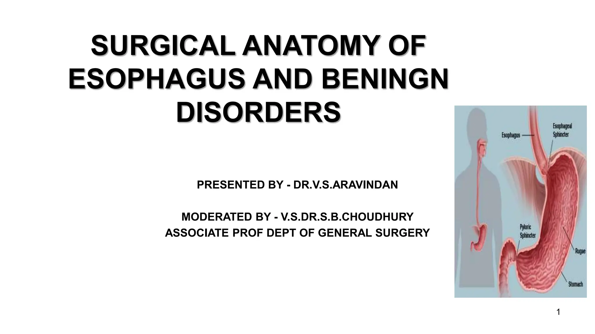
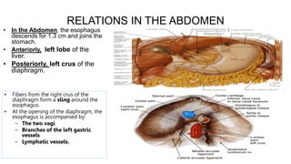
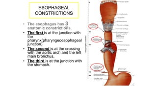
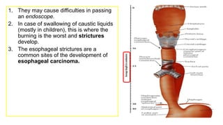
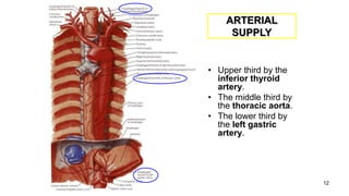
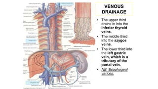
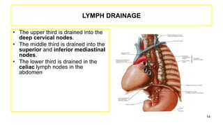
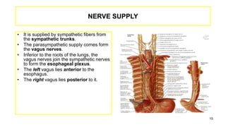
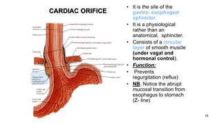
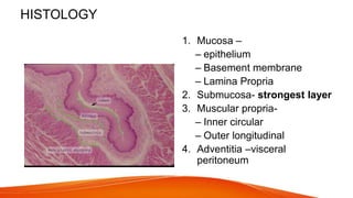
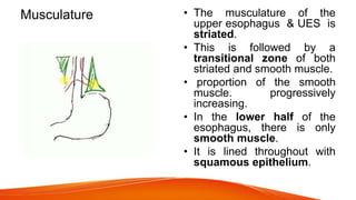
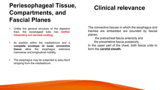
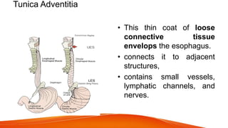
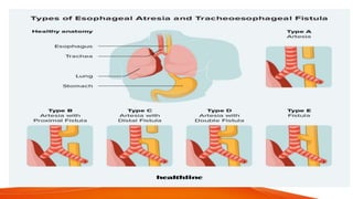
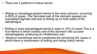
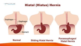
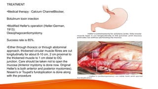
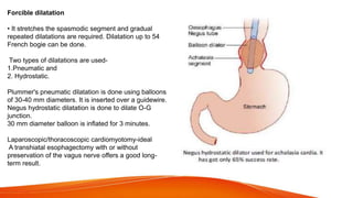
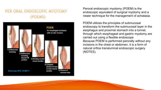
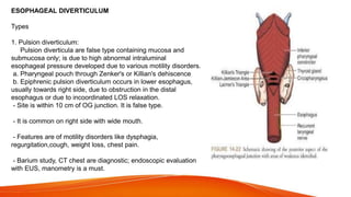
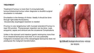
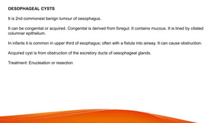
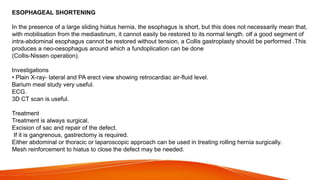
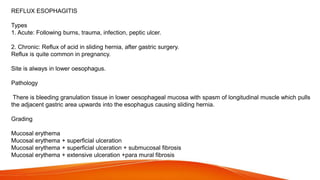
SURGICAL ANATOMY OF
ESOPHAGUS AND BENINGN
DISORDERS
PRESENTED BY - DR.V.S.ARAVINDAN
MODERATED BY - V.S.DR.S.B.CHOUDHURY
ASSOCIATE PROF DEPT OF GENERAL SURGERY
1
EMBRYOLOGY OF
OESOPHAGUS
• At the 4th week, a respiratory
diverticulum appears in the
ventral wall of the foregut at
the border with the pharynx.
• A tracheoesophageal septum
forms, gradually separating
the ventral respiratory
diverticulum from the dorsal
part of the fore gut hence,
giving rise to a ventral
Respiratory primodium and
dorsally the Oesophagus.
• With the descent of the heart
and oesophagus, the
oesophagus which was initially
short, lengthens rapidly.
ESOPHAGUS
• Oesophagus is the immediate continuation of
pharynx and conduit for food and fluids into
the stomach.
• 25 cm in length,2 cm in diameter, a flattened
muscular tube that extends from (lower
border of cricoid cartilage) C6 to cardiac
orifice of the stomach at T 10 level vertebrae.
• It runs vertically but inclines to the left from its
origin to thoracic inlet and again from T7 to
Oesophageal opening in the diaphragm.
• It is divided into 3 parts:
– 1- Cervical.
– 2- Thoracic.
– 3- Abdominal.
Abdominal
thoracic
Cervical
3
4
CERVICAL PART
• Posteriorly
• Post: Vertebral
column.
• Laterally:
• Lat: Lobes of the
thyroid gland.
• Anteriorly:
• Ant: Trachea and
the recurrent
laryngeal nerves.
RELATIONS
5
THORACIC PART
• In the thorax, it passes
downward and to the left
through superior then to
posterior mediastinum
• At the level of the sternal
angle, the aortic arch
pushes the esophagus again
to the midline.
6
ANTERIOR
RELATIONS
• Trachea
• Left recurrent
laryngeal
nerve
• Left principal
bronchus
• Pericardium
• Left atrium
Thoracic part
POSTERIOR RELATIONS – Thoracic duct
• Bodies of the thoracic vertebrae
• Thoracic duct
• Azygos vein
• Right posterior intercostal arteries
• Descending thoracic aorta (at the
lower end)
7
LATERAL RELATION
• On the Right side:
• Right mediastinal pleura
• Terminal part of the azygos vein.
• On the Left side:
• Left mediastinal pleura
• Left subclavian artery
• Aortic arch
• Thoracic duct
8
9
RELATIONS IN THE ABDOMEN
• In the Abdomen, the esophagus
descends for 1.3 cm and joins the
stomach.
• Anteriorly, left lobe of the
liver.
• Posteriorly, left crus of the
diaphragm.
• Fibers from the right crus of the
diaphragm form a sling around the
esophagus.
• At the opening of the diaphragm, the
esophagus is accompanied by:
– The two vagi
– Branches of the left gastric
vessels
– Lymphatic vessels.
ESOPHAGEAL
CONSTRICTIONS
• The esophagus has 3
anatomic constrictions.
• The first is at the junction with
the
pharynx(pharyngeoesophageal
junction).
• The second is at the crossing
with the aortic arch and the left
main bronchus.
• The third is at the junction with
the stomach.
1. They may cause difficulties in passing
an endoscope.
2. In case of swallowing of caustic liquids
(mostly in children), this is where the
burning is the worst and strictures
develop.
3. The esophageal strictures are a
common sites of the development of
esophageal carcinoma.
12
ARTERIAL
SUPPLY
• Upper third by the
inferior thyroid
artery.
• The middle third by
the thoracic aorta.
• The lower third by
the left gastric
artery.
VENOUS
DRAINAGE
• The upper third
drains in into the
inferior thyroid
veins.
• The middle third
into the azygos
veins.
• The lower third into
the left gastric
vein, which is a
tributary of the
portal vein.
• NB. Esophageal
varices.
LYMPH DRAINAGE
• The upper third is drained into the
deep cervical nodes.
• The middle third is drained into the
superior and inferior mediastinal
nodes.
• The lower third is drained in the
celiac lymph nodes in the
abdomen.
14
NERVE SUPPLY
• It is supplied by sympathetic fibers from
the sympathetic trunks.
• The parasympathetic supply comes form
the vagus nerves.
• Inferior to the roots of the lungs, the
vagus nerves join the sympathetic nerves
to form the esophageal plexus.
• The left vagus lies anterior to the
esophagus.
• The right vagus lies posterior to it.
15
16
CARDIAC ORIFICE
• It is the site of the
gastro- esophageal
sphincter.
• It is a physiological
rather than an
anatomical, sphincter.
• Consists of a circular
layer of smooth muscle
(under vagal and
hormonal control).
• Function:
• Prevents (GER)
regurgitation (reflux)
• NB. Notice the abrupt
mucosal transition from
esophagus to stomach
(Z- line)
HISTOLOGY
1. Mucosa –
– epithelium
– Basement membrane
– Lamina Propria
2. Submucosa- strongest layer
3. Muscular propria-
– Inner circular
– Outer longitudinal
4. Adventitia –visceral
peritoneum
Musculature • The musculature of the
upper esophagus & UES is
striated.
• This is followed by a
transitional zone of both
striated and smooth muscle.
• proportion of the smooth
muscle. progressively
increasing.
• In the lower half of the
esophagus, there is only
smooth muscle.
• It is lined throughout with
squamous epithelium.
Periesophageal Tissue,
Compartments, and
Fascial Planes
• Unlike the general structure of the digestive
tract, the esophageal tube has neither
mesentery nor serosal coating.
• Its position within the mediastinum and a
complete envelope of loose connective
tissue allow the esophagus extensive
transverse and longitudinal mobility.
• The esophagus may be subjected to easy blunt
stripping from the mediastinum.
Clinical relevance
The connective tissues in which the esophagus and
trachea are embedded are bounded by fascial
planes,
the pretracheal fascia anteriorly and
the prevertebral fascia posteriorly.
In the upper part of the chest, both fascia unite to
form the carotid sheath.
Tunica Adventitia
• This thin coat of loose
connective tissue
envelops the esophagus.
• connects it to adjacent
structures,
• contains small vessels,
lymphatic channels, and
nerves.
Tunica Muscularis • The tunica muscularis coats
the lumen of the esophagus
in two layers :
• the external muscle layer
parallels the longitudinal
axis of the tube,
• the muscle fibers of the
inner layer are arranged in
the horizontal axis.
• For this reason, these
muscle layers are classically
called longitudinal and
circular, respectively.
Tela Submucosa
• The submucosa is the connective
tissue layer that lies between the
muscular coat and the mucosa.
• It contains a meshwork of small
blood and lymph vessels, nerves,
and mucous glands.
• The duct of deep esophageal
glands pierce the muscularis
mucosae.
The mucous layer is composed of three
components:
the muscularis mucosae,
the tunica / lamina propria, and
the inner lining of nonkeratinizing stratified
squamous epithelium
Tunica Mucosa
Physiology
• The upper oesophageal sphincter starts at the upper
border of oesophagus and is about 3–5 cm in length and
functions during the act of swallowing.
• The lower oesophageal sphincter is situated at lower
portion of oesophagus. It is also 3–5 cm in length and
functions to prevent oesophageal reflux.Middle portion of
oesophagus shows active peristalsis. The waves are
weaker in the upper part, becoming gradually stronger
towards the lower portion.
ATRESIA
• Atresia is absence of opening (lumen)/ noncanalization.
• Atresia, in which a thin, noncanalized cord replaces a
segment of esophagus, is more common. Atresia occurs
most commonly at or near the tracheal bifurcation and
usually is associated with a fistula connecting the upper or
lower esophageal pouches to a bronchus or the trachea.
This abnormal connection can result in aspiration,
suffocation, pneumonia, or severe fluid and electrolyte
imbalances
STENOSIS
• The narrowing generally is caused by
- fibrous thickening of the submucosa, atrophy of the muscularis
propria, and secondary epithelial damage.
• Stenosis most often is due to inflammation and scarring, which
may be caused by chronic gastroesophageal reflux, irradiation, or
caustic (acid) injury.
• Stenosis associated dysphagia usually is progressive; difficulty
eating solids typically occurs long before problems with liquids.
HIATUS HERNIA
• Hiatus hernia is the herniation or protrusion of part of
the stomach through the oesophageal hiatus (opening)
of the diaphragm.
• Congenital hiatal hernias are recognized in infants and
children, but many are acquired in later life. Hiatal
hernia is symptomatic in fewer than 10% of adults, and
these cases are generally associated with other causes
of LES incompetence. Symptoms, including heartburn
and regurgitation of gastric juices, are similar to GERD.
• The basic defect is the failure of the muscle fibres of the diaphragm
that form the margin of the oesophageal hiatus. This occurs due to
shortening of the esophagus which may be congenital or acquired.
• More commonly, it is acquired due to secondary factors which
cause fibrous scarring of the oesophagus as follows:
• Degeneration of muscle due to aging.
• Increased intra-abdominal pressure such as in pregnancy,
abdominal tumours etc.
• Recurrent oesophageal regurgitation and spasm causing
inflammation and fibrosis.
• Increase in fatty tissue in obese people causing decreased
muscular elasticity of diaphragm
• There are 3 patterns in hiatus hernia :
• Sliding or oesophago-gastric hernia is the most common, occurring
in 85% of cases. The herniated part of the stomach appears as
supradiaphragmatic bell due to sliding up on both sides of the
oesophagus.
• Rolling or para-oesophageal hernia is seen in 10% of cases.This is a
true hernia in which cardiac end of the stomach rolls up para-
oesophageally, producing an intrathoracic sac.
• Mixed or transitional hernia constitutes the remaining 5% cases in
which there is combination of sliding and rolling hiatus hernia
Treatment
• Mainly it is surgical; the hernia is reduced and
diaphragmatic opening repaired. Early cases and those
unfit for surgery may be treated conservatively to reduce
reflux oesophagitis by measures such as (i) sleeping with
head and chest raised, (ii) avoidance of smoking, (iii) use
of drugs that reduce acidity (antacids and proton pump
inhibitors), (iv) reduction of obesity and (v) attention to the
causes which raises intra-abdominal pressure
ESOPHAGEAL VARICES
• Esophageal varices are tortuous, dilated and engorged
esophageal veins, seen along the longitudinal axis of esophagus.
They occur as a result of elevated pressure in the portal venous
system, most commonly in cirrhosis of the liver .
• Less common causes are: portal vein thrombosis, hepatic vein
thrombosis (Budd-Chiari syndrome) and pylephlebitis. The lesions
occur as a result of bypassing of portal venous blood from the
liver to the oesophageal venous plexus. The increased venous
pressure in the superficial veins of the esophagus may result in
ulceration and massive bleeding.
ESOPHAGEAL MOTILITY DISORDERS
MOTILITY DISORDERS
• Motility disorders of the esophagus run on a
continum from hypomotile to hypermotile
dysfunction, with intermediate types in between.
• There are both primary and secondary motor
disorders of the esophagus. Most esophageal
motility disorders fall into one of five primary
motor disorders: achalasia, DES, nutcracker
esophagus, hypertensive LES, and
ineffective esophageal motility (IEM)
.
• Secondary motor disorders of the esophagus
result from progression of other diseases such
as collagen vascular and neuromuscular
diseases.
MANOMETRY
Esophageal manometry is the most accurate method to assess the coordination and pressure of the lower
esophageal sphincter (LES) and the esophageal body.
Patients with GERD may have manometric findings of a defective LES or impaired esophageal motility.
Manometry is an important component in the preoperative workup of patients who are candidates for antireflux
surgery.
First, this form of testing excludes achalasia that may be occasionally misdiagnosed as GERD.
Second, esophageal manometry characterizes the esophageal motility, and this information will be used to
determine the surgical approach (Nissen or partial fundoplication).
Finally, manometry enables measurement of the precise location of the LES for accurate pH probe placement
Usually a train of five pressure transducers are bound together with the transducers placed at 5 cm intervals from
the tip and oriented radially at 72 degrees from each other around the circumference of the catheter. Recent
introduction and clinical application of high-resolution manometry (HRM) has made esophageal manometry
simple, fast,and accurate
Esophageal Manometry Analysis
Assessment of EGJ:
Resting pressure (Phigh or low)
Does EJG relax adequately with wet
swallows
Hiatus hernia
Esophageal body response to wet
swallows
Is peristalsis present or absent?
Peristaltic amplitude (high or low)
Is spasm present
Sphincters
Upper esophageal sphincter (UES)
3 cm long
Resting pressure: 20-60 mm Hg
Lower esophageal sphincter (LES)
Not an anatomically defined sphincter in man
Called "zone of high pressure"
Serves to reduce gastric regurgitation and
reflux
Located in the distal 3-5 cm of the esophagus
Normal resting pressure: 10-20 mm Hg
Esophageal Manometry Metrics
IRP (Integrated Relaxation Pressure)
DCI (Distal Contractile Integral)
Distal Latency
ACHALASIA
BACKGROUND
The literal meaning of achalasia is "failure to relax," which is said of any sphincter that
remains in a constant state of tone with periods of relaxation.
The incidence is 6 per 100,000 persons per year and is seen in young women and
middle-aged men and women alike.
Its pathogenesis is presumed to be idiopathic or infectious neurogenic degeneration.
Severe emotional stress, trauma, drastic weight reduction, and Chagas' disease
(parasitic infection with Trypanosoma cruzi) have also been implicated.
Prevailing theories support the model that the destruction of the nerves to the LES is
the primary pathology and that degeneration of the neuromuscular function of the body
of the esophagus is secondary.
This degeneration results in both hypertension of the LES and failure of the LES to relax on pharyngeal
swallowing, as well as pressurization of the esophagus, esophageal dilation, and a resultant loss of
progressive peristalsis.
Vigorous achalasia is seen in a subset of patients presenting with dysphagia. In these patients, the LES is
hypertensive and fails to relax, as seen in achalasia.
Achalasia is also known to be a premalignant condition of theesophagus. Over a 20-year period, a patient will
have up to an 8% chance of developing carcinoma.
SYMPTOMS AND DIAGNOSIS
The classic triad of presenting symptoms consists of dysphagia, regurgitation, and weight loss.
The dysphagia that patients experience begins with liquids and progresses to solids.
They eat slowly and use large volumes of water to help wash the food down into the stomach.
As the water builds up pressure, retrosternal chest pain is experienced and can be quite severe until the
LES opens and is quickly relieving. Regurgitation of undigested, foul-smelling foods is common, and with
progressive disease, aspiration can become life-threatening. Pneumonia, lung abscess, and
bronchiectasis often result from long-standing achalasia.
The diagnosis of achalasia is usually made from an esophagram and a motility study.
The esophagram will show a dilated
esophagus with a distal narrowing
referred to as the classic "bird's beak"
appearance of the barium-filled
esophagus.
Sphincter spasm and delayed
emptying through the LES, as well as
dilation of the esophageal body, are
observed.
A lack of peristaltic waves in the body
and failure of relaxation of the LES
are observed. Lack of a gastric air
bubble is a common finding on the
upright portion of the esophagram
Manometry is the gold standard test for diagnosis and will
help eliminate other potential esophageal motility disorders.
In typical achalasia, the manometry tracings show five classic
findings: two abnormalities of the LES and three of the
esophageal body.
The LES will be hypertensive with pressures usually above
35 mm Hg, but more importantly, it will fail to relax with
deglutition.
The body of the esophagus will have a pressure above
baseline (pressurization of the esophagus) from incomplete
air evacuation, simultaneous contractions with no evidence of
progressive peristalsis, and low-amplitude waveforms
indicating a lack of muscular tone.
These five findings provide a diagnosis of achalasia.
An endoscopy is performed to evaluate the mucosa for
evidence of esophagitis or cancer. It otherwise contributes
little to the diagnosis of achalasia.
TREATMENT
•Medical therapy : Calcium ChannelBlocker,
Botulinum toxin iniection
•Modified Heller's operation (Heller-German,
1913):
Oesophagocardiomyotomy.
Success rate is 85%.
•Either through thoracic or through abdominal
approach, thickened circular muscle fibres are cut
longitudinally for about 8-10 cm, 2 cm proximal to
the thickened muscle to 1 cm distal to OG
junction. Care should be taken not to open the
mucosa (Anterior myotomy is done now. Original
Heller's is both anterior and posterior myotomies).
Nissen's or Toupet's fundoplication is done along
with the procedure
Forcible dilatation
• It stretches the spasmodic segment and gradual
repeated dilatations are required. Dilatation up to 54
French bogie can be done.
Two types of dilatations are used-
1.Pneumatic and
2. Hydrostatic.
Plummer's pneumatic dilatation is done using balloons
of 30-40 mm diameters. It is inserted over a guidewire.
Negus hydrostatic dilatation is done to dilate O-G
junction.
30 mm diameter balloon is inflated for 3 minutes.
Laparoscopic/thoracoscopic cardiomyotomy-ideal
A transhiatal esophagectomy with or without
preservation of the vagus nerve offers a good long-
term result.
Peroral endoscopic myotomy (POEM) is the
endoscopic equivalent of surgical myotomy and a
newer technique for the management of achalasia.
POEM utilizes the principles of submucosal
endoscopy to transform the submucosal layer in the
esophagus and proximal stomach into a tunnel,
through which esophageal and gastric myotomy are
carried out using a flexible endoscope
Because POEM is performed perorally without any
incisions in the chest or abdomen, it is a form of
natural orifice transluminal endoscopic surgery
(NOTES).
ESOPHAGEAL DIVERTICULUM
Types
1. Pulsion diverticulum:
Pulsion diverticula are false type containing mucosa and
submucosa only; is due to high abnormal intraluminal
esophageal pressure developed due to various motility disorders.
a. Pharyngeal pouch through Zenker's or Killian's dehiscence
b. Epiphrenic pulsion diverticulum occurs in lower esophagus,
usually towards right side, due to obstruction in the distal
esophagus or due to incoordinated LOS relaxation.
- Site is within 10 cm of OG junction. It is false type.
- It is common on right side with wide mouth.
- Features are of motility disorders like dysphagia,
regurgitation,cough, weight loss, chest pain.
- Barium study, CT chest are diagnostic; endoscopic evaluation
with EUS, manometry is a must.
Treatment: Diverticulopex/diverticulectomv (excision)+ esophageal
myotomy (Heller's) + repair of associated hiatus hernia/antirefl ux
procedure.
2. Traction diverticulum:
Occurs in mid-oesophagus or in parabronchial region, is due to
mediastinal granulomatous disease like tuberculosis.
Traction diverticulum is a true type containing all layers in its wall and
is due to traction by the healing fibrosing mediastinal lymph nodes.
It is seen commonly towards right side. It has got wide mouth and it
rests on the spine.
Presentation is dysphagia, chest pain and regurgitation.
CT scan (chest), barium study, manometry, endoscopy to assess
mucosa with EUS, blood test for tuberculosis (ESR, peripheral smear)
are the investigations.
Treatment: Treating the cause like tuberculosis, histoplasmosis.
Diverticulum less than 2 cm is observed; progressive symptoms,size
> 2 cm needs surgery.
BENIGN TUMOURS OF THE OESOPHAGUS
Benign tumours of the oesophagus are rare (0.5-1% of all oesophageal tumours).
It grows slowly by expansion, compressing surrounding structures. It never infiltrates or spreads.
It can be solid, cystic, polypoid.
It is usually in submucosal plane.
It can cause obstruction/regurgitation/aspiration/ mediastinal compression.
It can be squamous papilloma/polyp/inflammatory pseudo tumour/ leiomvoma (commonest benign
tumour o oesophagus- 65%)/neurofibroma/ rhabdomvoma/ lipoma.
True adenoma in esophagus is very rare.
Features may be asymptomatic (85%-identified incidentally during contrast X-ray/endoscopy);
dysphagia/airway obstruction/pneumonia/spluttering during swallowing; stridor/regurgitation.
verticulopexy, long esophageal myotomy.
Leiomyoma (commonest -65%) is smooth, sessile, lobulated, firm, with grey-white whorled
appearance.
Only when leiomyoma reaches 5 cm in size it causes obstruction.
Multiple localised leiomyomas can occur which can be enucleated independently.
True diffuse leiomyomas can occur occasionally in females (4%) in lower oesophagus, as diffuse
hyperplasia
and thickening of both muscular layers; often as part of the Alport's syndrome which needs total
oesophagectomy with gastric pull up, even though benign. Benign leiomyoma of esophagus rarely
turns into
• leiomyosarcoma. 90% of esophageal leiomyomas occur in lower third © of the oesophagus.
Leiomyomas are common in men in 5th decade.
Leiomyoma which expresses the c-kit oncogene (CD117) is considered as GIST.
• Investigations-barium swallow X-ray (smooth circular outline/eccentric filling
defect)/oesophagoscopy/endosonography/
CT scan.
TREATMENT
Treatment-if tumour is more than 5 cm/symptomatic
tumour/intraluminal tumour when diagnosis is doubtful surgical
enucleation is indicated.
Enucleation is the therapy of choice. Ideally it should be done
through right-sided thoracotomy.
Occasionally esophageal resection is needed
If tumour is very large/tumour with mucosal ulceration/if tumour is
near OG junction. Thoracoscopic resection can be done. Leak,
empyema, sepsis and stricture are the occasional Complications
Unlike in the stomach and intestine (gastric leiomyoma more than
6 cm intestinal leiomyoma more than 4 cm are potentially
malignant) increased size of the oesophageal leiomyoma does not
predispose the malignant transformation.
OESOPHAGEAL CYSTS
It is 2nd commonest benign tumour of oesophagus.
It can be congenital or acquired. Congenital is derived from foregut. It contains mucous. It is lined by ciliated
columnar epithelium.
In infants it is common in upper third of esophagus; often with a fistula into airway. It can cause obstruction.
Acquired cyst is from obstruction of the excretory ducts of oesophageal glands.
Treatment: Enucleation or resection
ESOPHAGEAL SHORTENING
In the presence of a large sliding hiatus hernia, the esophagus is short, but this does not necessarily mean that,
with mobilisation from the mediastinum, it cannot easily be restored to its normal length. olf a good segment of
intra-abdominal esophagus cannot be restored without tension, a Collis gastroplasty should be performed .This
produces a neo-oesophagus around which a fundoplication can be done
(Collis-Nissen operation).
Investigations
• Plain X-ray- lateral and PA erect view showing retrocardiac air-fluid level.
Barium meal study very useful.
ECG.
3D CT scan is useful.
Treatment
Treatment is always surgical.
Excision of sac and repair of the defect.
If it is gangrenous, gastrectomy is required.
Either abdominal or thoracic or laparoscopic approach can be used in treating rolling hernia surgically.
Mesh reinforcement to hiatus to close the defect may be needed.
GERD(Gastro-Oesophageal Reflux)
• It is due to decreased function of lower oesophageal sphincter thus permitting
regurgitation of gastric contents into oesophagus. Other causes of gastro-
oesophageal reflux are pregnancy, hiatus hernia, scleroderma, excessive use of
tobacco and alcohol, and drugs that relax the smooth muscle (anticholinergic,
beta-adrenergic drugs and calcium-channel blockers).
• The symptoms of oesophageal reflux include substernal pain, heartburn and
regurgitation.
• The treatment consists of:1. Elevation of the head of bed at night.2. Avoiding food
at least 3 h before bedtime.3. Antacids.4. Drugs that increase tone of lower
oesophageal sphincter,e.g. metoclopramide.5. H2 receptor antagonists, e.g.
cimetidine and ranitidine.6. Avoiding smoking, alcohol, caffeine, chocolates,
mintsand carbonated drinks.7. Antireflux surgery, e.g. Nissen’s fundoplication.
REFLUX ESOPHAGITIS
Types
1. Acute: Following burns, trauma, infection, peptic ulcer.
2. Chronic: Reflux of acid in sliding hernia, after gastric surgery.
Reflux is quite common in pregnancy.
Site is always in lower oesophagus.
Pathology
There is bleeding granulation tissue in lower oesophageal mucosa with spasm of longitudinal muscle which pulls
the adjacent gastric area upwards into the esophagus causing sliding hernia.
Grading
Mucosal erythema
Mucosal erythema + superficial ulceration
Mucosal erythema + superficial ulceration + submucosal fibrosis
Mucosal erythema + extensive ulceration +para mural fibrosis
Clinical Features
It is a part of GORD.
Pain and burning sensation in retrosternal area often
referred to shoulder, neck, arm.
Heart burn is common.
Dysphagia.
Anaemia.
Diagnosis
Barium meal X-ray.
Gastroscopy and biopsy.
Barrett's ulcer is an ulcer with gastric (columnar)
metaplasia in lower oesophagus.
Treatment
Antacids
H2 blockers
Proton pump inhibitors-Main method and more
effective.
Omeprazole 20 mg BD one hour before food
(Morning) for 6 months
Lansoprazole 30 mg
Pantoprazole 40 mg
Esomeprazole 20 mg
• Rabeprazole 20 mg (can be given with food).
Prokinetic drugs like metochlopramide,
domperidone, cisapride, mosapride.
Treating GORD and associated causes. By
fundoplication and other surgeries.
Resection in severe cases.
BARRET'S ESOPHAGUS
Described by Norman Barrett, British in 1950
It is the metaplastic changes in the mucosa of the
esophagus as the result of GORD.
Squamous epithelium of lower end of the oesophagus is
replaced by diseased columnar epithelium columnar
metaplasia).
There is macroscopic visible length of columnar mucosa
with microscopic features of intestinal metaplasia.
It affects lower esophagus commonly often middle
esophagus
also.
Types (Based on Length)
If the length of metaplasia is more than 3 cm,
it is called as long segment Barrett's
oesophagus-classic Barrett type.
If the length is less than 3 cm, it is called as
short segment Barrett's oesophagus.
Histological Types
Gastric type: Contains chief and parietal cells.
Intestinal type: Contains goblet cells.
Junctional type: Contains mucous glands alike
of gastric cardia.
Clinical Features
Features of GORD.
Haematemesis.
Common in men; common in whites.
Complications of Barrett's esophagus
Ulcerations and stricture
Dysphagia
Bleeding
Perforation
Adeno carcinoma of O-G junction (25 times
more common)
MANAGEMENT
Regular endoscopic biopsy and surveillance for low grade dysplasia.
Ablation of Barrett's oesophagus by laser.
Photodynamic therapy-through endoscopy.
Argon beam coagulation.
Proton pump inhibitors-high dose for 3-6 months.
Antireflux treatment by surgery.
Resection-Always better choice-for high grade dysplasia.
Transhiatal oesophagectomy is preferred.
Endoscopic mucosal resection.
ESOPHAGEAL PERFORATION
Causes
Instrumental injuries-commonest cause, 75% commonest site is just above the level
of crico pharyngeus.
Foreign bodies.
Alkali injuries.
Carcinoma oesophagus 1%.
Boerhaave's syndrome 15%.
Trauma 9%.
Surgical trauma (Vagotomy, thyroidectomy, Heller's operation, pneumonectomy,
spine surgery).
Clinical Features
Chest pain, vomiting, shock, subcutaneous
emphysema.
Investigations
Chest X-ray-shows pneumomediastinum.
CT scan.
Complications
Mediastinitis.
Septicaemia.
Empyema, ARDS.
TREATMENT
Conservative treatment:
It is advocated in small perforations due to instrument where
there is minimal air leak and contamination of mediastinum with
less septic load. Crepitus should be absent; pleura should
be clear and without any obstruction.
Treatment is -antibiotics, nutrition (TPN/enteral through tube),
fluid management, proper observation and monitoring the
patient by repeated blood counts, and imaging.
Biodegradable removable self expanding stents also can be
used.
Thoracotomy, proper saline wash to pleura, mediastinum and
repair with buttressing the area using pedicled intercostal
musculopleural flap is done. Nasogastric tube for long duration,
jejunostomy tube for feeding, ICT for drainage is essential.
Often in late cases decortication of lung is needed to achieve
full expansion of the lung.
FOREIGN BODIES IN ESOPHAGUS
Common Foreign Bodies
Coins, metals, plastics.
Dentures.
Pins, toothpicks, batteries.
Fish or meat bones-dangerous-40%.
Food (meat-common/vegetables) impaction-45%.
Sites of Impaction in Oesophagus
Cervical constriction-C6.
Broncho-aortic constriction- T4.
Diaphragmatic constriction-T10.
Pre-existing malignancy or inflammatory stricture
site.
Features
• Sudden dysphagia with chest pain and breathlessness.
Later features of shock, sepsis, mediastinitis, empyema.
Management
X-ray shows site and level of the F/B.
Endoscopic removal can be tried.
Impacted large F/B should be removed by thoracotomy.
Antibiotics, jejunostomy, TPN are required.
PLUMMER-VINSON SYNDROME (PATERSON-KELLY
SYNDROME)
Here esophageal webs are seen in uppermost portion of
the esophagus with spasm of circular muscle fibres.
Common in patients with long standing iron deficiency
anaemia.
Common in females.
Superficial glossitis, cheilitis, koilonychia commonly seen.
Splenomegaly may be present.
In oesophageal webs, mucosa is hyperkeratotic, friable,
desquamated.
It is a premalignant condition and presents with severe
dysphagia.
Oesophagoscopy and biopsy is required to rule out
malignancy.
TREATMENT
Oral iron-ferrous sulphate 300 mg TDS with vitamins.
Blood transfusion is given when there is severe anaemia
© (Transfusion of packed cells).
© IV or IM iron therapy.
Once anaemia comes under control, webs will clear and
patient can swallow.
Follow-up endoscopy is a must.
Dilatation of web may be required.
CORROSIVE STRICTURE OF OESOPHAGUS
Corrosives are commonest cause of oesophageal stricture.
Mainly due to ingestion of alkali (sodium hydroxide), occasionally
due to acid (sulphuric acid, nitric acid).
Acid commonly damages the stomach.
It causes extensive inflammation of the mucosa with
perioesophagitis which, if not treated leads to multiple strictures in
esophagus.
Acute phase lasts for 3 weeks.
Damage is more in lower 1/3rd of oesophagus.
Alkali is odorless and tasteless and so large volume is ingested.
Alkali causes liquefaction, saponification and thrombosis of vessels
and later leading to fibrosis and stricture.
Acid causes intense pylorospasm, antral pooling of
acid,coagulation necrosis and eschar formation.
Severity depends on type of agent, its concentration and volume.
Features of esophageal corrosive lesion
Acute/immediate
- Severe pain, shock, laryngeal oedema -
Mediastinitis, septicaemia,
haemorrhage,perforation
Late/chronic
- Dysphagia
- Stricture-50%
- Severe malnutrition
- Recurrent respiratory infection
- Oesophageal shortening
- Malignant changes
- Tracheo-oesophageal fi stula formation
-Corrosive strictures can be multiple. Damage is
more in lower 1/3rdof oesophagus
TREATMENT
Acute phase management: Neutralisation with vinegar
or citrus food if it is alkali ingestion (If pH of the
solution is less than 11.5 then damage is less); it is
with antacids, milk, egg whites if it is acid ingestion.
Early endoscopy is needed to assess the severity and
extent.
Emetics and NaHCO3 are avoided as they may
precipitate perforation.
In 1st degree burns: 48 hours observation; oral feeds
are started once patient swallows saliva painlessly.
Regular follow-up endoscopy at 1st, 2nd and 8th
months.
2nd and 3rd degree burns: They are treated with fluid
therapy, antibiotics, nutrition, resuscitation, PPIs,
aerosolised steroids, fiberoptic guided airway
intubation if needed / tracheostomy;
endoscopic esophageal stenting, feeding jejunostomy,
laparoscopy for evaluation.
Unstable patients have high mortality. Laparotomy is done in such patients. If esophagus and stomach shows
full thickness necrosis, resection of these parts is done and end cervical oesophagostomy with jejunostomy is
done.
• When in doubt re-exploration for second look is done after 36-48 hours to assess the stomach.
Careful early gentle repeated endoscopy is mandatory.
Though advocated often for 2-3 weeks, use of steroids is controversial and under debate.
Antibiotics if there are chances of aspiration or perforation.
Regular esophageal dilatation is done for stricture.
Stricture is dilated endoscopically using guidewire.
Dilators are solid type with gradual increase in diameters.
Often radiologic C-ARM guidance is needed to pass the guide wire into the stomach.
Dilatation should be done up to minimum 16 mm diameter. Pneumatic or balloon dilatation is also practiced.
Gum elastic dilators, mercury weighted dilators, Eder-Puestow dilators, Savary-Gilliard dilators, balloon dilators
are other dilators used.
Earlier, blind dilatation using oesophageal bougies of increased diameters was the practice, which is followed
even now in many places, but chances of perforation is more.
Oesophageal resection in corrosive strictures is
technically difficult and may be hazardous.
Oesophageal bypass is better and easier, and
following later by regular endoscopic
surveillance for malignant transformation (5%).
Colon is used as replacing conduit as stomach
itself may be diseased in corrosive pathology.
In multiple strictures oesophageal resection
and colonic transposition may be advocated if
risk of malignancy is considered.
Malignancy can develop in corrosive strictures-
5%. It is 1000 fold greater than general
population.
OTHER CAUSES OF STRICTURE
OESOPHAGUS
Peptic stricture (oesophagitis induced)
Foreign body
Postsurgical, radiotherapy
Congenital
Infection like tuberculosis
Drugs like tetracycline, vitamin C
SCHATZKI'S RINGS
© They are semicircular protrusion of lower esophageal
mucosa located at or just above the oesophagogastric
junction (squamocolumnar junction). Its under-surface is
lined by columnar gastric epithelium.
They involve only the mucosa and submucosa of the
oesophagus, not the muscle.
They present with dysphagia and reflux.
Episodic aphagia can occur causing, food bolus or meat
bone to get impacted which requires emergency rigid
oesophagoscopy to remove the food.
© 5 ml of 2.5% oral papain every 30 minutes to digest food
protein along with 50 mg meperidine IV to dislodge the
impacted food bolus can be tried initially.
© Treatment
Intermittent esophageal bougienage.
Antireflux drugs.
Ring should not be excised.
BOERHAAVE'S SYNDROME
It is a tear in the lower third of the oesophagus which
occurs when a person vomits against a closed glottis,
causing leak into the mediastinum, pleural cavity and
peritoneum.
Site :2-10 cm of posterolateral part of lower esophagus.
Investigations
• Chest X-ray shows pneumomediastinum ('V' sign of
Naclerio).
MRI/CT chest.
• Total count.
Treatment
Nil by mouth.
Antibiotics, IV fluids, TPN.
Feeding by jejunostomy.
Most often surgery with resection may be required
(thoracotomy and гераїг).
• When severe mediastinitis occurs, condition has high
mortality.
MALLORY-WEISS SYNDROME
It is seen in adults with severe prolonged vomiting,
causing longitudinal tear in the mucosa of stomach
at and just below the cardia, leading to severe
haematemesis.
Violent vomiting often may be due to migraine or
vertigo or following a bout of alcohol.
Presents with severe vomiting and later hamate
mesis, with
features of shock.
It is common in one o'clock position.Only 10% of
cases involve lower esophageal mucosa.
Investigations
Gastroscopy, Hb%, PCV, blood grouping.
During gastroscopy, if stomach is not inflated properly, 50%
cases may be missed.
Differential diagnosis
Bleeding peptic ulcer
Oesophageal varices
Erosive gastritis
Carcinoma stomach
Treatment
Conservative, as it is only a mucosal tear.
Blood transfusion.
IV fluids.
Sedation.
Haemostatic agents like vasopressin.
Endoscopic iniection therapy
surgical anat eso final.pptx anatomy ppt

surgical anat eso final.pptx anatomy ppt

  • 1.
    SURGICAL ANATOMY OF ESOPHAGUSAND BENINGN DISORDERS PRESENTED BY - DR.V.S.ARAVINDAN MODERATED BY - V.S.DR.S.B.CHOUDHURY ASSOCIATE PROF DEPT OF GENERAL SURGERY 1
  • 2.
    EMBRYOLOGY OF OESOPHAGUS • Atthe 4th week, a respiratory diverticulum appears in the ventral wall of the foregut at the border with the pharynx. • A tracheoesophageal septum forms, gradually separating the ventral respiratory diverticulum from the dorsal part of the fore gut hence, giving rise to a ventral Respiratory primodium and dorsally the Oesophagus. • With the descent of the heart and oesophagus, the oesophagus which was initially short, lengthens rapidly.
  • 3.
    ESOPHAGUS • Oesophagus isthe immediate continuation of pharynx and conduit for food and fluids into the stomach. • 25 cm in length,2 cm in diameter, a flattened muscular tube that extends from (lower border of cricoid cartilage) C6 to cardiac orifice of the stomach at T 10 level vertebrae. • It runs vertically but inclines to the left from its origin to thoracic inlet and again from T7 to Oesophageal opening in the diaphragm. • It is divided into 3 parts: – 1- Cervical. – 2- Thoracic. – 3- Abdominal. Abdominal thoracic Cervical 3
  • 4.
    4 CERVICAL PART • Posteriorly •Post: Vertebral column. • Laterally: • Lat: Lobes of the thyroid gland. • Anteriorly: • Ant: Trachea and the recurrent laryngeal nerves. RELATIONS
  • 5.
    5 THORACIC PART • Inthe thorax, it passes downward and to the left through superior then to posterior mediastinum • At the level of the sternal angle, the aortic arch pushes the esophagus again to the midline.
  • 6.
    6 ANTERIOR RELATIONS • Trachea • Leftrecurrent laryngeal nerve • Left principal bronchus • Pericardium • Left atrium Thoracic part
  • 7.
    POSTERIOR RELATIONS –Thoracic duct • Bodies of the thoracic vertebrae • Thoracic duct • Azygos vein • Right posterior intercostal arteries • Descending thoracic aorta (at the lower end) 7
  • 8.
    LATERAL RELATION • Onthe Right side: • Right mediastinal pleura • Terminal part of the azygos vein. • On the Left side: • Left mediastinal pleura • Left subclavian artery • Aortic arch • Thoracic duct 8
  • 9.
    9 RELATIONS IN THEABDOMEN • In the Abdomen, the esophagus descends for 1.3 cm and joins the stomach. • Anteriorly, left lobe of the liver. • Posteriorly, left crus of the diaphragm. • Fibers from the right crus of the diaphragm form a sling around the esophagus. • At the opening of the diaphragm, the esophagus is accompanied by: – The two vagi – Branches of the left gastric vessels – Lymphatic vessels.
  • 10.
    ESOPHAGEAL CONSTRICTIONS • The esophagushas 3 anatomic constrictions. • The first is at the junction with the pharynx(pharyngeoesophageal junction). • The second is at the crossing with the aortic arch and the left main bronchus. • The third is at the junction with the stomach.
  • 11.
    1. They maycause difficulties in passing an endoscope. 2. In case of swallowing of caustic liquids (mostly in children), this is where the burning is the worst and strictures develop. 3. The esophageal strictures are a common sites of the development of esophageal carcinoma.
  • 12.
    12 ARTERIAL SUPPLY • Upper thirdby the inferior thyroid artery. • The middle third by the thoracic aorta. • The lower third by the left gastric artery.
  • 13.
    VENOUS DRAINAGE • The upperthird drains in into the inferior thyroid veins. • The middle third into the azygos veins. • The lower third into the left gastric vein, which is a tributary of the portal vein. • NB. Esophageal varices.
  • 14.
    LYMPH DRAINAGE • Theupper third is drained into the deep cervical nodes. • The middle third is drained into the superior and inferior mediastinal nodes. • The lower third is drained in the celiac lymph nodes in the abdomen. 14
  • 15.
    NERVE SUPPLY • Itis supplied by sympathetic fibers from the sympathetic trunks. • The parasympathetic supply comes form the vagus nerves. • Inferior to the roots of the lungs, the vagus nerves join the sympathetic nerves to form the esophageal plexus. • The left vagus lies anterior to the esophagus. • The right vagus lies posterior to it. 15
  • 16.
    16 CARDIAC ORIFICE • Itis the site of the gastro- esophageal sphincter. • It is a physiological rather than an anatomical, sphincter. • Consists of a circular layer of smooth muscle (under vagal and hormonal control). • Function: • Prevents (GER) regurgitation (reflux) • NB. Notice the abrupt mucosal transition from esophagus to stomach (Z- line)
  • 17.
    HISTOLOGY 1. Mucosa – –epithelium – Basement membrane – Lamina Propria 2. Submucosa- strongest layer 3. Muscular propria- – Inner circular – Outer longitudinal 4. Adventitia –visceral peritoneum
  • 18.
    Musculature • Themusculature of the upper esophagus & UES is striated. • This is followed by a transitional zone of both striated and smooth muscle. • proportion of the smooth muscle. progressively increasing. • In the lower half of the esophagus, there is only smooth muscle. • It is lined throughout with squamous epithelium.
  • 19.
    Periesophageal Tissue, Compartments, and FascialPlanes • Unlike the general structure of the digestive tract, the esophageal tube has neither mesentery nor serosal coating. • Its position within the mediastinum and a complete envelope of loose connective tissue allow the esophagus extensive transverse and longitudinal mobility. • The esophagus may be subjected to easy blunt stripping from the mediastinum. Clinical relevance The connective tissues in which the esophagus and trachea are embedded are bounded by fascial planes, the pretracheal fascia anteriorly and the prevertebral fascia posteriorly. In the upper part of the chest, both fascia unite to form the carotid sheath.
  • 20.
    Tunica Adventitia • Thisthin coat of loose connective tissue envelops the esophagus. • connects it to adjacent structures, • contains small vessels, lymphatic channels, and nerves.
  • 21.
    Tunica Muscularis •The tunica muscularis coats the lumen of the esophagus in two layers : • the external muscle layer parallels the longitudinal axis of the tube, • the muscle fibers of the inner layer are arranged in the horizontal axis. • For this reason, these muscle layers are classically called longitudinal and circular, respectively.
  • 22.
    Tela Submucosa • Thesubmucosa is the connective tissue layer that lies between the muscular coat and the mucosa. • It contains a meshwork of small blood and lymph vessels, nerves, and mucous glands. • The duct of deep esophageal glands pierce the muscularis mucosae. The mucous layer is composed of three components: the muscularis mucosae, the tunica / lamina propria, and the inner lining of nonkeratinizing stratified squamous epithelium Tunica Mucosa
  • 23.
    Physiology • The upperoesophageal sphincter starts at the upper border of oesophagus and is about 3–5 cm in length and functions during the act of swallowing. • The lower oesophageal sphincter is situated at lower portion of oesophagus. It is also 3–5 cm in length and functions to prevent oesophageal reflux.Middle portion of oesophagus shows active peristalsis. The waves are weaker in the upper part, becoming gradually stronger towards the lower portion.
  • 24.
    ATRESIA • Atresia isabsence of opening (lumen)/ noncanalization. • Atresia, in which a thin, noncanalized cord replaces a segment of esophagus, is more common. Atresia occurs most commonly at or near the tracheal bifurcation and usually is associated with a fistula connecting the upper or lower esophageal pouches to a bronchus or the trachea. This abnormal connection can result in aspiration, suffocation, pneumonia, or severe fluid and electrolyte imbalances
  • 26.
    STENOSIS • The narrowinggenerally is caused by - fibrous thickening of the submucosa, atrophy of the muscularis propria, and secondary epithelial damage. • Stenosis most often is due to inflammation and scarring, which may be caused by chronic gastroesophageal reflux, irradiation, or caustic (acid) injury. • Stenosis associated dysphagia usually is progressive; difficulty eating solids typically occurs long before problems with liquids.
  • 27.
    HIATUS HERNIA • Hiatushernia is the herniation or protrusion of part of the stomach through the oesophageal hiatus (opening) of the diaphragm. • Congenital hiatal hernias are recognized in infants and children, but many are acquired in later life. Hiatal hernia is symptomatic in fewer than 10% of adults, and these cases are generally associated with other causes of LES incompetence. Symptoms, including heartburn and regurgitation of gastric juices, are similar to GERD.
  • 28.
    • The basicdefect is the failure of the muscle fibres of the diaphragm that form the margin of the oesophageal hiatus. This occurs due to shortening of the esophagus which may be congenital or acquired. • More commonly, it is acquired due to secondary factors which cause fibrous scarring of the oesophagus as follows: • Degeneration of muscle due to aging. • Increased intra-abdominal pressure such as in pregnancy, abdominal tumours etc. • Recurrent oesophageal regurgitation and spasm causing inflammation and fibrosis. • Increase in fatty tissue in obese people causing decreased muscular elasticity of diaphragm
  • 29.
    • There are3 patterns in hiatus hernia : • Sliding or oesophago-gastric hernia is the most common, occurring in 85% of cases. The herniated part of the stomach appears as supradiaphragmatic bell due to sliding up on both sides of the oesophagus. • Rolling or para-oesophageal hernia is seen in 10% of cases.This is a true hernia in which cardiac end of the stomach rolls up para- oesophageally, producing an intrathoracic sac. • Mixed or transitional hernia constitutes the remaining 5% cases in which there is combination of sliding and rolling hiatus hernia
  • 31.
    Treatment • Mainly itis surgical; the hernia is reduced and diaphragmatic opening repaired. Early cases and those unfit for surgery may be treated conservatively to reduce reflux oesophagitis by measures such as (i) sleeping with head and chest raised, (ii) avoidance of smoking, (iii) use of drugs that reduce acidity (antacids and proton pump inhibitors), (iv) reduction of obesity and (v) attention to the causes which raises intra-abdominal pressure
  • 32.
    ESOPHAGEAL VARICES • Esophagealvarices are tortuous, dilated and engorged esophageal veins, seen along the longitudinal axis of esophagus. They occur as a result of elevated pressure in the portal venous system, most commonly in cirrhosis of the liver . • Less common causes are: portal vein thrombosis, hepatic vein thrombosis (Budd-Chiari syndrome) and pylephlebitis. The lesions occur as a result of bypassing of portal venous blood from the liver to the oesophageal venous plexus. The increased venous pressure in the superficial veins of the esophagus may result in ulceration and massive bleeding.
  • 33.
    ESOPHAGEAL MOTILITY DISORDERS MOTILITYDISORDERS • Motility disorders of the esophagus run on a continum from hypomotile to hypermotile dysfunction, with intermediate types in between. • There are both primary and secondary motor disorders of the esophagus. Most esophageal motility disorders fall into one of five primary motor disorders: achalasia, DES, nutcracker esophagus, hypertensive LES, and ineffective esophageal motility (IEM) . • Secondary motor disorders of the esophagus result from progression of other diseases such as collagen vascular and neuromuscular diseases.
  • 34.
    MANOMETRY Esophageal manometry isthe most accurate method to assess the coordination and pressure of the lower esophageal sphincter (LES) and the esophageal body. Patients with GERD may have manometric findings of a defective LES or impaired esophageal motility. Manometry is an important component in the preoperative workup of patients who are candidates for antireflux surgery. First, this form of testing excludes achalasia that may be occasionally misdiagnosed as GERD. Second, esophageal manometry characterizes the esophageal motility, and this information will be used to determine the surgical approach (Nissen or partial fundoplication). Finally, manometry enables measurement of the precise location of the LES for accurate pH probe placement Usually a train of five pressure transducers are bound together with the transducers placed at 5 cm intervals from the tip and oriented radially at 72 degrees from each other around the circumference of the catheter. Recent introduction and clinical application of high-resolution manometry (HRM) has made esophageal manometry simple, fast,and accurate
  • 35.
    Esophageal Manometry Analysis Assessmentof EGJ: Resting pressure (Phigh or low) Does EJG relax adequately with wet swallows Hiatus hernia Esophageal body response to wet swallows Is peristalsis present or absent? Peristaltic amplitude (high or low) Is spasm present Sphincters Upper esophageal sphincter (UES) 3 cm long Resting pressure: 20-60 mm Hg Lower esophageal sphincter (LES) Not an anatomically defined sphincter in man Called "zone of high pressure" Serves to reduce gastric regurgitation and reflux Located in the distal 3-5 cm of the esophagus Normal resting pressure: 10-20 mm Hg
  • 36.
    Esophageal Manometry Metrics IRP(Integrated Relaxation Pressure) DCI (Distal Contractile Integral) Distal Latency
  • 37.
    ACHALASIA BACKGROUND The literal meaningof achalasia is "failure to relax," which is said of any sphincter that remains in a constant state of tone with periods of relaxation. The incidence is 6 per 100,000 persons per year and is seen in young women and middle-aged men and women alike. Its pathogenesis is presumed to be idiopathic or infectious neurogenic degeneration. Severe emotional stress, trauma, drastic weight reduction, and Chagas' disease (parasitic infection with Trypanosoma cruzi) have also been implicated. Prevailing theories support the model that the destruction of the nerves to the LES is the primary pathology and that degeneration of the neuromuscular function of the body of the esophagus is secondary.
  • 38.
    This degeneration resultsin both hypertension of the LES and failure of the LES to relax on pharyngeal swallowing, as well as pressurization of the esophagus, esophageal dilation, and a resultant loss of progressive peristalsis. Vigorous achalasia is seen in a subset of patients presenting with dysphagia. In these patients, the LES is hypertensive and fails to relax, as seen in achalasia. Achalasia is also known to be a premalignant condition of theesophagus. Over a 20-year period, a patient will have up to an 8% chance of developing carcinoma.
  • 39.
    SYMPTOMS AND DIAGNOSIS Theclassic triad of presenting symptoms consists of dysphagia, regurgitation, and weight loss. The dysphagia that patients experience begins with liquids and progresses to solids. They eat slowly and use large volumes of water to help wash the food down into the stomach. As the water builds up pressure, retrosternal chest pain is experienced and can be quite severe until the LES opens and is quickly relieving. Regurgitation of undigested, foul-smelling foods is common, and with progressive disease, aspiration can become life-threatening. Pneumonia, lung abscess, and bronchiectasis often result from long-standing achalasia. The diagnosis of achalasia is usually made from an esophagram and a motility study.
  • 40.
    The esophagram willshow a dilated esophagus with a distal narrowing referred to as the classic "bird's beak" appearance of the barium-filled esophagus. Sphincter spasm and delayed emptying through the LES, as well as dilation of the esophageal body, are observed. A lack of peristaltic waves in the body and failure of relaxation of the LES are observed. Lack of a gastric air bubble is a common finding on the upright portion of the esophagram
  • 41.
    Manometry is thegold standard test for diagnosis and will help eliminate other potential esophageal motility disorders. In typical achalasia, the manometry tracings show five classic findings: two abnormalities of the LES and three of the esophageal body. The LES will be hypertensive with pressures usually above 35 mm Hg, but more importantly, it will fail to relax with deglutition. The body of the esophagus will have a pressure above baseline (pressurization of the esophagus) from incomplete air evacuation, simultaneous contractions with no evidence of progressive peristalsis, and low-amplitude waveforms indicating a lack of muscular tone. These five findings provide a diagnosis of achalasia. An endoscopy is performed to evaluate the mucosa for evidence of esophagitis or cancer. It otherwise contributes little to the diagnosis of achalasia.
  • 43.
    TREATMENT •Medical therapy :Calcium ChannelBlocker, Botulinum toxin iniection •Modified Heller's operation (Heller-German, 1913): Oesophagocardiomyotomy. Success rate is 85%. •Either through thoracic or through abdominal approach, thickened circular muscle fibres are cut longitudinally for about 8-10 cm, 2 cm proximal to the thickened muscle to 1 cm distal to OG junction. Care should be taken not to open the mucosa (Anterior myotomy is done now. Original Heller's is both anterior and posterior myotomies). Nissen's or Toupet's fundoplication is done along with the procedure
  • 44.
    Forcible dilatation • Itstretches the spasmodic segment and gradual repeated dilatations are required. Dilatation up to 54 French bogie can be done. Two types of dilatations are used- 1.Pneumatic and 2. Hydrostatic. Plummer's pneumatic dilatation is done using balloons of 30-40 mm diameters. It is inserted over a guidewire. Negus hydrostatic dilatation is done to dilate O-G junction. 30 mm diameter balloon is inflated for 3 minutes. Laparoscopic/thoracoscopic cardiomyotomy-ideal A transhiatal esophagectomy with or without preservation of the vagus nerve offers a good long- term result.
  • 45.
    Peroral endoscopic myotomy(POEM) is the endoscopic equivalent of surgical myotomy and a newer technique for the management of achalasia. POEM utilizes the principles of submucosal endoscopy to transform the submucosal layer in the esophagus and proximal stomach into a tunnel, through which esophageal and gastric myotomy are carried out using a flexible endoscope Because POEM is performed perorally without any incisions in the chest or abdomen, it is a form of natural orifice transluminal endoscopic surgery (NOTES).
  • 46.
    ESOPHAGEAL DIVERTICULUM Types 1. Pulsiondiverticulum: Pulsion diverticula are false type containing mucosa and submucosa only; is due to high abnormal intraluminal esophageal pressure developed due to various motility disorders. a. Pharyngeal pouch through Zenker's or Killian's dehiscence b. Epiphrenic pulsion diverticulum occurs in lower esophagus, usually towards right side, due to obstruction in the distal esophagus or due to incoordinated LOS relaxation. - Site is within 10 cm of OG junction. It is false type. - It is common on right side with wide mouth. - Features are of motility disorders like dysphagia, regurgitation,cough, weight loss, chest pain. - Barium study, CT chest are diagnostic; endoscopic evaluation with EUS, manometry is a must.
  • 47.
    Treatment: Diverticulopex/diverticulectomv (excision)+esophageal myotomy (Heller's) + repair of associated hiatus hernia/antirefl ux procedure. 2. Traction diverticulum: Occurs in mid-oesophagus or in parabronchial region, is due to mediastinal granulomatous disease like tuberculosis. Traction diverticulum is a true type containing all layers in its wall and is due to traction by the healing fibrosing mediastinal lymph nodes. It is seen commonly towards right side. It has got wide mouth and it rests on the spine. Presentation is dysphagia, chest pain and regurgitation. CT scan (chest), barium study, manometry, endoscopy to assess mucosa with EUS, blood test for tuberculosis (ESR, peripheral smear) are the investigations. Treatment: Treating the cause like tuberculosis, histoplasmosis. Diverticulum less than 2 cm is observed; progressive symptoms,size > 2 cm needs surgery.
  • 49.
    BENIGN TUMOURS OFTHE OESOPHAGUS Benign tumours of the oesophagus are rare (0.5-1% of all oesophageal tumours). It grows slowly by expansion, compressing surrounding structures. It never infiltrates or spreads. It can be solid, cystic, polypoid. It is usually in submucosal plane. It can cause obstruction/regurgitation/aspiration/ mediastinal compression. It can be squamous papilloma/polyp/inflammatory pseudo tumour/ leiomvoma (commonest benign tumour o oesophagus- 65%)/neurofibroma/ rhabdomvoma/ lipoma. True adenoma in esophagus is very rare. Features may be asymptomatic (85%-identified incidentally during contrast X-ray/endoscopy); dysphagia/airway obstruction/pneumonia/spluttering during swallowing; stridor/regurgitation. verticulopexy, long esophageal myotomy.
  • 50.
    Leiomyoma (commonest -65%)is smooth, sessile, lobulated, firm, with grey-white whorled appearance. Only when leiomyoma reaches 5 cm in size it causes obstruction. Multiple localised leiomyomas can occur which can be enucleated independently. True diffuse leiomyomas can occur occasionally in females (4%) in lower oesophagus, as diffuse hyperplasia and thickening of both muscular layers; often as part of the Alport's syndrome which needs total oesophagectomy with gastric pull up, even though benign. Benign leiomyoma of esophagus rarely turns into • leiomyosarcoma. 90% of esophageal leiomyomas occur in lower third © of the oesophagus. Leiomyomas are common in men in 5th decade. Leiomyoma which expresses the c-kit oncogene (CD117) is considered as GIST. • Investigations-barium swallow X-ray (smooth circular outline/eccentric filling defect)/oesophagoscopy/endosonography/ CT scan.
  • 51.
    TREATMENT Treatment-if tumour ismore than 5 cm/symptomatic tumour/intraluminal tumour when diagnosis is doubtful surgical enucleation is indicated. Enucleation is the therapy of choice. Ideally it should be done through right-sided thoracotomy. Occasionally esophageal resection is needed If tumour is very large/tumour with mucosal ulceration/if tumour is near OG junction. Thoracoscopic resection can be done. Leak, empyema, sepsis and stricture are the occasional Complications Unlike in the stomach and intestine (gastric leiomyoma more than 6 cm intestinal leiomyoma more than 4 cm are potentially malignant) increased size of the oesophageal leiomyoma does not predispose the malignant transformation.
  • 52.
    OESOPHAGEAL CYSTS It is2nd commonest benign tumour of oesophagus. It can be congenital or acquired. Congenital is derived from foregut. It contains mucous. It is lined by ciliated columnar epithelium. In infants it is common in upper third of esophagus; often with a fistula into airway. It can cause obstruction. Acquired cyst is from obstruction of the excretory ducts of oesophageal glands. Treatment: Enucleation or resection
  • 53.
    ESOPHAGEAL SHORTENING In thepresence of a large sliding hiatus hernia, the esophagus is short, but this does not necessarily mean that, with mobilisation from the mediastinum, it cannot easily be restored to its normal length. olf a good segment of intra-abdominal esophagus cannot be restored without tension, a Collis gastroplasty should be performed .This produces a neo-oesophagus around which a fundoplication can be done (Collis-Nissen operation). Investigations • Plain X-ray- lateral and PA erect view showing retrocardiac air-fluid level. Barium meal study very useful. ECG. 3D CT scan is useful. Treatment Treatment is always surgical. Excision of sac and repair of the defect. If it is gangrenous, gastrectomy is required. Either abdominal or thoracic or laparoscopic approach can be used in treating rolling hernia surgically. Mesh reinforcement to hiatus to close the defect may be needed.
  • 54.
    GERD(Gastro-Oesophageal Reflux) • Itis due to decreased function of lower oesophageal sphincter thus permitting regurgitation of gastric contents into oesophagus. Other causes of gastro- oesophageal reflux are pregnancy, hiatus hernia, scleroderma, excessive use of tobacco and alcohol, and drugs that relax the smooth muscle (anticholinergic, beta-adrenergic drugs and calcium-channel blockers). • The symptoms of oesophageal reflux include substernal pain, heartburn and regurgitation. • The treatment consists of:1. Elevation of the head of bed at night.2. Avoiding food at least 3 h before bedtime.3. Antacids.4. Drugs that increase tone of lower oesophageal sphincter,e.g. metoclopramide.5. H2 receptor antagonists, e.g. cimetidine and ranitidine.6. Avoiding smoking, alcohol, caffeine, chocolates, mintsand carbonated drinks.7. Antireflux surgery, e.g. Nissen’s fundoplication.
  • 55.
    REFLUX ESOPHAGITIS Types 1. Acute:Following burns, trauma, infection, peptic ulcer. 2. Chronic: Reflux of acid in sliding hernia, after gastric surgery. Reflux is quite common in pregnancy. Site is always in lower oesophagus. Pathology There is bleeding granulation tissue in lower oesophageal mucosa with spasm of longitudinal muscle which pulls the adjacent gastric area upwards into the esophagus causing sliding hernia. Grading Mucosal erythema Mucosal erythema + superficial ulceration Mucosal erythema + superficial ulceration + submucosal fibrosis Mucosal erythema + extensive ulceration +para mural fibrosis
  • 56.
    Clinical Features It isa part of GORD. Pain and burning sensation in retrosternal area often referred to shoulder, neck, arm. Heart burn is common. Dysphagia. Anaemia. Diagnosis Barium meal X-ray. Gastroscopy and biopsy. Barrett's ulcer is an ulcer with gastric (columnar) metaplasia in lower oesophagus. Treatment Antacids H2 blockers Proton pump inhibitors-Main method and more effective. Omeprazole 20 mg BD one hour before food (Morning) for 6 months Lansoprazole 30 mg Pantoprazole 40 mg Esomeprazole 20 mg • Rabeprazole 20 mg (can be given with food). Prokinetic drugs like metochlopramide, domperidone, cisapride, mosapride. Treating GORD and associated causes. By fundoplication and other surgeries. Resection in severe cases.
  • 57.
    BARRET'S ESOPHAGUS Described byNorman Barrett, British in 1950 It is the metaplastic changes in the mucosa of the esophagus as the result of GORD. Squamous epithelium of lower end of the oesophagus is replaced by diseased columnar epithelium columnar metaplasia). There is macroscopic visible length of columnar mucosa with microscopic features of intestinal metaplasia. It affects lower esophagus commonly often middle esophagus also.
  • 58.
    Types (Based onLength) If the length of metaplasia is more than 3 cm, it is called as long segment Barrett's oesophagus-classic Barrett type. If the length is less than 3 cm, it is called as short segment Barrett's oesophagus. Histological Types Gastric type: Contains chief and parietal cells. Intestinal type: Contains goblet cells. Junctional type: Contains mucous glands alike of gastric cardia. Clinical Features Features of GORD. Haematemesis. Common in men; common in whites. Complications of Barrett's esophagus Ulcerations and stricture Dysphagia Bleeding Perforation Adeno carcinoma of O-G junction (25 times more common)
  • 59.
    MANAGEMENT Regular endoscopic biopsyand surveillance for low grade dysplasia. Ablation of Barrett's oesophagus by laser. Photodynamic therapy-through endoscopy. Argon beam coagulation. Proton pump inhibitors-high dose for 3-6 months. Antireflux treatment by surgery. Resection-Always better choice-for high grade dysplasia. Transhiatal oesophagectomy is preferred. Endoscopic mucosal resection.
  • 60.
    ESOPHAGEAL PERFORATION Causes Instrumental injuries-commonestcause, 75% commonest site is just above the level of crico pharyngeus. Foreign bodies. Alkali injuries. Carcinoma oesophagus 1%. Boerhaave's syndrome 15%. Trauma 9%. Surgical trauma (Vagotomy, thyroidectomy, Heller's operation, pneumonectomy, spine surgery).
  • 61.
    Clinical Features Chest pain,vomiting, shock, subcutaneous emphysema. Investigations Chest X-ray-shows pneumomediastinum. CT scan. Complications Mediastinitis. Septicaemia. Empyema, ARDS. TREATMENT Conservative treatment: It is advocated in small perforations due to instrument where there is minimal air leak and contamination of mediastinum with less septic load. Crepitus should be absent; pleura should be clear and without any obstruction. Treatment is -antibiotics, nutrition (TPN/enteral through tube), fluid management, proper observation and monitoring the patient by repeated blood counts, and imaging. Biodegradable removable self expanding stents also can be used. Thoracotomy, proper saline wash to pleura, mediastinum and repair with buttressing the area using pedicled intercostal musculopleural flap is done. Nasogastric tube for long duration, jejunostomy tube for feeding, ICT for drainage is essential. Often in late cases decortication of lung is needed to achieve full expansion of the lung.
  • 62.
    FOREIGN BODIES INESOPHAGUS Common Foreign Bodies Coins, metals, plastics. Dentures. Pins, toothpicks, batteries. Fish or meat bones-dangerous-40%. Food (meat-common/vegetables) impaction-45%. Sites of Impaction in Oesophagus Cervical constriction-C6. Broncho-aortic constriction- T4. Diaphragmatic constriction-T10. Pre-existing malignancy or inflammatory stricture site. Features • Sudden dysphagia with chest pain and breathlessness. Later features of shock, sepsis, mediastinitis, empyema. Management X-ray shows site and level of the F/B. Endoscopic removal can be tried. Impacted large F/B should be removed by thoracotomy. Antibiotics, jejunostomy, TPN are required.
  • 63.
    PLUMMER-VINSON SYNDROME (PATERSON-KELLY SYNDROME) Hereesophageal webs are seen in uppermost portion of the esophagus with spasm of circular muscle fibres. Common in patients with long standing iron deficiency anaemia. Common in females. Superficial glossitis, cheilitis, koilonychia commonly seen. Splenomegaly may be present. In oesophageal webs, mucosa is hyperkeratotic, friable, desquamated. It is a premalignant condition and presents with severe dysphagia. Oesophagoscopy and biopsy is required to rule out malignancy.
  • 64.
    TREATMENT Oral iron-ferrous sulphate300 mg TDS with vitamins. Blood transfusion is given when there is severe anaemia © (Transfusion of packed cells). © IV or IM iron therapy. Once anaemia comes under control, webs will clear and patient can swallow. Follow-up endoscopy is a must. Dilatation of web may be required.
  • 65.
    CORROSIVE STRICTURE OFOESOPHAGUS Corrosives are commonest cause of oesophageal stricture. Mainly due to ingestion of alkali (sodium hydroxide), occasionally due to acid (sulphuric acid, nitric acid). Acid commonly damages the stomach. It causes extensive inflammation of the mucosa with perioesophagitis which, if not treated leads to multiple strictures in esophagus. Acute phase lasts for 3 weeks. Damage is more in lower 1/3rd of oesophagus. Alkali is odorless and tasteless and so large volume is ingested. Alkali causes liquefaction, saponification and thrombosis of vessels and later leading to fibrosis and stricture. Acid causes intense pylorospasm, antral pooling of acid,coagulation necrosis and eschar formation. Severity depends on type of agent, its concentration and volume. Features of esophageal corrosive lesion Acute/immediate - Severe pain, shock, laryngeal oedema - Mediastinitis, septicaemia, haemorrhage,perforation Late/chronic - Dysphagia - Stricture-50% - Severe malnutrition - Recurrent respiratory infection - Oesophageal shortening - Malignant changes - Tracheo-oesophageal fi stula formation -Corrosive strictures can be multiple. Damage is more in lower 1/3rdof oesophagus
  • 66.
    TREATMENT Acute phase management:Neutralisation with vinegar or citrus food if it is alkali ingestion (If pH of the solution is less than 11.5 then damage is less); it is with antacids, milk, egg whites if it is acid ingestion. Early endoscopy is needed to assess the severity and extent. Emetics and NaHCO3 are avoided as they may precipitate perforation. In 1st degree burns: 48 hours observation; oral feeds are started once patient swallows saliva painlessly. Regular follow-up endoscopy at 1st, 2nd and 8th months. 2nd and 3rd degree burns: They are treated with fluid therapy, antibiotics, nutrition, resuscitation, PPIs, aerosolised steroids, fiberoptic guided airway intubation if needed / tracheostomy; endoscopic esophageal stenting, feeding jejunostomy, laparoscopy for evaluation.
  • 67.
    Unstable patients havehigh mortality. Laparotomy is done in such patients. If esophagus and stomach shows full thickness necrosis, resection of these parts is done and end cervical oesophagostomy with jejunostomy is done. • When in doubt re-exploration for second look is done after 36-48 hours to assess the stomach. Careful early gentle repeated endoscopy is mandatory. Though advocated often for 2-3 weeks, use of steroids is controversial and under debate. Antibiotics if there are chances of aspiration or perforation. Regular esophageal dilatation is done for stricture. Stricture is dilated endoscopically using guidewire. Dilators are solid type with gradual increase in diameters. Often radiologic C-ARM guidance is needed to pass the guide wire into the stomach. Dilatation should be done up to minimum 16 mm diameter. Pneumatic or balloon dilatation is also practiced. Gum elastic dilators, mercury weighted dilators, Eder-Puestow dilators, Savary-Gilliard dilators, balloon dilators are other dilators used. Earlier, blind dilatation using oesophageal bougies of increased diameters was the practice, which is followed even now in many places, but chances of perforation is more.
  • 68.
    Oesophageal resection incorrosive strictures is technically difficult and may be hazardous. Oesophageal bypass is better and easier, and following later by regular endoscopic surveillance for malignant transformation (5%). Colon is used as replacing conduit as stomach itself may be diseased in corrosive pathology. In multiple strictures oesophageal resection and colonic transposition may be advocated if risk of malignancy is considered. Malignancy can develop in corrosive strictures- 5%. It is 1000 fold greater than general population. OTHER CAUSES OF STRICTURE OESOPHAGUS Peptic stricture (oesophagitis induced) Foreign body Postsurgical, radiotherapy Congenital Infection like tuberculosis Drugs like tetracycline, vitamin C
  • 69.
    SCHATZKI'S RINGS © Theyare semicircular protrusion of lower esophageal mucosa located at or just above the oesophagogastric junction (squamocolumnar junction). Its under-surface is lined by columnar gastric epithelium. They involve only the mucosa and submucosa of the oesophagus, not the muscle. They present with dysphagia and reflux. Episodic aphagia can occur causing, food bolus or meat bone to get impacted which requires emergency rigid oesophagoscopy to remove the food. © 5 ml of 2.5% oral papain every 30 minutes to digest food protein along with 50 mg meperidine IV to dislodge the impacted food bolus can be tried initially. © Treatment Intermittent esophageal bougienage. Antireflux drugs. Ring should not be excised.
  • 70.
    BOERHAAVE'S SYNDROME It isa tear in the lower third of the oesophagus which occurs when a person vomits against a closed glottis, causing leak into the mediastinum, pleural cavity and peritoneum. Site :2-10 cm of posterolateral part of lower esophagus. Investigations • Chest X-ray shows pneumomediastinum ('V' sign of Naclerio). MRI/CT chest. • Total count. Treatment Nil by mouth. Antibiotics, IV fluids, TPN. Feeding by jejunostomy. Most often surgery with resection may be required (thoracotomy and гераїг). • When severe mediastinitis occurs, condition has high mortality.
  • 71.
    MALLORY-WEISS SYNDROME It isseen in adults with severe prolonged vomiting, causing longitudinal tear in the mucosa of stomach at and just below the cardia, leading to severe haematemesis. Violent vomiting often may be due to migraine or vertigo or following a bout of alcohol. Presents with severe vomiting and later hamate mesis, with features of shock. It is common in one o'clock position.Only 10% of cases involve lower esophageal mucosa. Investigations Gastroscopy, Hb%, PCV, blood grouping. During gastroscopy, if stomach is not inflated properly, 50% cases may be missed. Differential diagnosis Bleeding peptic ulcer Oesophageal varices Erosive gastritis Carcinoma stomach Treatment Conservative, as it is only a mucosal tear. Blood transfusion. IV fluids. Sedation. Haemostatic agents like vasopressin. Endoscopic iniection therapy