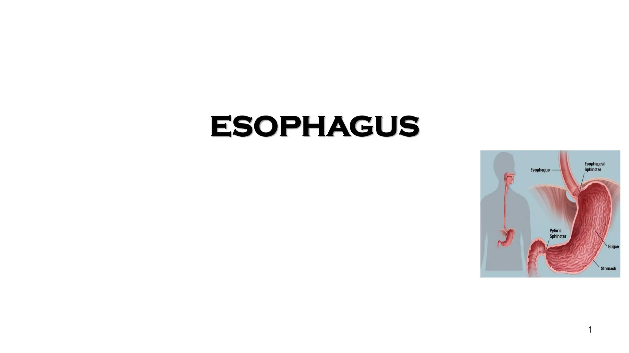
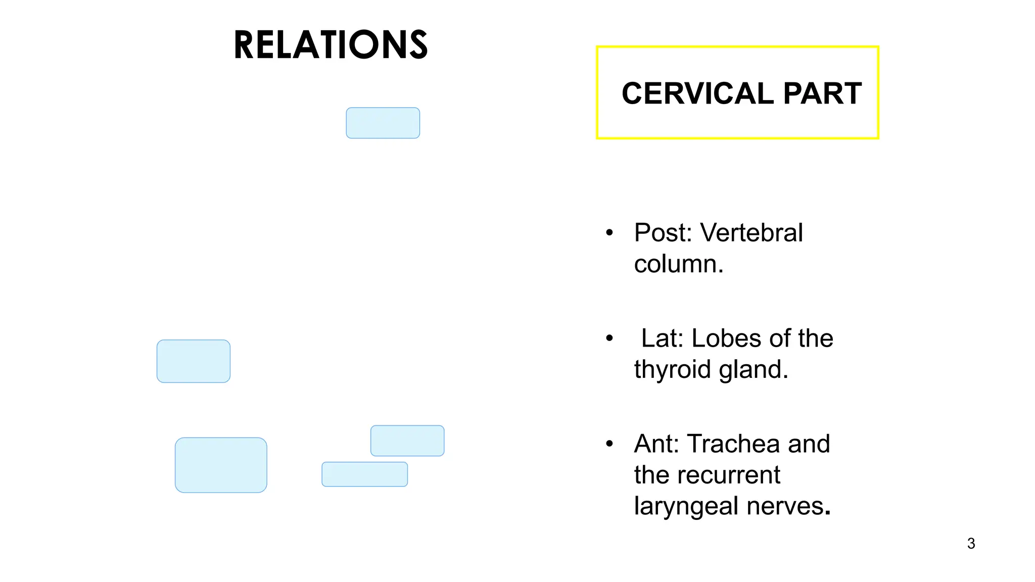
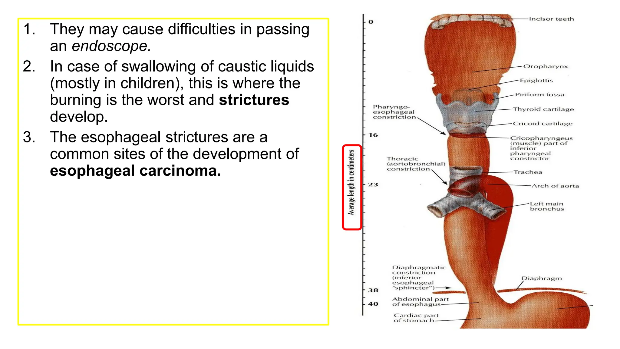
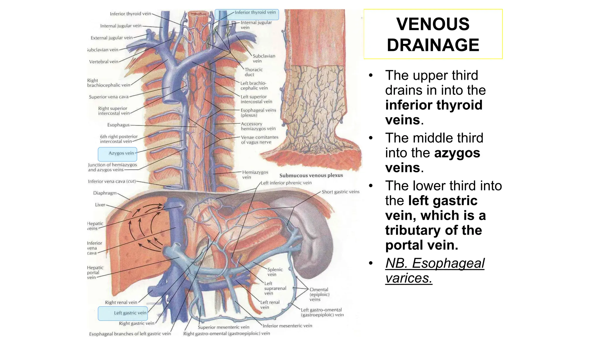
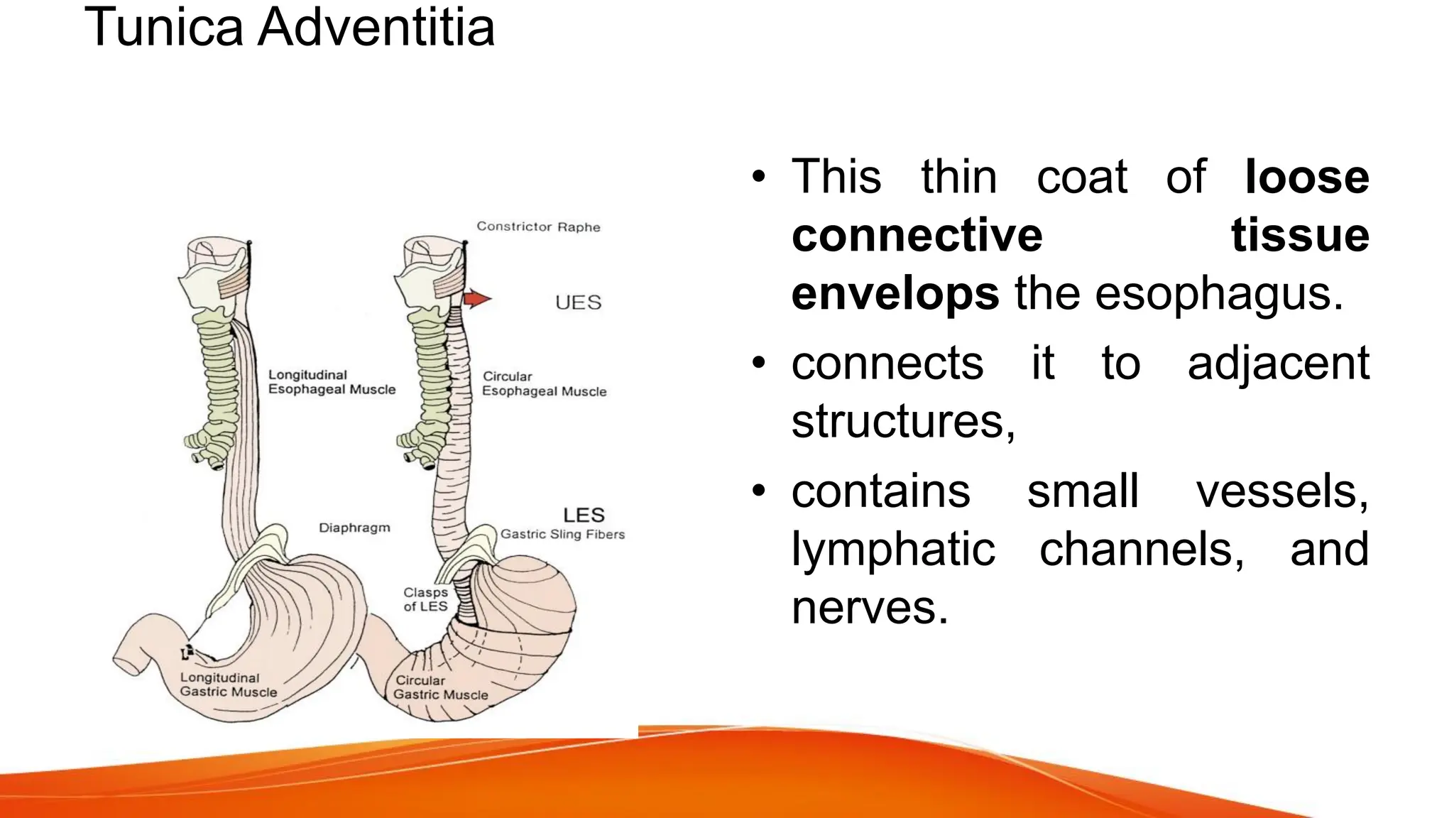
ESOPHAGUS
1
ESOPHAGUS
• Oesophagus is the immediate continuation of
pharynx and conduit for food and fluids into
the stomach.
• 25 cm in length,2 cm in diameter, a flattened
muscular tube that extends from (lower
border of cricoid cartilage) C6 to cardiac
orifice of the stomach at T 10 level vertebrae.
• It runs vertically but inclines to the left from its
origin to thoracic inlet and again from T7 to
Oesophageal opening in the diaphragm.
• It is divided into 3 parts:
– 1- Cervical.
– 2- Thoracic.
– 3- Abdominal.
• Proximal and distal- oncosurgery
Abdominal
thoracic
Cervical
2
3
CERVICAL PART
• Posteriorly
• Post: Vertebral
column.
• Laterally:
• Lat: Lobes of the
thyroid gland.
• Anteriorly:
• Ant: Trachea and
the recurrent
laryngeal nerves.
RELATIONS
4
THORACIC PART
• In the thorax, it passes
downward and to the left
through superior then to
posterior mediastinum
• At the level of the sternal
angle, the aortic arch
pushes the esophagus again
to the midline.
5
ANTERIOR
RELATIONS
• Trachea
• Left recurrent
laryngeal
nerve
• Left principal
bronchus
• Pericardium
• Left atrium
Thoracic part
POSTERIOR RELATIONS – Thoracic duct
• Bodies of the thoracic vertebrae
• Thoracic duct
• Azygos vein
• Right posterior intercostal arteries
• Descending thoracic aorta (at the
lower end)
6
LATERAL RELATION
• On the Right side:
• Right mediastinal pleura
• Terminal part of the azygos vein.
• On the Left side:
• Left mediastinal pleura
• Left subclavian artery
• Aortic arch
• Thoracic duct
7
8
RELATIONS IN THE ABDOMEN
• In the Abdomen, the esophagus
descends for 1.3 cm and joins the
stomach.
• Anteriorly, left lobe of the
liver.
• Posteriorly, left crus of the
diaphragm.
• Fibers from the right crus of the
diaphragm form a sling around
the esophagus.
• At the opening of the diaphragm,
the esophagus is accompanied
by:
– The two vagi
– Branches of the left gastric
vessels
– Lymphatic vessels.
ESOPHAGEAL
CONSTRICTIONS
• The esophagus has 3
anatomic constrictions.
• The first is at the junction with
the
pharynx(pharyngeoesophageal
junction).
• The second is at the crossing
with the aortic arch and the left
main bronchus.
• The third is at the junction with
the stomach.
1. They may cause difficulties in passing
an endoscope.
2. In case of swallowing of caustic liquids
(mostly in children), this is where the
burning is the worst and strictures
develop.
3. The esophageal strictures are a
common sites of the development of
esophageal carcinoma.
11
ARTERIAL
SUPPLY
• Upper third by the
inferior thyroid
artery.
• The middle third by
the thoracic aorta.
• The lower third by
the left gastric
artery.
VENOUS
DRAINAGE
• The upper third
drains in into the
inferior thyroid
veins.
• The middle third
into the azygos
veins.
• The lower third into
the left gastric
vein, which is a
tributary of the
portal vein.
• NB. Esophageal
varices.
LYMPH DRAINAGE
• The upper third is drained into the
deep cervical nodes.
• The middle third is drained into the
superior and inferior mediastinal
nodes.
• The lower third is drained in the
celiac lymph nodes in the
abdomen.
13
NERVE SUPPLY
• It is supplied by sympathetic fibers from
the sympathetic trunks.
• The parasympathetic supply comes form
the vagus nerves.
• Inferior to the roots of the lungs, the
vagus nerves join the sympathetic nerves
to form the esophageal plexus.
• The left vagus lies anterior to the
esophagus.
• The right vagus lies posterior to it.
14
15
CARDIAC ORIFICE
• It is the site of the
gastro- esophageal
sphincter.
• It is a physiological
rather than an
anatomical, sphincter.
• Consists of a circular
layer of smooth muscle
(under vagal and
hormonal control).
• Function:
• Prevents (GER)
regurgitation (reflux)
• NB. Notice the abrupt
mucosal transition from
esophagus to stomach
(Z- line)
HISTOLOGY
1. Mucosa –
– epithelium
– Basement membrane
– Lamina Propria
2. Submucosa- strongest layer
3. Muscular propria-
– Inner circular
– Outer longitudinal
4. Adventitia –visceral
peritoneum
Musculature
• The musculature of the
upper esophagus & UES is
striated.
• This is followed by a
transitional zone of both
striated and smooth muscle.
• proportion of the smooth
muscle. progressively
increasing.
• In the lower half of the
esophagus, there is only
smooth muscle.
• It is lined throughout with
Periesophageal Tissue, Compartments, and
Fascial Planes
• Unlike the general structure of the digestive tract, the
esophageal tube has neither mesentery nor serosal
coating.
• Its position within the mediastinum and a complete
envelope of loose connective tissue allow the
esophagus extensive transverse and longitudinal mobility.
• The esophagus may be subjected to easy blunt stripping
from the mediastinum.
Clinical relevance
• The connective tissues in which the esophagus and
trachea are embedded are bounded by fascial planes,
– the pretracheal fascia anteriorly and
– the prevertebral fascia posteriorly.
• In the upper part of the chest, both fascia unite to form the
carotid sheath.
Tunica Adventitia
• This thin coat of loose
connective tissue
envelops the esophagus.
• connects it to adjacent
structures,
• contains small vessels,
lymphatic channels, and
nerves.
Tunica Muscularis
• The tunica muscularis coats
the lumen of the esophagus
in two layers :
• the external muscle layer
parallels the longitudinal
axis of the tube,
• the muscle fibers of the
inner layer are arranged in
the horizontal axis.
• For this reason, these
muscle layers are classically
Tela Submucosa
• The submucosa is the connective tissue layer that lies
between the muscular coat and the mucosa.
• It contains a meshwork of small blood and lymph vessels,
nerves, and mucous glands.
• The duct of deep esophageal glands pierce the
muscularis mucosae.
Tunica Mucosa
• The mucous layer is composed of three components:
– the muscularis mucosae,
– the tunica / lamina propria, and
– the inner lining of nonkeratinizing stratified squamous
epithelium .
EMBRYOLOGY OF
OESOPHAGUS
• At the 4th week, a respirator
diverticulum appears in the
ventral wall of the foregut at
the border with the pharynx.
• A tracheoesophageal septum
forms, gradually separating
the ventral respiratory
diverticulum from the dorsal
part of the fore gut hence,
giving rise to a ventral
Respiratory primodium and
dorsally the Oesophagus.
• With the descent of the heart
and oesophagus, the
oesophagus which was initially
short, lengthens rapidly.
Physiology
• The upper oesophageal sphincter starts at the upper
border of oesophagus and is about 3–5 cm in length and
functions during the act of swallowing.
• The lower oesophageal sphincter is situated at lower
portion of oesophagus. It is also 3–5 cm in length and
functions to prevent oesophageal reflux.Middle portion of
oesophagus shows active peristalsis. The waves are
weaker in the upper part, becoming gradually stronger
towards the lower portion.
The act of swallowing is divided into three phases:
1. Oral or buccal phase: The food which is placed in the mouth
is chewed, lubricated with saliva, converted into a bolus and
then propelled into the pharynx by elevation of the tongue
against the palate.
2. Pharyngeal phase: It is initiated when the bolus of food
comes into contact with pharyngeal mucosa. A series of reflex
actions take place carrying the food past oro and
laryngopharynx into the oesophagus. The communications
into nasopharynx, oral cavity and larynx are cut off.
(a)Closure of nasopharynx:Soft palate contracts against the
Passavant’s ridge on the posterior pharyngeal wall and
completely cuts off the nasopharynx from the oropharynx
(b)Closure of oropharyngeal isthmus:The entry of food back into
oral cavity is prevented by contraction of tongue against the
palate and sphincteric action of palatoglossal muscles
(c) Closure of larynx:Aspiration into the larynx is prevented by
temporary cessation of respiration, closure of laryngeal inlet by
contraction of aryepiglottic folds, closure of false and true cords,
and rising of larynx under the base of tongue.
(d) Contraction of pharyngeal muscles and relaxation of
cricopharyngeus: Relaxation of cricopharyngeus muscles is so
timed and synchronous that food passes from pharynx into the
ATRESIA
• Atresia is absence of opening (lumen)/ noncanalization.
• Atresia, in which a thin, noncanalized cord replaces a
segment of esophagus, is more common. Atresia occurs
most commonly at or near the tracheal bifurcation and
usually is associated with a fistula connecting the upper or
lower esophageal pouches to a bronchus or the trachea.
This abnormal connection can result in aspiration,
suffocation, pneumonia, or severe fluid and electrolyte
imbalances
STENOSIS
• Abnormal narrowing of the esophageal lumen is called
esophageal stenosis. Passage of food can be impeded by
esophageal stenosis.
• The narrowing generally is caused by fibrous thickening of
the submucosa, atrophy of the muscularis propria, and
secondary epithelial damage. Stenosis most often is due
to inflammation and scarring, which may be caused by
chronic gastroesophageal reflux, irradiation, or caustic
(acid) injury.
• Stenosis associated dysphagia usually is progressive;
difficulty eating solids typically occurs long before
HIATUS HERNIA
• Hiatus hernia is the herniation or protrusion of part of
the stomach through the oesophageal hiatus (opening)
of the diaphragm.
• Congenital hiatal hernias are recognized in infants and
children, but many are acquired in later life. Hiatal
hernia is symptomatic in fewer than 10% of adults, and
these cases are generally associated with other causes
of LES incompetence. Symptoms, including heartburn
and regurgitation of gastric juices, are similar to GERD.
• The basic defect is the failure of the muscle fibres of the
diaphragm that form the margin of the oesophageal hiatus.
This occurs due to shortening of the esophagus which may
be congenital or acquired.
• More commonly, it is acquired due to secondary factors
which cause fibrous scarring of the oesophagus as follows:
• Degeneration of muscle due to aging.
• Increased intra-abdominal pressure such as in pregnancy,
abdominal tumours etc.
• Recurrent oesophageal regurgitation and spasm causing
inflammation and fibrosis.
• Increase in fatty tissue in obese people causing decreased
• There are 3 patterns in hiatus hernia :
• Sliding or oesophago-gastric hernia is the most common, occurring
in 85% of cases. The herniated part of the stomach appears as
supradiaphragmatic bell due to sliding up on both sides of the
oesophagus.
• Rolling or para-oesophageal hernia is seen in 10% of cases.This is a
true hernia in which cardiac end of the stomach rolls up para-
oesophageally, producing an intrathoracic sac.
• Mixed or transitional hernia constitutes the remaining 5% cases in
which there is combination of sliding and rolling hiatus hernia
Treatment
• Mainly it is surgical; the hernia is reduced and
diaphragmatic opening repaired. Early cases and those
unfit for surgery may be treated conservatively to reduce
reflux oesophagitis by measures such as (i) sleeping with
head and chest raised, (ii) avoidance of smoking, (iii) use
of drugs that reduce acidity (antacids and proton pump
inhibitors), (iv) reduction of obesity and (v) attention to the
causes which raises intra-abdominal pressure
ESOPHAGEAL VARICES
• Esophageal varices are tortuous, dilated and engorged
esophageal veins, seen along the longitudinal axis of
esophagus. They occur as a result of elevated pressure in
the portal venous system, most commonly in cirrhosis of
the liver .
• Less common causes are: portal vein thrombosis, hepatic
vein thrombosis (Budd-Chiari syndrome) and
pylephlebitis. The lesions occur as a result of bypassing
of portal venous blood from the liver to the oesophageal
venous plexus. The increased venous pressure in the
superficial veins of the esophagus may result in ulceration
• Instead of returning directly to the heart, venous blood
from the gastrointestinal tract is delivered to the liver
via the portal vein before reaching the inferior vena
cava. This circulatory pattern is responsible for the first-
pass effect, in which drugs and other materials
absorbed in the intestines are processed by the liver
before entering the systemic circulation. Diseases that
impede this flow cause portal hypertension, which can
lead to the development of esophageal varices, an
important cause of esophageal bleeding.
• One of the few sites where the splanchnic (visceral) and
systemic venous circulations can communicate is the
esophagus. Thus, portal hypertension induces development of
collateral channels that allow portal blood to shunt (divert) into
the caval system.
• However, these collateral veins enlarge the subepithelial and
submucosal venous plexi within the distal esophagus. These
vessels, termed varices, develop in 90% of cirrhotic patients,
most commonly in association with alcoholic liver disease.
Worldwide, hepatic schistosomiasis is the second most common
cause of varices.
• Varices can be detected by angiography and appear as tortuous
dilated veins lying primarily within the submucosa of the distal
GERD(Gastro-Oesophageal Reflux)
• It is due to decreased function of lower oesophageal sphincter thus
permitting regurgitation of gastric contents into oesophagus. Other
causes of gastro-oesophageal reflux are pregnancy, hiatus hernia,
scleroderma, excessive use of tobacco and alcohol, and drugs that relax
the smooth muscle (anticholinergic, beta-adrenergic drugs and calcium-
channel blockers).
• The symptoms of oesophageal reflux include substernal pain, heartburn
and regurgitation.
• The treatment consists of:1. Elevation of the head of bed at night.2.
Avoiding food at least 3 h before bedtime.3. Antacids.4. Drugs that
increase tone of lower oesophageal sphincter,e.g. metoclopramide.5. H2
receptor antagonists, e.g. cimetidine and ranitidine.6. Avoiding smoking,
alcohol, caffeine, chocolates, mintsand carbonated drinks.7. Antireflux
Neoplasms Of Oesophagus
BENIGN NEOPLASMS
Benign neoplasms are rare compared to malignant ones
• Leiomyoma is the most common and accounts for two- thirds of all the benign
neoplasms. It arises from the smooth muscle and grows in the wall of
oesophagus. Dysphagia is produced when tumour exceeds the diameter of 5
cm. Barium swallow shows an ovoid filling defect. Endoscopy reveals a
submucosal swelling. Biopsy should not be taken. Treatment is enucleation of
the tumour by thoracotomy.
• Mucosal polyps, lipomas, fibromas and haemangiomas are other benign
tumours. They are often pedunculated and present in the oesophageal lumen.
Endoscopic removal is avoided because of the danger of oesophageal
perforation. Treatment is surgical excision by oesophagotomy
FINAL SURG ANAT ESOPHAGUS.ppt full slides

FINAL SURG ANAT ESOPHAGUS.ppt full slides

  • 1.
  • 2.
    ESOPHAGUS • Oesophagus isthe immediate continuation of pharynx and conduit for food and fluids into the stomach. • 25 cm in length,2 cm in diameter, a flattened muscular tube that extends from (lower border of cricoid cartilage) C6 to cardiac orifice of the stomach at T 10 level vertebrae. • It runs vertically but inclines to the left from its origin to thoracic inlet and again from T7 to Oesophageal opening in the diaphragm. • It is divided into 3 parts: – 1- Cervical. – 2- Thoracic. – 3- Abdominal. • Proximal and distal- oncosurgery Abdominal thoracic Cervical 2
  • 3.
    3 CERVICAL PART • Posteriorly •Post: Vertebral column. • Laterally: • Lat: Lobes of the thyroid gland. • Anteriorly: • Ant: Trachea and the recurrent laryngeal nerves. RELATIONS
  • 4.
    4 THORACIC PART • Inthe thorax, it passes downward and to the left through superior then to posterior mediastinum • At the level of the sternal angle, the aortic arch pushes the esophagus again to the midline.
  • 5.
    5 ANTERIOR RELATIONS • Trachea • Leftrecurrent laryngeal nerve • Left principal bronchus • Pericardium • Left atrium Thoracic part
  • 6.
    POSTERIOR RELATIONS –Thoracic duct • Bodies of the thoracic vertebrae • Thoracic duct • Azygos vein • Right posterior intercostal arteries • Descending thoracic aorta (at the lower end) 6
  • 7.
    LATERAL RELATION • Onthe Right side: • Right mediastinal pleura • Terminal part of the azygos vein. • On the Left side: • Left mediastinal pleura • Left subclavian artery • Aortic arch • Thoracic duct 7
  • 8.
    8 RELATIONS IN THEABDOMEN • In the Abdomen, the esophagus descends for 1.3 cm and joins the stomach. • Anteriorly, left lobe of the liver. • Posteriorly, left crus of the diaphragm. • Fibers from the right crus of the diaphragm form a sling around the esophagus. • At the opening of the diaphragm, the esophagus is accompanied by: – The two vagi – Branches of the left gastric vessels – Lymphatic vessels.
  • 9.
    ESOPHAGEAL CONSTRICTIONS • The esophagushas 3 anatomic constrictions. • The first is at the junction with the pharynx(pharyngeoesophageal junction). • The second is at the crossing with the aortic arch and the left main bronchus. • The third is at the junction with the stomach.
  • 10.
    1. They maycause difficulties in passing an endoscope. 2. In case of swallowing of caustic liquids (mostly in children), this is where the burning is the worst and strictures develop. 3. The esophageal strictures are a common sites of the development of esophageal carcinoma.
  • 11.
    11 ARTERIAL SUPPLY • Upper thirdby the inferior thyroid artery. • The middle third by the thoracic aorta. • The lower third by the left gastric artery.
  • 12.
    VENOUS DRAINAGE • The upperthird drains in into the inferior thyroid veins. • The middle third into the azygos veins. • The lower third into the left gastric vein, which is a tributary of the portal vein. • NB. Esophageal varices.
  • 13.
    LYMPH DRAINAGE • Theupper third is drained into the deep cervical nodes. • The middle third is drained into the superior and inferior mediastinal nodes. • The lower third is drained in the celiac lymph nodes in the abdomen. 13
  • 14.
    NERVE SUPPLY • Itis supplied by sympathetic fibers from the sympathetic trunks. • The parasympathetic supply comes form the vagus nerves. • Inferior to the roots of the lungs, the vagus nerves join the sympathetic nerves to form the esophageal plexus. • The left vagus lies anterior to the esophagus. • The right vagus lies posterior to it. 14
  • 15.
    15 CARDIAC ORIFICE • Itis the site of the gastro- esophageal sphincter. • It is a physiological rather than an anatomical, sphincter. • Consists of a circular layer of smooth muscle (under vagal and hormonal control). • Function: • Prevents (GER) regurgitation (reflux) • NB. Notice the abrupt mucosal transition from esophagus to stomach (Z- line)
  • 16.
    HISTOLOGY 1. Mucosa – –epithelium – Basement membrane – Lamina Propria 2. Submucosa- strongest layer 3. Muscular propria- – Inner circular – Outer longitudinal 4. Adventitia –visceral peritoneum
  • 17.
    Musculature • The musculatureof the upper esophagus & UES is striated. • This is followed by a transitional zone of both striated and smooth muscle. • proportion of the smooth muscle. progressively increasing. • In the lower half of the esophagus, there is only smooth muscle. • It is lined throughout with
  • 18.
    Periesophageal Tissue, Compartments,and Fascial Planes • Unlike the general structure of the digestive tract, the esophageal tube has neither mesentery nor serosal coating. • Its position within the mediastinum and a complete envelope of loose connective tissue allow the esophagus extensive transverse and longitudinal mobility. • The esophagus may be subjected to easy blunt stripping from the mediastinum.
  • 19.
    Clinical relevance • Theconnective tissues in which the esophagus and trachea are embedded are bounded by fascial planes, – the pretracheal fascia anteriorly and – the prevertebral fascia posteriorly. • In the upper part of the chest, both fascia unite to form the carotid sheath.
  • 20.
    Tunica Adventitia • Thisthin coat of loose connective tissue envelops the esophagus. • connects it to adjacent structures, • contains small vessels, lymphatic channels, and nerves.
  • 21.
    Tunica Muscularis • Thetunica muscularis coats the lumen of the esophagus in two layers : • the external muscle layer parallels the longitudinal axis of the tube, • the muscle fibers of the inner layer are arranged in the horizontal axis. • For this reason, these muscle layers are classically
  • 22.
    Tela Submucosa • Thesubmucosa is the connective tissue layer that lies between the muscular coat and the mucosa. • It contains a meshwork of small blood and lymph vessels, nerves, and mucous glands. • The duct of deep esophageal glands pierce the muscularis mucosae.
  • 23.
    Tunica Mucosa • Themucous layer is composed of three components: – the muscularis mucosae, – the tunica / lamina propria, and – the inner lining of nonkeratinizing stratified squamous epithelium .
  • 24.
    EMBRYOLOGY OF OESOPHAGUS • Atthe 4th week, a respirator diverticulum appears in the ventral wall of the foregut at the border with the pharynx. • A tracheoesophageal septum forms, gradually separating the ventral respiratory diverticulum from the dorsal part of the fore gut hence, giving rise to a ventral Respiratory primodium and dorsally the Oesophagus. • With the descent of the heart and oesophagus, the oesophagus which was initially short, lengthens rapidly.
  • 25.
    Physiology • The upperoesophageal sphincter starts at the upper border of oesophagus and is about 3–5 cm in length and functions during the act of swallowing. • The lower oesophageal sphincter is situated at lower portion of oesophagus. It is also 3–5 cm in length and functions to prevent oesophageal reflux.Middle portion of oesophagus shows active peristalsis. The waves are weaker in the upper part, becoming gradually stronger towards the lower portion.
  • 26.
    The act ofswallowing is divided into three phases: 1. Oral or buccal phase: The food which is placed in the mouth is chewed, lubricated with saliva, converted into a bolus and then propelled into the pharynx by elevation of the tongue against the palate. 2. Pharyngeal phase: It is initiated when the bolus of food comes into contact with pharyngeal mucosa. A series of reflex actions take place carrying the food past oro and laryngopharynx into the oesophagus. The communications into nasopharynx, oral cavity and larynx are cut off.
  • 27.
    (a)Closure of nasopharynx:Softpalate contracts against the Passavant’s ridge on the posterior pharyngeal wall and completely cuts off the nasopharynx from the oropharynx (b)Closure of oropharyngeal isthmus:The entry of food back into oral cavity is prevented by contraction of tongue against the palate and sphincteric action of palatoglossal muscles (c) Closure of larynx:Aspiration into the larynx is prevented by temporary cessation of respiration, closure of laryngeal inlet by contraction of aryepiglottic folds, closure of false and true cords, and rising of larynx under the base of tongue. (d) Contraction of pharyngeal muscles and relaxation of cricopharyngeus: Relaxation of cricopharyngeus muscles is so timed and synchronous that food passes from pharynx into the
  • 28.
    ATRESIA • Atresia isabsence of opening (lumen)/ noncanalization. • Atresia, in which a thin, noncanalized cord replaces a segment of esophagus, is more common. Atresia occurs most commonly at or near the tracheal bifurcation and usually is associated with a fistula connecting the upper or lower esophageal pouches to a bronchus or the trachea. This abnormal connection can result in aspiration, suffocation, pneumonia, or severe fluid and electrolyte imbalances
  • 30.
    STENOSIS • Abnormal narrowingof the esophageal lumen is called esophageal stenosis. Passage of food can be impeded by esophageal stenosis. • The narrowing generally is caused by fibrous thickening of the submucosa, atrophy of the muscularis propria, and secondary epithelial damage. Stenosis most often is due to inflammation and scarring, which may be caused by chronic gastroesophageal reflux, irradiation, or caustic (acid) injury. • Stenosis associated dysphagia usually is progressive; difficulty eating solids typically occurs long before
  • 31.
    HIATUS HERNIA • Hiatushernia is the herniation or protrusion of part of the stomach through the oesophageal hiatus (opening) of the diaphragm. • Congenital hiatal hernias are recognized in infants and children, but many are acquired in later life. Hiatal hernia is symptomatic in fewer than 10% of adults, and these cases are generally associated with other causes of LES incompetence. Symptoms, including heartburn and regurgitation of gastric juices, are similar to GERD.
  • 32.
    • The basicdefect is the failure of the muscle fibres of the diaphragm that form the margin of the oesophageal hiatus. This occurs due to shortening of the esophagus which may be congenital or acquired. • More commonly, it is acquired due to secondary factors which cause fibrous scarring of the oesophagus as follows: • Degeneration of muscle due to aging. • Increased intra-abdominal pressure such as in pregnancy, abdominal tumours etc. • Recurrent oesophageal regurgitation and spasm causing inflammation and fibrosis. • Increase in fatty tissue in obese people causing decreased
  • 33.
    • There are3 patterns in hiatus hernia : • Sliding or oesophago-gastric hernia is the most common, occurring in 85% of cases. The herniated part of the stomach appears as supradiaphragmatic bell due to sliding up on both sides of the oesophagus. • Rolling or para-oesophageal hernia is seen in 10% of cases.This is a true hernia in which cardiac end of the stomach rolls up para- oesophageally, producing an intrathoracic sac. • Mixed or transitional hernia constitutes the remaining 5% cases in which there is combination of sliding and rolling hiatus hernia
  • 35.
    Treatment • Mainly itis surgical; the hernia is reduced and diaphragmatic opening repaired. Early cases and those unfit for surgery may be treated conservatively to reduce reflux oesophagitis by measures such as (i) sleeping with head and chest raised, (ii) avoidance of smoking, (iii) use of drugs that reduce acidity (antacids and proton pump inhibitors), (iv) reduction of obesity and (v) attention to the causes which raises intra-abdominal pressure
  • 36.
    ESOPHAGEAL VARICES • Esophagealvarices are tortuous, dilated and engorged esophageal veins, seen along the longitudinal axis of esophagus. They occur as a result of elevated pressure in the portal venous system, most commonly in cirrhosis of the liver . • Less common causes are: portal vein thrombosis, hepatic vein thrombosis (Budd-Chiari syndrome) and pylephlebitis. The lesions occur as a result of bypassing of portal venous blood from the liver to the oesophageal venous plexus. The increased venous pressure in the superficial veins of the esophagus may result in ulceration
  • 37.
    • Instead ofreturning directly to the heart, venous blood from the gastrointestinal tract is delivered to the liver via the portal vein before reaching the inferior vena cava. This circulatory pattern is responsible for the first- pass effect, in which drugs and other materials absorbed in the intestines are processed by the liver before entering the systemic circulation. Diseases that impede this flow cause portal hypertension, which can lead to the development of esophageal varices, an important cause of esophageal bleeding.
  • 38.
    • One ofthe few sites where the splanchnic (visceral) and systemic venous circulations can communicate is the esophagus. Thus, portal hypertension induces development of collateral channels that allow portal blood to shunt (divert) into the caval system. • However, these collateral veins enlarge the subepithelial and submucosal venous plexi within the distal esophagus. These vessels, termed varices, develop in 90% of cirrhotic patients, most commonly in association with alcoholic liver disease. Worldwide, hepatic schistosomiasis is the second most common cause of varices. • Varices can be detected by angiography and appear as tortuous dilated veins lying primarily within the submucosa of the distal
  • 83.
    GERD(Gastro-Oesophageal Reflux) • Itis due to decreased function of lower oesophageal sphincter thus permitting regurgitation of gastric contents into oesophagus. Other causes of gastro-oesophageal reflux are pregnancy, hiatus hernia, scleroderma, excessive use of tobacco and alcohol, and drugs that relax the smooth muscle (anticholinergic, beta-adrenergic drugs and calcium- channel blockers). • The symptoms of oesophageal reflux include substernal pain, heartburn and regurgitation. • The treatment consists of:1. Elevation of the head of bed at night.2. Avoiding food at least 3 h before bedtime.3. Antacids.4. Drugs that increase tone of lower oesophageal sphincter,e.g. metoclopramide.5. H2 receptor antagonists, e.g. cimetidine and ranitidine.6. Avoiding smoking, alcohol, caffeine, chocolates, mintsand carbonated drinks.7. Antireflux
  • 120.
    Neoplasms Of Oesophagus BENIGNNEOPLASMS Benign neoplasms are rare compared to malignant ones • Leiomyoma is the most common and accounts for two- thirds of all the benign neoplasms. It arises from the smooth muscle and grows in the wall of oesophagus. Dysphagia is produced when tumour exceeds the diameter of 5 cm. Barium swallow shows an ovoid filling defect. Endoscopy reveals a submucosal swelling. Biopsy should not be taken. Treatment is enucleation of the tumour by thoracotomy. • Mucosal polyps, lipomas, fibromas and haemangiomas are other benign tumours. They are often pedunculated and present in the oesophageal lumen. Endoscopic removal is avoided because of the danger of oesophageal perforation. Treatment is surgical excision by oesophagotomy