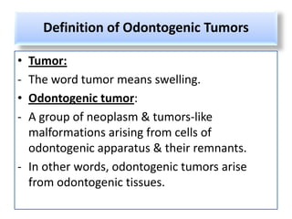
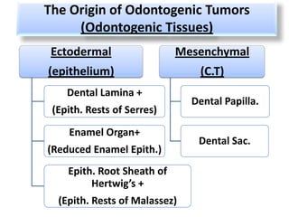
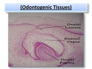
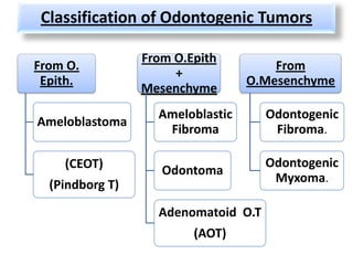
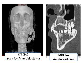
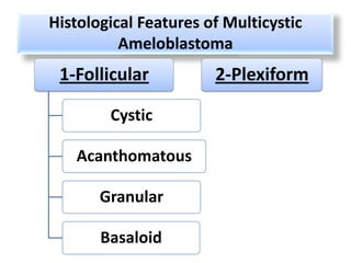
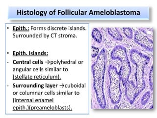
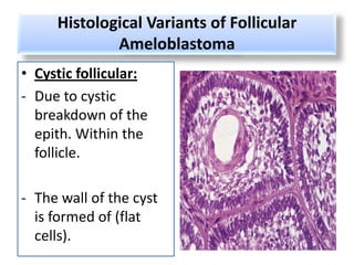
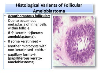
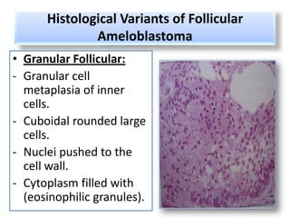
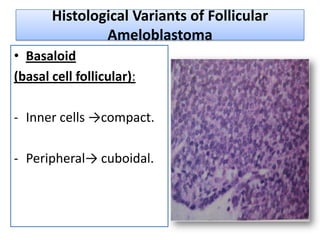
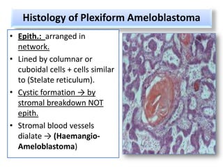
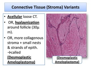
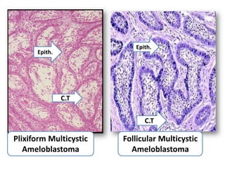
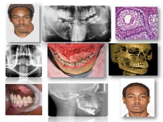
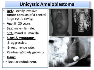
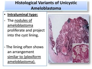
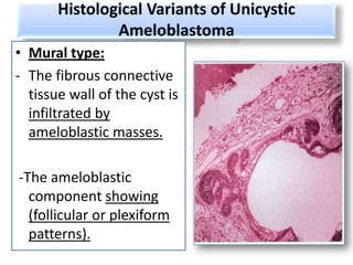
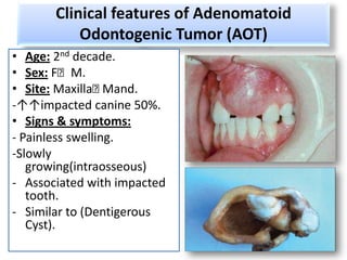
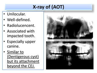
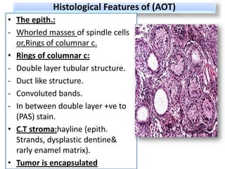
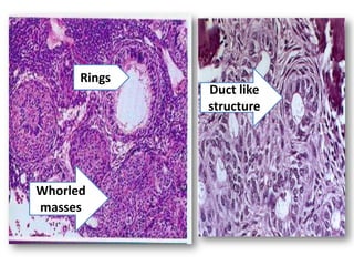
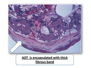
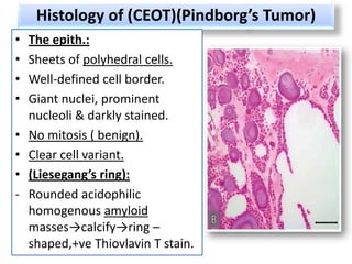
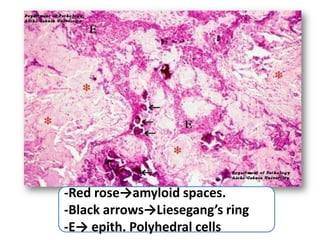
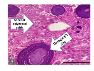
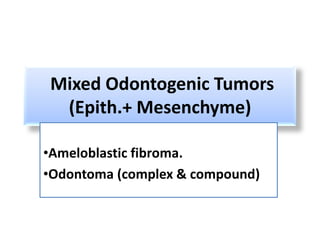
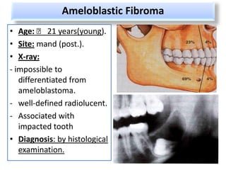
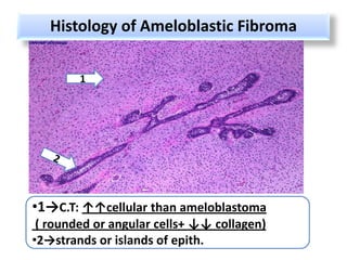
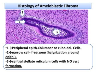
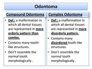
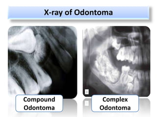
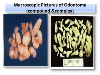
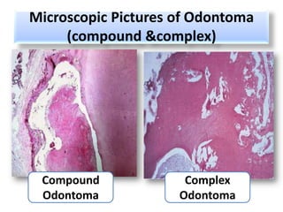
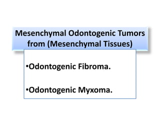
Odontogenic Tumors
Definition of Odontogenic Tumors

• Tumor:
- The word tumor means swelling.
• Odontogenic tumor:
- A group of neoplasm & tumors-like
  malformations arising from cells of
  odontogenic apparatus & their remnants.
- In other words, odontogenic tumors arise
  from odontogenic tissues.
The Origin of Odontogenic Tumors
     (Odontogenic Tissues)
 Ectodermal                 Mesenchymal
(epithelium)                    (C.T)
    Dental Lamina +
                               Dental Papilla.
 (Epith. Rests of Serres)

    Enamel Organ+
                                Dental Sac.
(Reduced Enamel Epith.)
    Epith. Root Sheath of
         Hertwig’s +
  (Epith. Rests of Malassez)
(Odontogenic Tissues)
Classification of Odontogenic Tumors

                 From O.Epith
From O.                               From
                      +
 Epith.                           O.Mesenchyme
                 Mesenchyme
                   Ameloblastic      Odontogenic
Ameloblastoma
                    Fibroma           Fibroma.

    (CEOT)                           Odontogenic
                    Odontoma
  (Pindborg T)                        Myxoma.

                   Adenomatoid O.T
                         (AOT)
Origin of Ameloblastoma

Several origins have been suggested:
• Dental Lamina + its remnants( epith.
  Rests of Serres).
• Enamel organ + its remnants ( reduced
  enamel epith.).
• Epith. Lining of Dentigerous cyst.
• Basal layer of the oral mucosa.
Types of Ameloblastoma
• 1- Simple, Solid,(Multicystic
   Ameloblastoma), with its histological variants:
-Follicular A.
-Plexiform A.
• 2-(Unicystic Ameloblastoma), with its
   histological variants:
-Luminal Unicystic A.
-Intraluminal Unicystic A.
-Mural Unicystic A.
• 3-Peripheral (S.T) ameloblastoma.
• 4-Desmoplastic ameloblastoma.
Solid, Multicystic Ameloblastoma
• Def.:
- It is a benign but locally invasive neoplasm
  consisting of epithelium lying in a fibrous
  stroma.
- It is one of the few neoplasms that arise from
  odontogenic epith.
- It has ↑ recurrence rate than other types of
  Ameloblastoma.
Clinical Features of Multicystic
                Ameloblastoma
•  Age: 4th- 5th decade.
•  Sex: male = female.
•  Race: ↑ in African.
•  Site: ↑mand. Molar
   area.
• Signs & symptoms:
-painless
-Slowly growing.
-gradula facial asymmetry.
-Looseness of teeth.
-NO metastasis (benign).
Macroscopic pictures of Multicystic, Solid
             Ameloblastoma
• Starts in cancellous bone &
  enlarge slowly.
• Extends buccal & lingual
  plate of bone.
• There is always a continous
  sheet of paper thin bone
  covering the tumor.
• May be solid or cystic.
• Cystic tumor may be one
  large (unicystic) or microcysts
  (multicystic).
X-ray of Solid, Multicystic Ameloblastoma
• Multilocular
  Radiolucency.
• (Honeycomb) or (soap-
  bubble) appearance.
• Recently diagnosed by:
- Computed tomography
  (CT) scan.
- Magnetic resonance
  imaging (MRI).
C.T (3d)           MRI for
scan for Ameloblastoma   Ameloblastoma
Histological Features of Multicystic
          Ameloblastoma
 1-Follicular          2-Plexiform

        Cystic

   Acanthomatous

       Granular

       Basaloid
Histology of Follicular Ameloblastoma
• Epith.: Forms discrete islands.
  Surrounded by CT stroma.

• Epith. Islands:
- Central cells →polyhedral or
  angular cells similar to
  (stellate reticulum).
- Surrounding layer →cuboidal
  or columnar cells similar to
  (internal enamel
  epith.)(preameloblasts).
Histological Variants of Follicular
             Ameloblastoma
• Cystic follicular:
- Due to cystic
  breakdown of the
  epith. Within the
  follicle.

- The wall of the cyst
  is formed of (flat
  cells).
Histological Variants of Follicular
              Ameloblastoma
• Acanthomatous follicular:
- Due to squamous
  metaplasia of inner cells
  within follicle.
- If ↑ keratin →(kerato
  ameloblastoma).
- If some keratinised +
- another microcysts with
  non-keratinised epith.+
- papillary forms→
  (papilliferous kerato-
  ameloblastoma).
Histological Variants of Follicular
              Ameloblastoma
• Granular Follicular:
- Granular cell
  metaplasia of inner
  cells.
- Cuboidal rounded large
  cells.
- Nuclei pushed to the
  cell wall.
- Cytoplasm filled with
  (eosinophilic granules).
Histological Variants of Follicular
               Ameloblastoma
• Basaloid
(basal cell follicular):

- Inner cells →compact.

- Peripheral→ cuboidal.
Histology of Plexiform Ameloblastoma
• Epith.: arranged in
  network.
• Lined by columnar or
  cuboidal cells + cells similar
  to (Stelate reticulum).
• Cystic formation → by
  stromal breakdown NOT
  epith.
• Stromal blood vessels
  dialate → (Haemangio-
  Ameloblastoma)
Connective Tissue (Stroma) Variants

• Acellular loose CT.
• OR, hyalaynization
  around follicle (30µ
  m).
• OR, more collagenous
  stroma + small nests
  & strands of epith.
  →called
  (Desmoplastic            (Desmoplastic
  Ameloplastoma)          Ameloplastoma)
Epith.
      Epith.


               C.T

                              C.T

Plixiform Multicystic   Follicular Multicystic
   Ameloblastoma          Ameloblastoma
Unicystic Ameloblastoma
• Def.: Locally invasive
  tumor consists of a central
  large cystic cavity.
• Age: ˂20 years.
• Sex: male= female.
• Site: mand.˂ maxilla.
• Signs & symptoms:
- ↓ aggressive.
- ↓ recurrence rate.
- Painless &Slowly growing.
• X-ray:
Unilocular radiolucent.
(3D) CT scan of Unictystic
     Ameloblastoma
Histology of Unicystic Ameloplastoma


  One large
expansile cyst
Histological Variants of Unicystic
        Ameloblastoma


             1- Luminal


       2-
 Intraluminal           3-Mural
Histological Variants of Unicystic
                 Ameloblastoma
• Luminal type:
- The change is confined
  within the luminal surface
  of the cyst.
- Part of the epithelial lining
  changes into ameloblastic
  epithelium→ (columnar or
  cuboidal c)+hyperchromatic
  nuclei + reverse polarity.
- basilar cytoplasmic
  vacoulation.
- The overlying epithelial
  cells consist of loose
  stellate reticulum like cells.
Histological Variants of Unicystic
              Ameloblastoma
• Intraluminal type:
- The nodules of
  ameloblastoma
  proliferate and project
  into the cyst lining.

- The lining often shows
   an arrangement
   similar to (plexiform
   ameloblastoma).
Histological Variants of Unicystic
               Ameloblastoma
• Mural type:
- The fibrous connective
  tissue wall of the cyst is
  infiltrated by
  ameloblastic masses.

-The ameloblastic
  component showing
  (follicular or plexiform
  patterns).
Adenomatoid Odontogenic Tumor (AOT)
• Def.:
- A tumor of odontogenic epith.with duct
  structures & with varing degrees of inductive
  changes in the C.T stroma.
- Its different clinically & histologically from
  ameloblastoma.
- Its believed that the lesion is a (Hamartomatous)
  in nature.
• Origin:
-Enamel organ & its remnants(Reduced E.E)
Clinical features of Adenomatoid
           Odontogenic Tumor (AOT)
• Age: 2nd decade.
• Sex: F˂M.
• Site: Maxilla˂  Mand.
-↑↑impacted canine 50%.
• Signs & symptoms:
- Painless swelling.
-Slowly
   growing(intraosseous)
- Associated with impacted
   tooth.
- Similar to (Dentigerous
   Cyst).
X-ray of (AOT)
• Unilocular.
• Well-defined.
• Radiolucencent.
• Associated with
  impacted tooth.
• Especially upper
  canine.
• Similar to
  (Dentigerous cyst)
  but its attachment
  beyond the CEJ.
Histological Features of (AOT)
• The epith.:
- Whorled masses of spindle cells
  or,Rings of columnar c.
• Rings of columnar c:
- Double layer tubular structure.
- Duct like structure.
- Convoluted bands.
- In between double layer +ve to
  (PAS) stain.
• C.T stroma:hayline (epith.
  Strands, dysplastic dentine&
  rarly enamel matrix).
• Tumor is encapsulated
Rings
              Duct like
              structure


Whorled
masses
AOT is encapsulated with thick
         fibrous band
Calcifying Epith. Odontogenic Tumor (CEOT)
             (Pindborg’s Tumor)
• Def.:
- It is a locally invasive epith. Neoplasm
   characterized by the development of intra-
   epith. Structure probably of an amyloid-like
   nature.
- This amyloid-like structure may become
   calcified & liberated into the surrounding
   stroma.
• Origin (Histogenesis):
- (Reduced E.E) of associated unerupted tooth.
Clinical Features of (CEOT)(Pindborg’s
                      Tumor)
•   Age: between 20-60 years.
•   Sex: M=F
•   Site:
-   Intraosseous →mand˂max
    (post.).
-   Extraosseous→anterior.
•   Signs & symptoms:
-   painless.
-   Slowly growing.
-   Associated with impacted
    tooth.
X-ray of (CEOT)(Pindborg’s Tumor)

• Irregular
  radiolucent area.

• Containing
  radioopaque
  masses close to
  unerupted tooth
(3D) CT scan of Pindborg’s
      Tumor (CEOT)
Histology of (CEOT)(Pindborg’s Tumor)
•   The epith.:
•   Sheets of polyhedral cells.
•   Well-defined cell border.
•   Giant nuclei, prominent
    nucleoli & darkly stained.
•   No mitosis ( benign).
•   Clear cell variant.
•   (Liesegang’s ring):
-   Rounded acidophilic
    homogenous amyloid
    masses→calcify→ring –
    shaped,+ve Thiovlavin T stain.
-Red rose→amyloid spaces.
-Black arrows→Liesegang’s ring
-E→ epith. Polyhedral cells
Sheet of
polyhedral
  epith.
Mixed Odontogenic Tumors
 (Epith.+ Mesenchyme)

•Ameloblastic fibroma.
•Odontoma (complex & compound)
Ameloblastic Fibroma
• Age: ˂ 21 years(young).
• Site: mand (post.).
• X-ray:
- impossible to
   differentiated from
   ameloblastoma.
- well-defined radiolucent.
- Associated with
   impacted tooth
• Diagnosis: by histological
   examination.
Histology of Ameloblastic Fibroma

        1


•1→C.T: ↑↑cellular than ameloblastoma
 ( rounded or angular cells+ ↓↓ collagen)
•2→strands or islands of epith.
Histology of Ameloblastic Fibroma
            2

        1


•1→Peripheral epith.Columnar or cuboidal. Cells.
•2→narrow cell- free zone (halynization around
epith.).
•3→central stellate reticulum cells with NO cyst
formation.
Odontoma
 Compound Odontoma           Complex Odontoma
• Def.: a malformation in    • Def.: a malformation in
  which all dental tissues     which all dental tissues
  are represented in more      are represented in more
  orderly pattern than         disorderly pattern.
  comlex.                    • Contains many
• Contains many tooth-         disordered tooth-like
  like structures.             structures.
• Don’t resemble the         • Don’t resemble the
  normal tooth                 normal tooth
  morphologically .            morphologically .
X-ray of Odontoma


Compound           Complex
Odontoma          Odontoma
Macroscopic Pictures of Odontoma
     (compound &complex)
Microscopic Pictures of Odontoma
     (compound &complex)


 Compound             Complex
 Odontoma            Odontoma
Mesenchymal Odontogenic Tumors
  from (Mesenchymal Tissues)

  •Odontogenic Fibroma.

  •Odontogenic Myxoma.
Odontogenic Fibroma

• Def.:
A fibroblastic neoplasm containing:
- Inactive odontogenic epith.
- More cellular fibrous tissue.
- Hard tissue (dysplastic cementum or bone).
• Origin:
-Tooth follicle (dental sac) →fibroblastic
   elements or,
- Its derivative ( periodontal ligament)
X-ray of Odontogenic Fibroma


    Well-defined (unilocular)
    monolocular radioucency
Histology of Odontogenic Fibroma
• Primitive cellular
  fibroblastic tissue.
• Strands or islands of
  inactive odontogenic
  epith.
• Islands of ostoid or
  cement-like tissue
  scattered in the
  fibroblastic mass.
• Invariable collagen.
• The tumor is
  encapsulated.
3
1


      2


•1→fibroblastic tissue
•2→ostoid islands
•3→ inactive epith. strands
Odontogenic Myxoma
• Def.:
It is a locally invasive neoplasm resembling dental
    follicle in location & structure & containing:
 - Angular spindle cells.
- Lying in mucoid stroma.
• Origin:
- Odontogenic mesenchyme because:
1- frequent inclusion of epith. Rests.
2- missing associated tooth.
3- no similar tumor in any bone type( only jaw).
4- similar histologically with embryonic mesenchyme.
Odontogenic Myxoma Clinically
• Age: young.
• Sex:F=M
• Site:
-only jaw bone ↑mand.
- In place of missing tooth.
• Signs & symptoms:
- Fusiform swelling.
- Rapidly growing.
- Difficult complete removal
   (little encapsulation)
- may invade antrum.
Odontogenic Myxoma Radiographically


   •Multilocular radiolucencies
   •Soap-bubbles appearance.
Odontogenic Myxoma Histologically

1

                                3


    2


    •1→myxoma filling marrow spaces.
    •2→bony trabaculea.
    •3→spindle cells in mucoid matrix.
Thanks
    Presented by:
Marwa Assem Salama

Odontogenic tumors

  • 1.
  • 2.
    Definition of OdontogenicTumors • Tumor: - The word tumor means swelling. • Odontogenic tumor: - A group of neoplasm & tumors-like malformations arising from cells of odontogenic apparatus & their remnants. - In other words, odontogenic tumors arise from odontogenic tissues.
  • 3.
    The Origin ofOdontogenic Tumors (Odontogenic Tissues) Ectodermal Mesenchymal (epithelium) (C.T) Dental Lamina + Dental Papilla. (Epith. Rests of Serres) Enamel Organ+ Dental Sac. (Reduced Enamel Epith.) Epith. Root Sheath of Hertwig’s + (Epith. Rests of Malassez)
  • 4.
  • 5.
    Classification of OdontogenicTumors From O.Epith From O. From + Epith. O.Mesenchyme Mesenchyme Ameloblastic Odontogenic Ameloblastoma Fibroma Fibroma. (CEOT) Odontogenic Odontoma (Pindborg T) Myxoma. Adenomatoid O.T (AOT)
  • 6.
    Origin of Ameloblastoma Severalorigins have been suggested: • Dental Lamina + its remnants( epith. Rests of Serres). • Enamel organ + its remnants ( reduced enamel epith.). • Epith. Lining of Dentigerous cyst. • Basal layer of the oral mucosa.
  • 7.
    Types of Ameloblastoma •1- Simple, Solid,(Multicystic Ameloblastoma), with its histological variants: -Follicular A. -Plexiform A. • 2-(Unicystic Ameloblastoma), with its histological variants: -Luminal Unicystic A. -Intraluminal Unicystic A. -Mural Unicystic A. • 3-Peripheral (S.T) ameloblastoma. • 4-Desmoplastic ameloblastoma.
  • 8.
    Solid, Multicystic Ameloblastoma •Def.: - It is a benign but locally invasive neoplasm consisting of epithelium lying in a fibrous stroma. - It is one of the few neoplasms that arise from odontogenic epith. - It has ↑ recurrence rate than other types of Ameloblastoma.
  • 9.
    Clinical Features ofMulticystic Ameloblastoma • Age: 4th- 5th decade. • Sex: male = female. • Race: ↑ in African. • Site: ↑mand. Molar area. • Signs & symptoms: -painless -Slowly growing. -gradula facial asymmetry. -Looseness of teeth. -NO metastasis (benign).
  • 10.
    Macroscopic pictures ofMulticystic, Solid Ameloblastoma • Starts in cancellous bone & enlarge slowly. • Extends buccal & lingual plate of bone. • There is always a continous sheet of paper thin bone covering the tumor. • May be solid or cystic. • Cystic tumor may be one large (unicystic) or microcysts (multicystic).
  • 11.
    X-ray of Solid,Multicystic Ameloblastoma • Multilocular Radiolucency. • (Honeycomb) or (soap- bubble) appearance. • Recently diagnosed by: - Computed tomography (CT) scan. - Magnetic resonance imaging (MRI).
  • 12.
    C.T (3d) MRI for scan for Ameloblastoma Ameloblastoma
  • 13.
    Histological Features ofMulticystic Ameloblastoma 1-Follicular 2-Plexiform Cystic Acanthomatous Granular Basaloid
  • 14.
    Histology of FollicularAmeloblastoma • Epith.: Forms discrete islands. Surrounded by CT stroma. • Epith. Islands: - Central cells →polyhedral or angular cells similar to (stellate reticulum). - Surrounding layer →cuboidal or columnar cells similar to (internal enamel epith.)(preameloblasts).
  • 15.
    Histological Variants ofFollicular Ameloblastoma • Cystic follicular: - Due to cystic breakdown of the epith. Within the follicle. - The wall of the cyst is formed of (flat cells).
  • 16.
    Histological Variants ofFollicular Ameloblastoma • Acanthomatous follicular: - Due to squamous metaplasia of inner cells within follicle. - If ↑ keratin →(kerato ameloblastoma). - If some keratinised + - another microcysts with non-keratinised epith.+ - papillary forms→ (papilliferous kerato- ameloblastoma).
  • 17.
    Histological Variants ofFollicular Ameloblastoma • Granular Follicular: - Granular cell metaplasia of inner cells. - Cuboidal rounded large cells. - Nuclei pushed to the cell wall. - Cytoplasm filled with (eosinophilic granules).
  • 18.
    Histological Variants ofFollicular Ameloblastoma • Basaloid (basal cell follicular): - Inner cells →compact. - Peripheral→ cuboidal.
  • 19.
    Histology of PlexiformAmeloblastoma • Epith.: arranged in network. • Lined by columnar or cuboidal cells + cells similar to (Stelate reticulum). • Cystic formation → by stromal breakdown NOT epith. • Stromal blood vessels dialate → (Haemangio- Ameloblastoma)
  • 20.
    Connective Tissue (Stroma)Variants • Acellular loose CT. • OR, hyalaynization around follicle (30µ m). • OR, more collagenous stroma + small nests & strands of epith. →called (Desmoplastic (Desmoplastic Ameloplastoma) Ameloplastoma)
  • 21.
    Epith. Epith. C.T C.T Plixiform Multicystic Follicular Multicystic Ameloblastoma Ameloblastoma
  • 23.
    Unicystic Ameloblastoma • Def.:Locally invasive tumor consists of a central large cystic cavity. • Age: ˂20 years. • Sex: male= female. • Site: mand.˂ maxilla. • Signs & symptoms: - ↓ aggressive. - ↓ recurrence rate. - Painless &Slowly growing. • X-ray: Unilocular radiolucent.
  • 24.
    (3D) CT scanof Unictystic Ameloblastoma
  • 25.
    Histology of UnicysticAmeloplastoma One large expansile cyst
  • 26.
    Histological Variants ofUnicystic Ameloblastoma 1- Luminal 2- Intraluminal 3-Mural
  • 27.
    Histological Variants ofUnicystic Ameloblastoma • Luminal type: - The change is confined within the luminal surface of the cyst. - Part of the epithelial lining changes into ameloblastic epithelium→ (columnar or cuboidal c)+hyperchromatic nuclei + reverse polarity. - basilar cytoplasmic vacoulation. - The overlying epithelial cells consist of loose stellate reticulum like cells.
  • 28.
    Histological Variants ofUnicystic Ameloblastoma • Intraluminal type: - The nodules of ameloblastoma proliferate and project into the cyst lining. - The lining often shows an arrangement similar to (plexiform ameloblastoma).
  • 29.
    Histological Variants ofUnicystic Ameloblastoma • Mural type: - The fibrous connective tissue wall of the cyst is infiltrated by ameloblastic masses. -The ameloblastic component showing (follicular or plexiform patterns).
  • 30.
    Adenomatoid Odontogenic Tumor(AOT) • Def.: - A tumor of odontogenic epith.with duct structures & with varing degrees of inductive changes in the C.T stroma. - Its different clinically & histologically from ameloblastoma. - Its believed that the lesion is a (Hamartomatous) in nature. • Origin: -Enamel organ & its remnants(Reduced E.E)
  • 31.
    Clinical features ofAdenomatoid Odontogenic Tumor (AOT) • Age: 2nd decade. • Sex: F˂M. • Site: Maxilla˂ Mand. -↑↑impacted canine 50%. • Signs & symptoms: - Painless swelling. -Slowly growing(intraosseous) - Associated with impacted tooth. - Similar to (Dentigerous Cyst).
  • 32.
    X-ray of (AOT) •Unilocular. • Well-defined. • Radiolucencent. • Associated with impacted tooth. • Especially upper canine. • Similar to (Dentigerous cyst) but its attachment beyond the CEJ.
  • 33.
    Histological Features of(AOT) • The epith.: - Whorled masses of spindle cells or,Rings of columnar c. • Rings of columnar c: - Double layer tubular structure. - Duct like structure. - Convoluted bands. - In between double layer +ve to (PAS) stain. • C.T stroma:hayline (epith. Strands, dysplastic dentine& rarly enamel matrix). • Tumor is encapsulated
  • 34.
    Rings Duct like structure Whorled masses
  • 35.
    AOT is encapsulatedwith thick fibrous band
  • 36.
    Calcifying Epith. OdontogenicTumor (CEOT) (Pindborg’s Tumor) • Def.: - It is a locally invasive epith. Neoplasm characterized by the development of intra- epith. Structure probably of an amyloid-like nature. - This amyloid-like structure may become calcified & liberated into the surrounding stroma. • Origin (Histogenesis): - (Reduced E.E) of associated unerupted tooth.
  • 37.
    Clinical Features of(CEOT)(Pindborg’s Tumor) • Age: between 20-60 years. • Sex: M=F • Site: - Intraosseous →mand˂max (post.). - Extraosseous→anterior. • Signs & symptoms: - painless. - Slowly growing. - Associated with impacted tooth.
  • 38.
    X-ray of (CEOT)(Pindborg’sTumor) • Irregular radiolucent area. • Containing radioopaque masses close to unerupted tooth
  • 39.
    (3D) CT scanof Pindborg’s Tumor (CEOT)
  • 40.
    Histology of (CEOT)(Pindborg’sTumor) • The epith.: • Sheets of polyhedral cells. • Well-defined cell border. • Giant nuclei, prominent nucleoli & darkly stained. • No mitosis ( benign). • Clear cell variant. • (Liesegang’s ring): - Rounded acidophilic homogenous amyloid masses→calcify→ring – shaped,+ve Thiovlavin T stain.
  • 41.
    -Red rose→amyloid spaces. -Blackarrows→Liesegang’s ring -E→ epith. Polyhedral cells
  • 42.
  • 43.
    Mixed Odontogenic Tumors (Epith.+ Mesenchyme) •Ameloblastic fibroma. •Odontoma (complex & compound)
  • 44.
    Ameloblastic Fibroma • Age:˂ 21 years(young). • Site: mand (post.). • X-ray: - impossible to differentiated from ameloblastoma. - well-defined radiolucent. - Associated with impacted tooth • Diagnosis: by histological examination.
  • 45.
    Histology of AmeloblasticFibroma 1 •1→C.T: ↑↑cellular than ameloblastoma ( rounded or angular cells+ ↓↓ collagen) •2→strands or islands of epith.
  • 46.
    Histology of AmeloblasticFibroma 2 1 •1→Peripheral epith.Columnar or cuboidal. Cells. •2→narrow cell- free zone (halynization around epith.). •3→central stellate reticulum cells with NO cyst formation.
  • 47.
    Odontoma Compound Odontoma Complex Odontoma • Def.: a malformation in • Def.: a malformation in which all dental tissues which all dental tissues are represented in more are represented in more orderly pattern than disorderly pattern. comlex. • Contains many • Contains many tooth- disordered tooth-like like structures. structures. • Don’t resemble the • Don’t resemble the normal tooth normal tooth morphologically . morphologically .
  • 48.
    X-ray of Odontoma Compound Complex Odontoma Odontoma
  • 49.
    Macroscopic Pictures ofOdontoma (compound &complex)
  • 50.
    Microscopic Pictures ofOdontoma (compound &complex) Compound Complex Odontoma Odontoma
  • 51.
    Mesenchymal Odontogenic Tumors from (Mesenchymal Tissues) •Odontogenic Fibroma. •Odontogenic Myxoma.
  • 52.
    Odontogenic Fibroma • Def.: Afibroblastic neoplasm containing: - Inactive odontogenic epith. - More cellular fibrous tissue. - Hard tissue (dysplastic cementum or bone). • Origin: -Tooth follicle (dental sac) →fibroblastic elements or, - Its derivative ( periodontal ligament)
  • 53.
    X-ray of OdontogenicFibroma Well-defined (unilocular) monolocular radioucency
  • 54.
    Histology of OdontogenicFibroma • Primitive cellular fibroblastic tissue. • Strands or islands of inactive odontogenic epith. • Islands of ostoid or cement-like tissue scattered in the fibroblastic mass. • Invariable collagen. • The tumor is encapsulated.
  • 55.
    3 1 2 •1→fibroblastic tissue •2→ostoid islands •3→ inactive epith. strands
  • 56.
    Odontogenic Myxoma • Def.: Itis a locally invasive neoplasm resembling dental follicle in location & structure & containing: - Angular spindle cells. - Lying in mucoid stroma. • Origin: - Odontogenic mesenchyme because: 1- frequent inclusion of epith. Rests. 2- missing associated tooth. 3- no similar tumor in any bone type( only jaw). 4- similar histologically with embryonic mesenchyme.
  • 57.
    Odontogenic Myxoma Clinically •Age: young. • Sex:F=M • Site: -only jaw bone ↑mand. - In place of missing tooth. • Signs & symptoms: - Fusiform swelling. - Rapidly growing. - Difficult complete removal (little encapsulation) - may invade antrum.
  • 58.
    Odontogenic Myxoma Radiographically •Multilocular radiolucencies •Soap-bubbles appearance.
  • 59.
    Odontogenic Myxoma Histologically 1 3 2 •1→myxoma filling marrow spaces. •2→bony trabaculea. •3→spindle cells in mucoid matrix.
  • 60.
    Thanks Presented by: Marwa Assem Salama