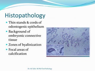
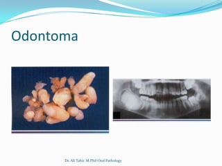
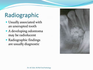
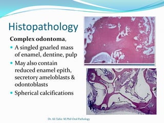
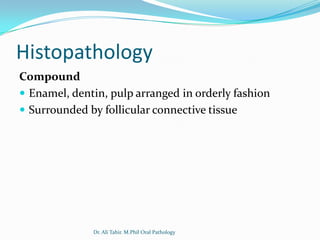
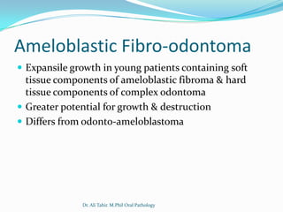
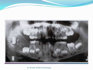
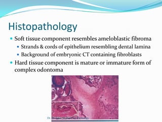
Derived from Ectomesenchyme


              Dr. Ali Tahir. M.Phil Oral Pathology
Ameloblastic Fibroma
 A circumscribed lesion located over un-erupted
 molars in young patients consisting of odontogenic
 epithelium & connective tissue


              Dr. Ali Tahir. M.Phil Oral Pathology
Clinical & radiographical features
 Younger patients, average age of 14 yrs
 Slow growing
 Common in mandible, molar areas, 75% associated
  with un-erupted tooth
 Small are asymptomatic, larger ones cause swelling
 Well defined Unilocular/Multilocular radiolucency


               Dr. Ali Tahir. M.Phil Oral Pathology
Histopathology
 Thin stands & cords of
  odontogenic epithelium
 Background of
  embryonic connective
  tissue
 Zones of hyalinization
 Focal areas of
  calcification


              Dr. Ali Tahir. M.Phil Oral Pathology
Odontoma
 Most common odontogenic tumour in west
 Hamartomatous (not true neoplasm) lesion commonly
 associated with unerupted teeth & composed of
 enamel, dentin, pulp & cementum in either
 recognizable tooth shapes (compound) or a solid,
 gnarled mass (complex)


             Dr. Ali Tahir. M.Phil Oral Pathology
Odontoma


      Dr. Ali Tahir. M.Phil Oral Pathology
Clinical Features
 First & second decade, mean age 14 yrs
 Majority asymptomatic
 More common in maxilla
 Compound are common in anterior jaws
 Complex is common in post jaws


              Dr. Ali Tahir. M.Phil Oral Pathology
Radiographic
 Compound
    Unilocular, containing multiple radiopaque structures
     resembling miniature teeth
    May contain 2-3 or upto 20-30 tooth like structures
 Complex:
    Unilocular,usually small but may grow upto 10cm
    Solid radiopaque mass
    Surrounded by thin zone of radiolucency
    Cortication


               Dr. Ali Tahir. M.Phil Oral Pathology
Radiographic
 Usually associated with
  an unerupted tooth
 A developing odontoma
  may be radiolucent
 Radiographic findings
  are usually diagnostic


              Dr. Ali Tahir. M.Phil Oral Pathology
Histopathology
Complex odontoma,
 A singled gnarled mass
  of enamel, dentine, pulp
 May also contain
  reduced enamel epith,
  secretory ameloblasts &
  odontoblasts
 Spherical calcifications


              Dr. Ali Tahir. M.Phil Oral Pathology
Histopathology
Compound
 Enamel, dentin, pulp arranged in orderly fashion
 Surrounded by follicular connective tissue


              Dr. Ali Tahir. M.Phil Oral Pathology
Ameloblastic Fibro-odontoma
 Expansile growth in young patients containing soft
  tissue components of ameloblastic fibroma & hard
  tissue components of complex odontoma
 Greater potential for growth & destruction
 Differs from odonto-ameloblastoma


              Dr. Ali Tahir. M.Phil Oral Pathology
Clinical Features
 First & second decade
 Common in post mandible
 Presents as slow growing swelling
 Usually in area of un-erupted tooth
 Pain is rare


                 Dr. Ali Tahir. M.Phil Oral Pathology
Radiographic
 Unilocular, well circumscribed, mixed radiopaque &
  radiolucent lesion
 Opacities are usually diffuse & nodular
 May contain an impacted tooth
 Variable amount of calcifications with radio-density of
  a tooth structure


               Dr. Ali Tahir. M.Phil Oral Pathology
Dr. Ali Tahir. M.Phil Oral Pathology
Histopathology
 Soft tissue component resembles ameloblastic fibroma
    Strands & cords of epithelium resembling dental lamina
    Background of embryonic CT containing fibroblasts
 Hard tissue component is mature or immature form of
  complex odontoma


               Dr. Ali Tahir. M.Phil Oral Pathology
Histopathology


       Dr. Ali Tahir. M.Phil Oral Pathology
Odontogenic Malignancies


        Dr. Ali Tahir. M.Phil Oral Pathology
Malignant Ameloblastoma and
Ameloblastic Carcinoma
 Less than 1 % of the ameloblastomas show malignant
  behavior with the development of metastases.
 Malignant ameloblastoma is a tumor that shows
  histologic features of the typical (benign) ameloblastoma
  in both the primary and secondary deposits.
 Ameloblastic carcinoma is a tumor that shows cytologic
  features of malignancy in the primary tumor, in recurrence
  and any metastases.


               Dr. Ali Tahir. M.Phil Oral Pathology
Radiographic
 With the malignant ameloblastoma, the appearance is
  similar to the typical solid/multicystic ameloblastoma.
 The ameloblastic carcinoma is often more aggressive
  with the lesion appearing as an ill-defined
  radiolucency with cortical destruction


               Dr. Ali Tahir. M.Phil Oral Pathology
Dr. Ali Tahir. M.Phil Oral Pathology
Ameloblastic Carcinoma


       Dr. Ali Tahir. M.Phil Oral Pathology
Clear Cell Odontogenic Carcinoma


        Dr. Ali Tahir. M.Phil Oral Pathology
Clear Cell Odontogenic Carcinoma

 An aggressive & destructive intra-osseous lesion
 consisting of poorly differentiated epithelial cells and
 clear cells


                Dr. Ali Tahir. M.Phil Oral Pathology
CCOC
Clinical Features
 Uncommon
 Painful swelling of anterior mandible
 5th-7th decade, mean age 58 years
 Female predilection
 Loosening of teeth
 Potentially aggressive, capable of frequent recurrences
  & metastasis
 Features indicative of odontogenic origin

               Dr. Ali Tahir. M.Phil Oral Pathology
CCOC
 Radiographically shows
 honeycomb poorly
 defined radiolucency


              Dr. Ali Tahir. M.Phil Oral Pathology
Histopathology
 Biphasic
 Mono-phasic
 Ameloblastomatous


             Dr. Ali Tahir. M.Phil Oral Pathology
Histopathology
Biphasic
 Nests of cells with clear cytoplasm mixed with cells
  containing eosinophilic cytoplasm
Monophasic
 Only clear cells
Ameloblastomatous
 Nests of cells showing central cystic change &
  squamous differentiation
 Peripheral nuclear palisading with reverse polarity

               Dr. Ali Tahir. M.Phil Oral Pathology
Histopathology


       Dr. Ali Tahir. M.Phil Oral Pathology
Primary Intra-osseous Carcinoma


         Dr. Ali Tahir. M.Phil Oral Pathology
Primary Intra-osseous Carcinoma
WHO Definition
A squamous cell carcinoma arising within the jaw, having no
  initial connection with the oral mucosa, and presumably
  developing from residues of the odontogenic epithelium
 Two possible origins
    Lining of odontogenic cysts
    From remnants of odontogenic epithelium (arising de novo)
 2/3rd cases arise from odontogenic cysts
 PIOC occurs only in the jaw bones


                 Dr. Ali Tahir. M.Phil Oral Pathology
Primary Intra-osseous Carcinoma
Clinical & Radiographic features
 Male to female ration is 2.2:1
 Mostly in elderly patients above 60 yrs
 Painful swelling
 Bony expansion may be present
 Destroys large areas of bone
 Root resorption
 Sensory disturbances/Neural involvement
 Local/regional metastasizes
             Dr. Ali Tahir. M.Phil Oral Pathology
Primary Intra-osseous Carcinoma


        Dr. Ali Tahir. M.Phil Oral Pathology
D.D (histological)
 Acanthomatous ameloblastoma
 Ameloblastic carcinoma
 Squamous odontogenic tumour
 Mucoepidermoid carcinoma


             Dr. Ali Tahir. M.Phil Oral Pathology
Diagnosis
 Clinical findings
 Ruling out the extension from oral, gingival or sinus
  epithelium
 Radiograph/CT
 Histopathology


               Dr. Ali Tahir. M.Phil Oral Pathology
Forgive your enemy...

          ...but remember the bastard’s name


            Dr. Ali Tahir. M.Phil Oral Pathology

Odontogenic tumours part 4

  • 1.
    Derived from Ectomesenchyme Dr. Ali Tahir. M.Phil Oral Pathology
  • 2.
    Ameloblastic Fibroma  Acircumscribed lesion located over un-erupted molars in young patients consisting of odontogenic epithelium & connective tissue Dr. Ali Tahir. M.Phil Oral Pathology
  • 3.
    Clinical & radiographicalfeatures  Younger patients, average age of 14 yrs  Slow growing  Common in mandible, molar areas, 75% associated with un-erupted tooth  Small are asymptomatic, larger ones cause swelling  Well defined Unilocular/Multilocular radiolucency Dr. Ali Tahir. M.Phil Oral Pathology
  • 4.
    Histopathology  Thin stands& cords of odontogenic epithelium  Background of embryonic connective tissue  Zones of hyalinization  Focal areas of calcification Dr. Ali Tahir. M.Phil Oral Pathology
  • 5.
    Odontoma  Most commonodontogenic tumour in west  Hamartomatous (not true neoplasm) lesion commonly associated with unerupted teeth & composed of enamel, dentin, pulp & cementum in either recognizable tooth shapes (compound) or a solid, gnarled mass (complex) Dr. Ali Tahir. M.Phil Oral Pathology
  • 6.
    Odontoma Dr. Ali Tahir. M.Phil Oral Pathology
  • 7.
    Clinical Features  First& second decade, mean age 14 yrs  Majority asymptomatic  More common in maxilla  Compound are common in anterior jaws  Complex is common in post jaws Dr. Ali Tahir. M.Phil Oral Pathology
  • 8.
    Radiographic  Compound  Unilocular, containing multiple radiopaque structures resembling miniature teeth  May contain 2-3 or upto 20-30 tooth like structures  Complex:  Unilocular,usually small but may grow upto 10cm  Solid radiopaque mass  Surrounded by thin zone of radiolucency  Cortication Dr. Ali Tahir. M.Phil Oral Pathology
  • 9.
    Radiographic  Usually associatedwith an unerupted tooth  A developing odontoma may be radiolucent  Radiographic findings are usually diagnostic Dr. Ali Tahir. M.Phil Oral Pathology
  • 10.
    Histopathology Complex odontoma,  Asingled gnarled mass of enamel, dentine, pulp  May also contain reduced enamel epith, secretory ameloblasts & odontoblasts  Spherical calcifications Dr. Ali Tahir. M.Phil Oral Pathology
  • 11.
    Histopathology Compound  Enamel, dentin,pulp arranged in orderly fashion  Surrounded by follicular connective tissue Dr. Ali Tahir. M.Phil Oral Pathology
  • 12.
    Ameloblastic Fibro-odontoma  Expansilegrowth in young patients containing soft tissue components of ameloblastic fibroma & hard tissue components of complex odontoma  Greater potential for growth & destruction  Differs from odonto-ameloblastoma Dr. Ali Tahir. M.Phil Oral Pathology
  • 13.
    Clinical Features  First& second decade  Common in post mandible  Presents as slow growing swelling  Usually in area of un-erupted tooth  Pain is rare Dr. Ali Tahir. M.Phil Oral Pathology
  • 14.
    Radiographic  Unilocular, wellcircumscribed, mixed radiopaque & radiolucent lesion  Opacities are usually diffuse & nodular  May contain an impacted tooth  Variable amount of calcifications with radio-density of a tooth structure Dr. Ali Tahir. M.Phil Oral Pathology
  • 15.
    Dr. Ali Tahir.M.Phil Oral Pathology
  • 16.
    Histopathology  Soft tissuecomponent resembles ameloblastic fibroma  Strands & cords of epithelium resembling dental lamina  Background of embryonic CT containing fibroblasts  Hard tissue component is mature or immature form of complex odontoma Dr. Ali Tahir. M.Phil Oral Pathology
  • 17.
    Histopathology Dr. Ali Tahir. M.Phil Oral Pathology
  • 18.
    Odontogenic Malignancies Dr. Ali Tahir. M.Phil Oral Pathology
  • 19.
    Malignant Ameloblastoma and AmeloblasticCarcinoma  Less than 1 % of the ameloblastomas show malignant behavior with the development of metastases.  Malignant ameloblastoma is a tumor that shows histologic features of the typical (benign) ameloblastoma in both the primary and secondary deposits.  Ameloblastic carcinoma is a tumor that shows cytologic features of malignancy in the primary tumor, in recurrence and any metastases. Dr. Ali Tahir. M.Phil Oral Pathology
  • 20.
    Radiographic  With themalignant ameloblastoma, the appearance is similar to the typical solid/multicystic ameloblastoma.  The ameloblastic carcinoma is often more aggressive with the lesion appearing as an ill-defined radiolucency with cortical destruction Dr. Ali Tahir. M.Phil Oral Pathology
  • 21.
    Dr. Ali Tahir.M.Phil Oral Pathology
  • 22.
    Ameloblastic Carcinoma Dr. Ali Tahir. M.Phil Oral Pathology
  • 23.
    Clear Cell OdontogenicCarcinoma Dr. Ali Tahir. M.Phil Oral Pathology
  • 24.
    Clear Cell OdontogenicCarcinoma  An aggressive & destructive intra-osseous lesion consisting of poorly differentiated epithelial cells and clear cells Dr. Ali Tahir. M.Phil Oral Pathology
  • 25.
    CCOC Clinical Features  Uncommon Painful swelling of anterior mandible  5th-7th decade, mean age 58 years  Female predilection  Loosening of teeth  Potentially aggressive, capable of frequent recurrences & metastasis  Features indicative of odontogenic origin Dr. Ali Tahir. M.Phil Oral Pathology
  • 26.
    CCOC  Radiographically shows honeycomb poorly defined radiolucency Dr. Ali Tahir. M.Phil Oral Pathology
  • 27.
    Histopathology  Biphasic  Mono-phasic Ameloblastomatous Dr. Ali Tahir. M.Phil Oral Pathology
  • 28.
    Histopathology Biphasic  Nests ofcells with clear cytoplasm mixed with cells containing eosinophilic cytoplasm Monophasic  Only clear cells Ameloblastomatous  Nests of cells showing central cystic change & squamous differentiation  Peripheral nuclear palisading with reverse polarity Dr. Ali Tahir. M.Phil Oral Pathology
  • 29.
    Histopathology Dr. Ali Tahir. M.Phil Oral Pathology
  • 30.
    Primary Intra-osseous Carcinoma Dr. Ali Tahir. M.Phil Oral Pathology
  • 31.
    Primary Intra-osseous Carcinoma WHODefinition A squamous cell carcinoma arising within the jaw, having no initial connection with the oral mucosa, and presumably developing from residues of the odontogenic epithelium  Two possible origins  Lining of odontogenic cysts  From remnants of odontogenic epithelium (arising de novo)  2/3rd cases arise from odontogenic cysts  PIOC occurs only in the jaw bones Dr. Ali Tahir. M.Phil Oral Pathology
  • 32.
    Primary Intra-osseous Carcinoma Clinical& Radiographic features  Male to female ration is 2.2:1  Mostly in elderly patients above 60 yrs  Painful swelling  Bony expansion may be present  Destroys large areas of bone  Root resorption  Sensory disturbances/Neural involvement  Local/regional metastasizes Dr. Ali Tahir. M.Phil Oral Pathology
  • 33.
    Primary Intra-osseous Carcinoma Dr. Ali Tahir. M.Phil Oral Pathology
  • 34.
    D.D (histological)  Acanthomatousameloblastoma  Ameloblastic carcinoma  Squamous odontogenic tumour  Mucoepidermoid carcinoma Dr. Ali Tahir. M.Phil Oral Pathology
  • 35.
    Diagnosis  Clinical findings Ruling out the extension from oral, gingival or sinus epithelium  Radiograph/CT  Histopathology Dr. Ali Tahir. M.Phil Oral Pathology
  • 36.
    Forgive your enemy... ...but remember the bastard’s name Dr. Ali Tahir. M.Phil Oral Pathology