Download as PDF, PPTX
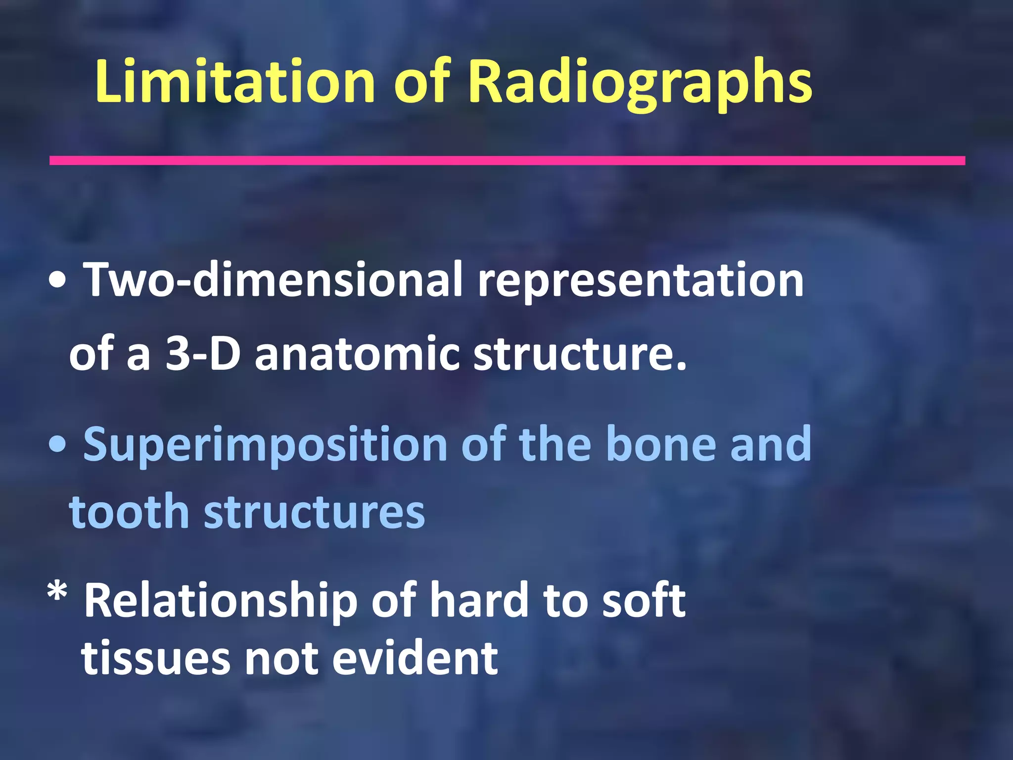
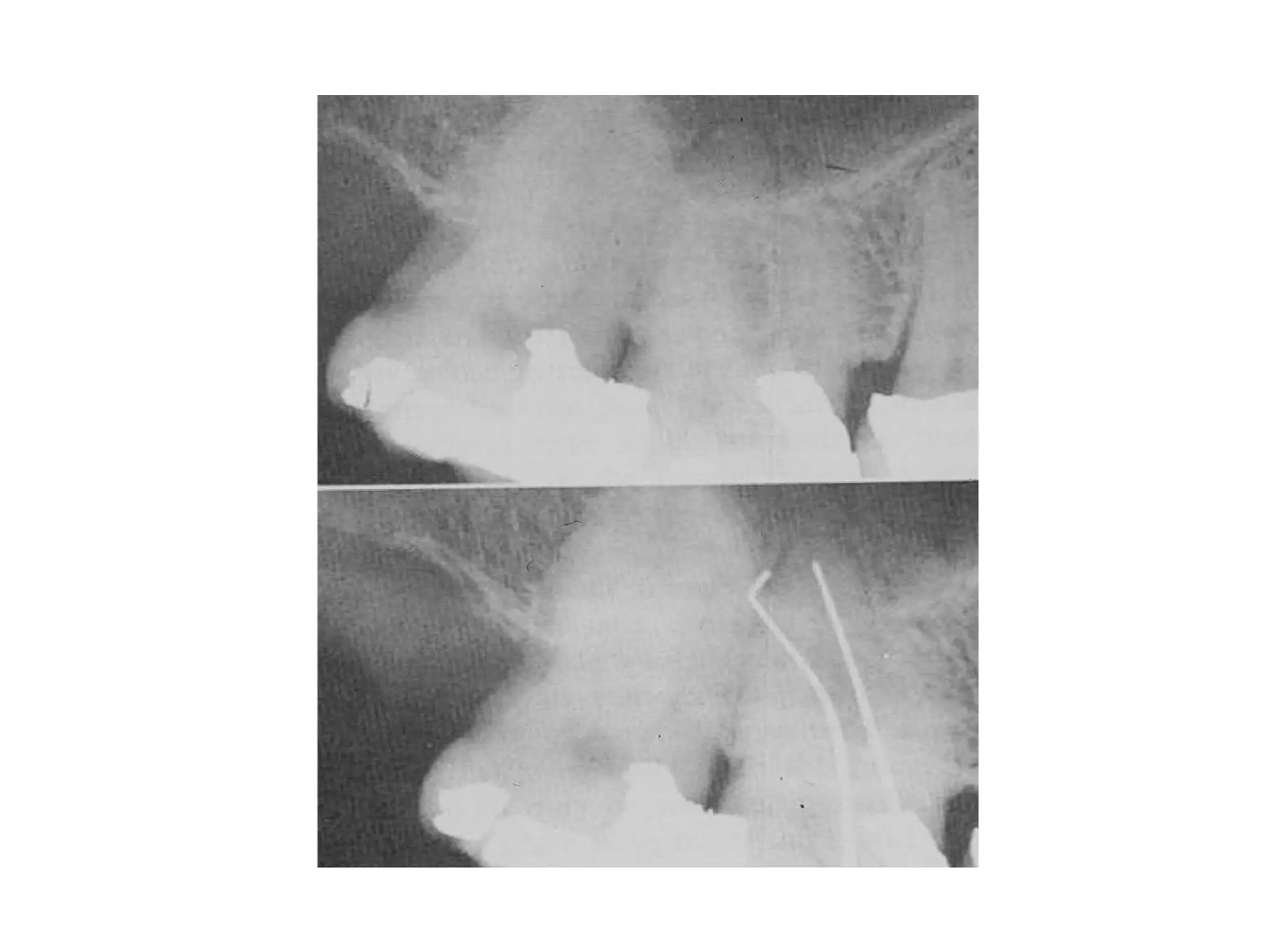
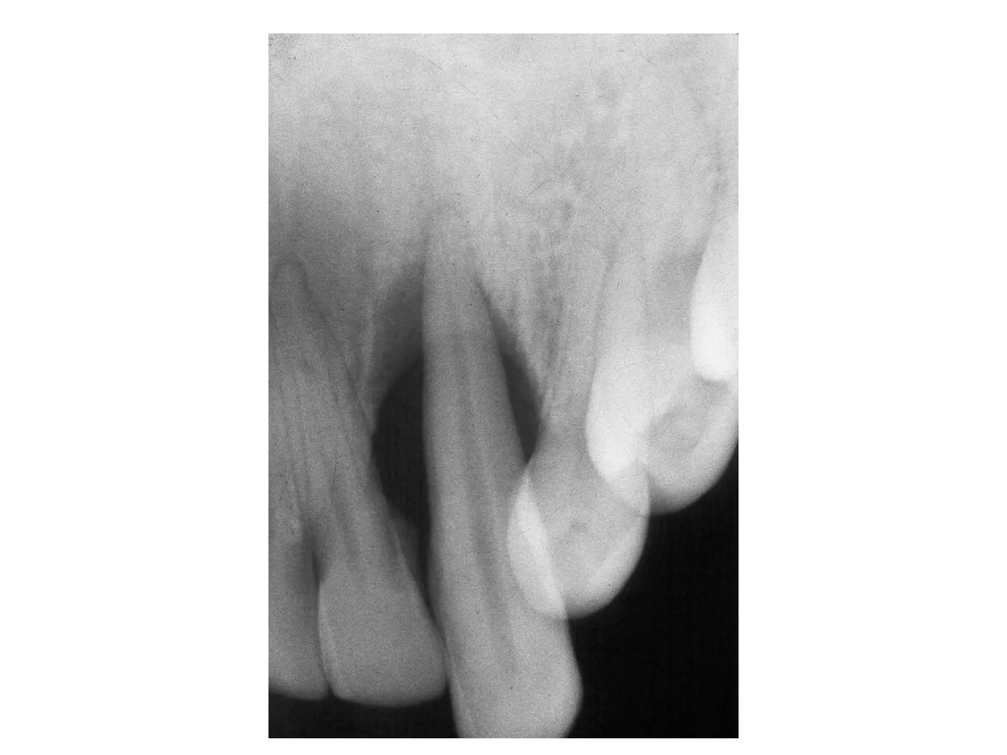
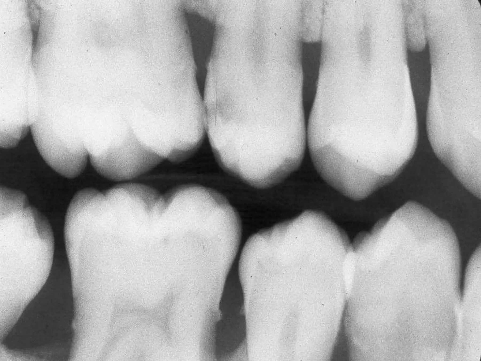
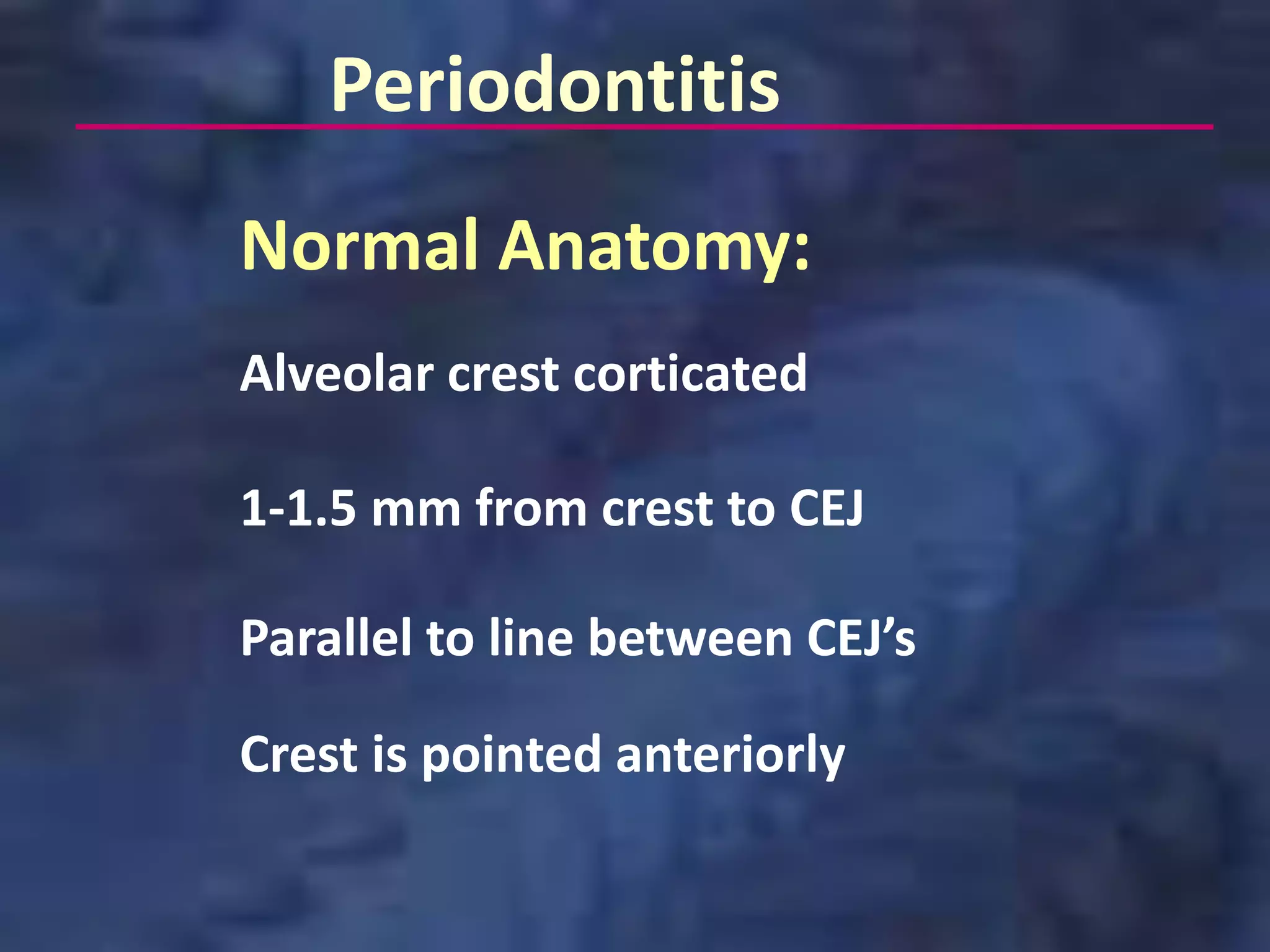
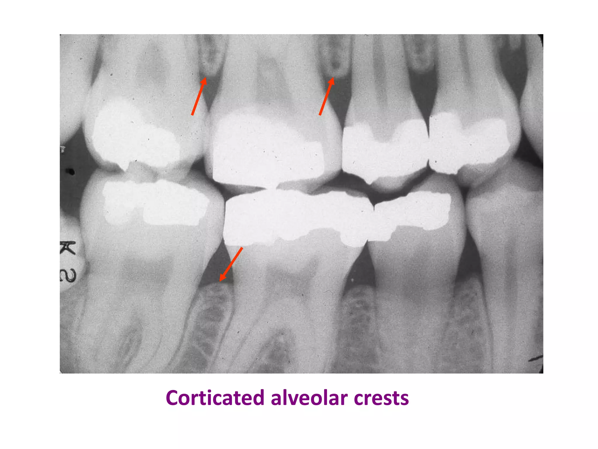
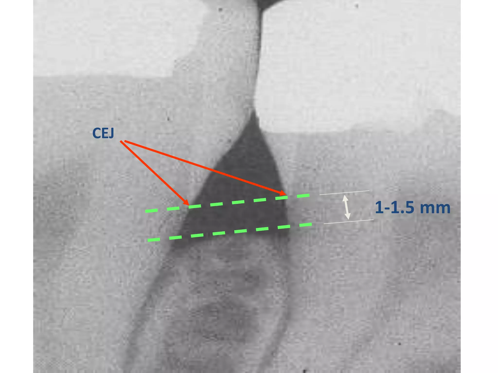
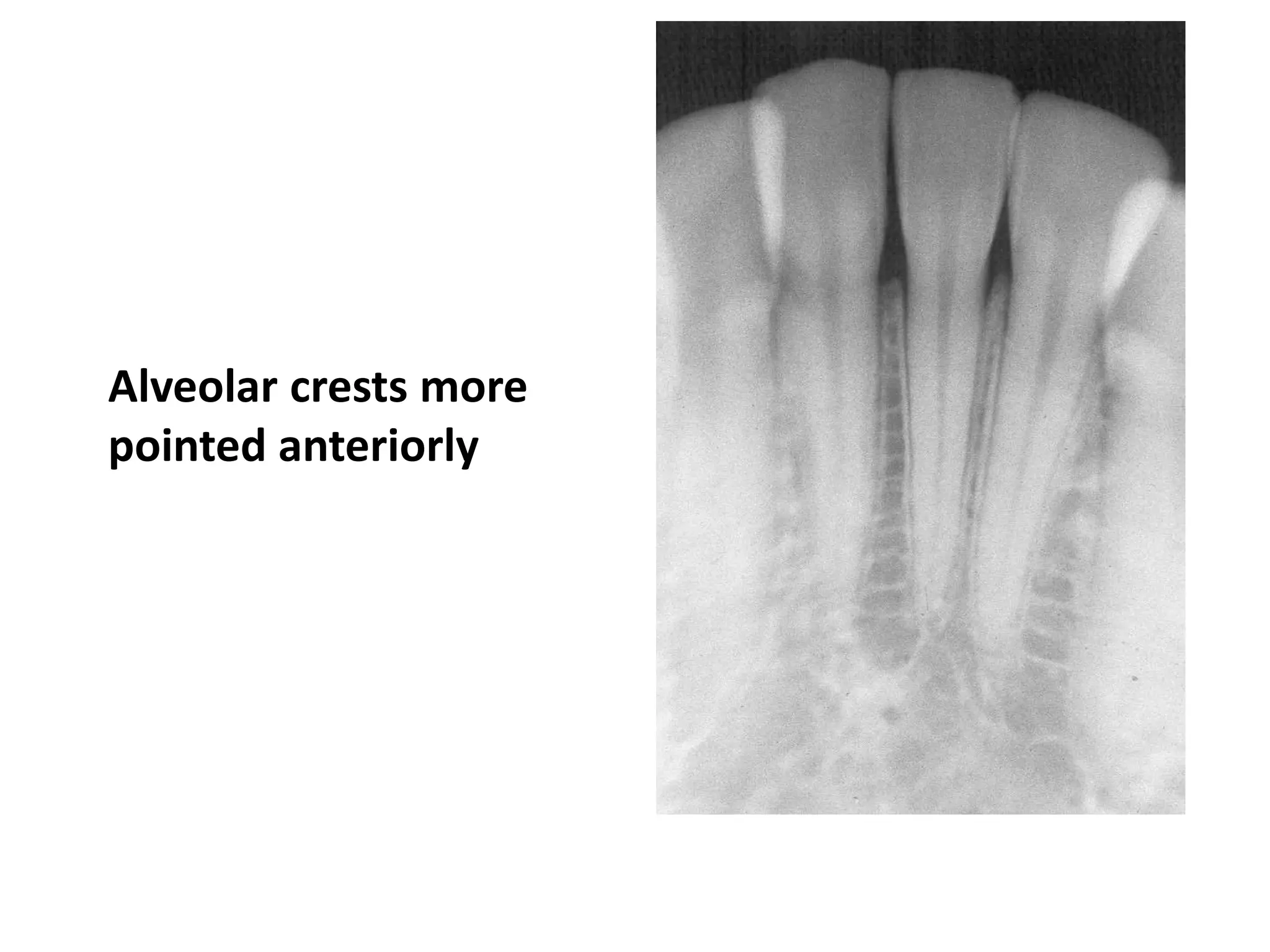
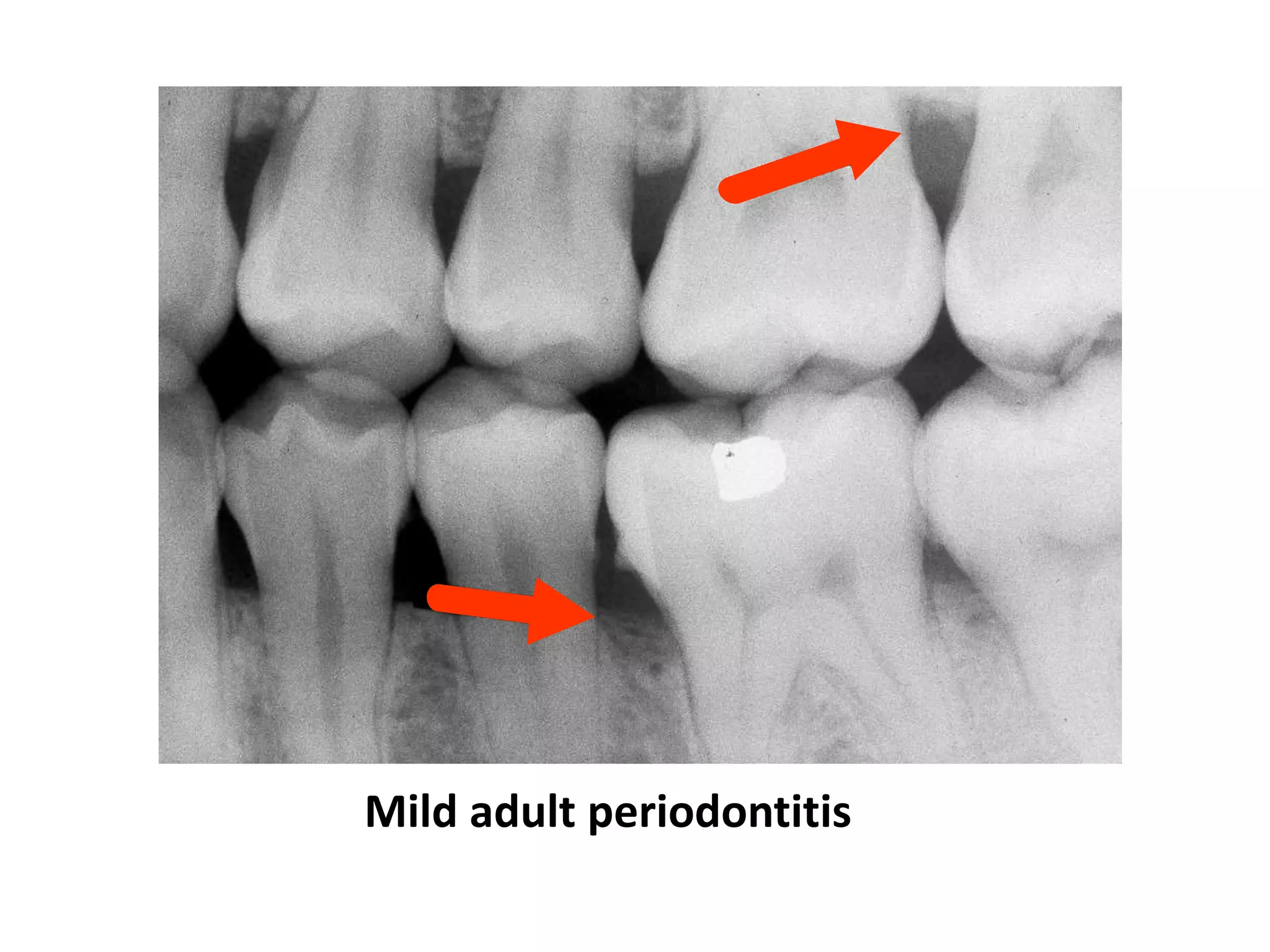
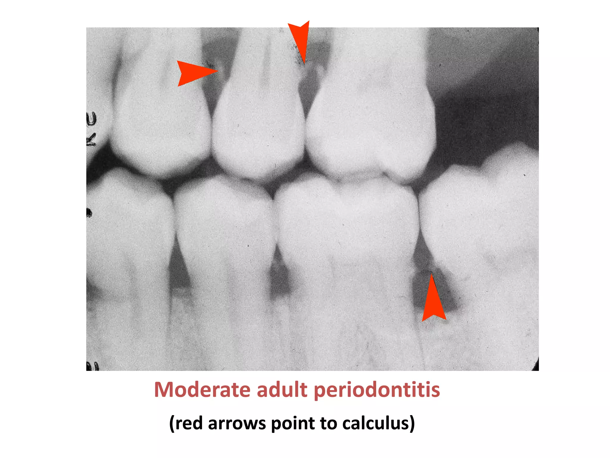
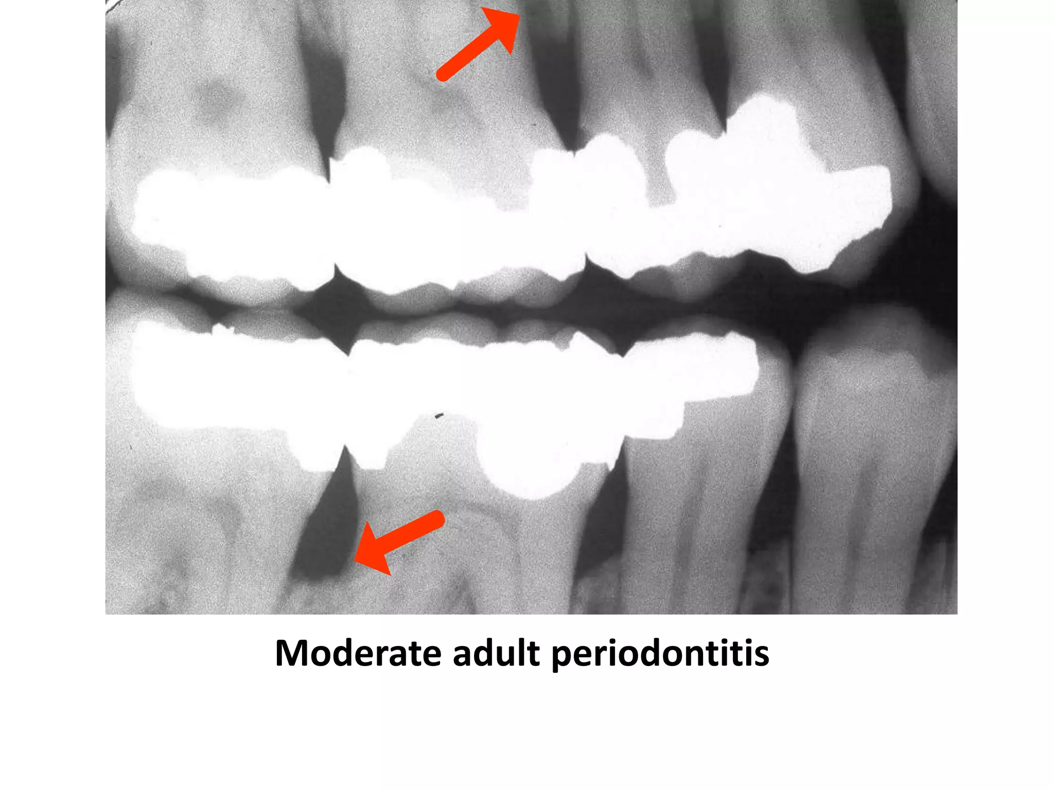
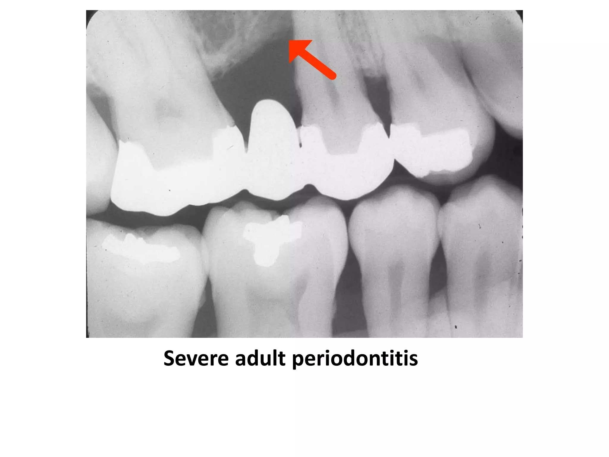
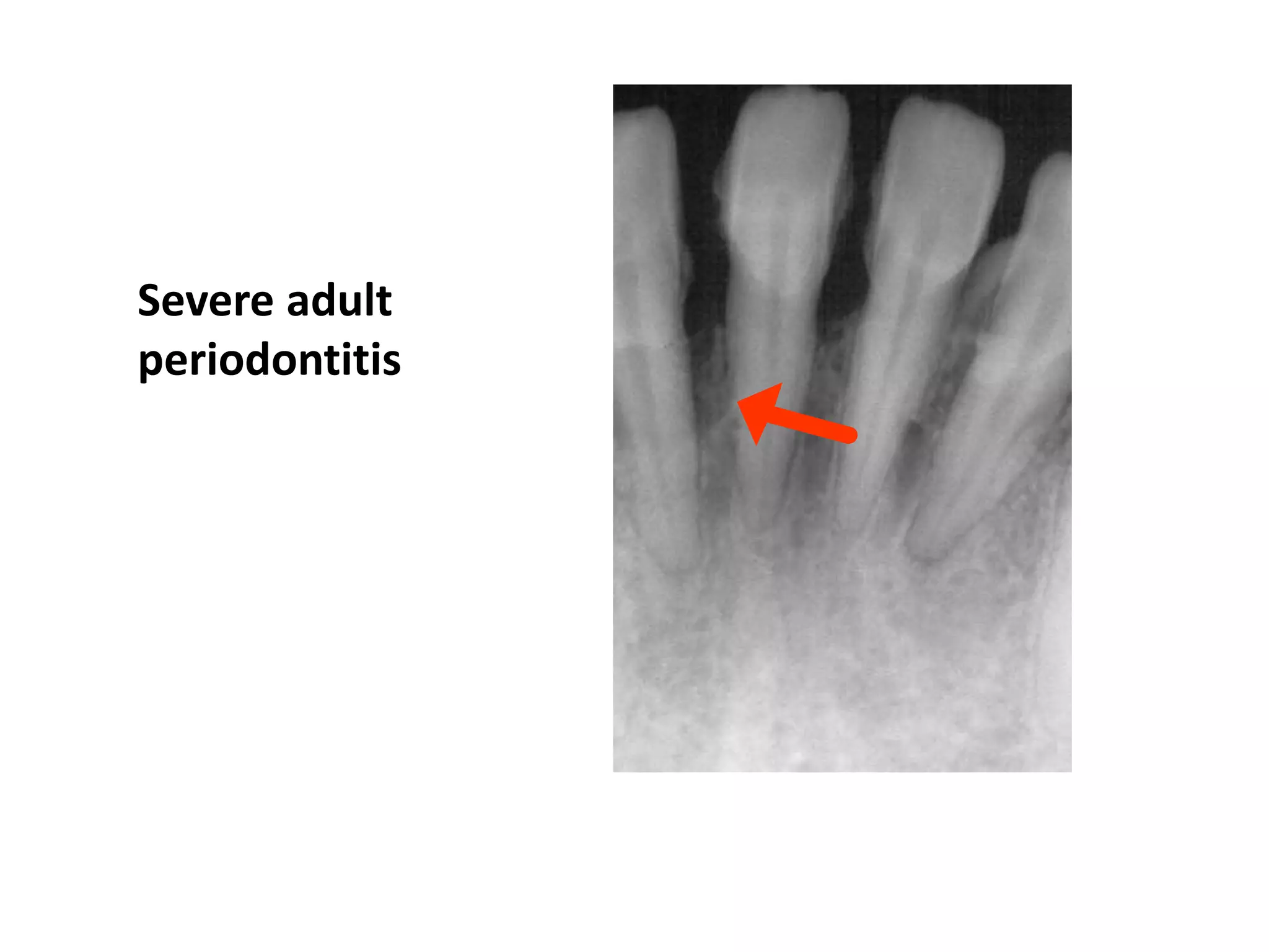
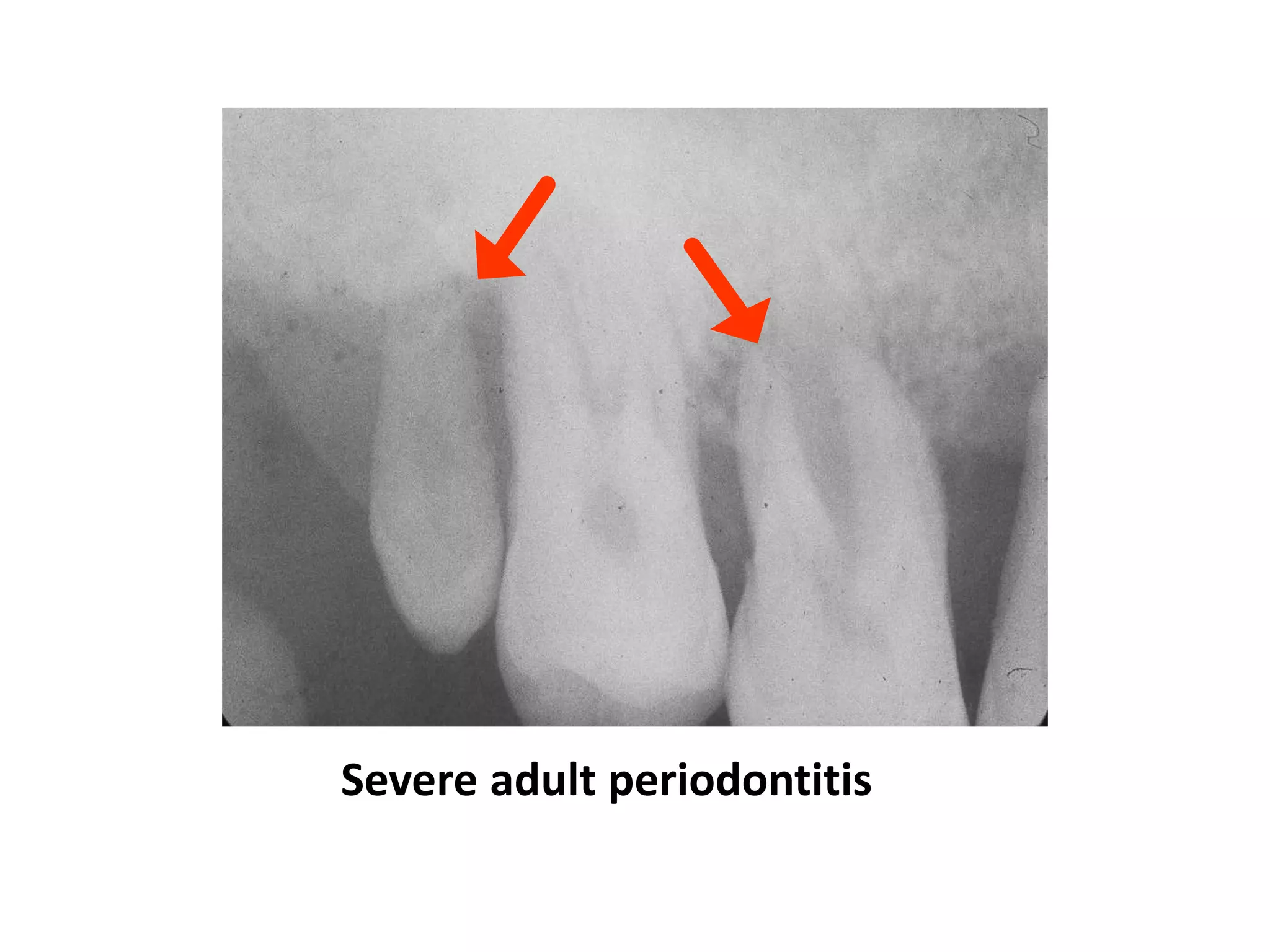
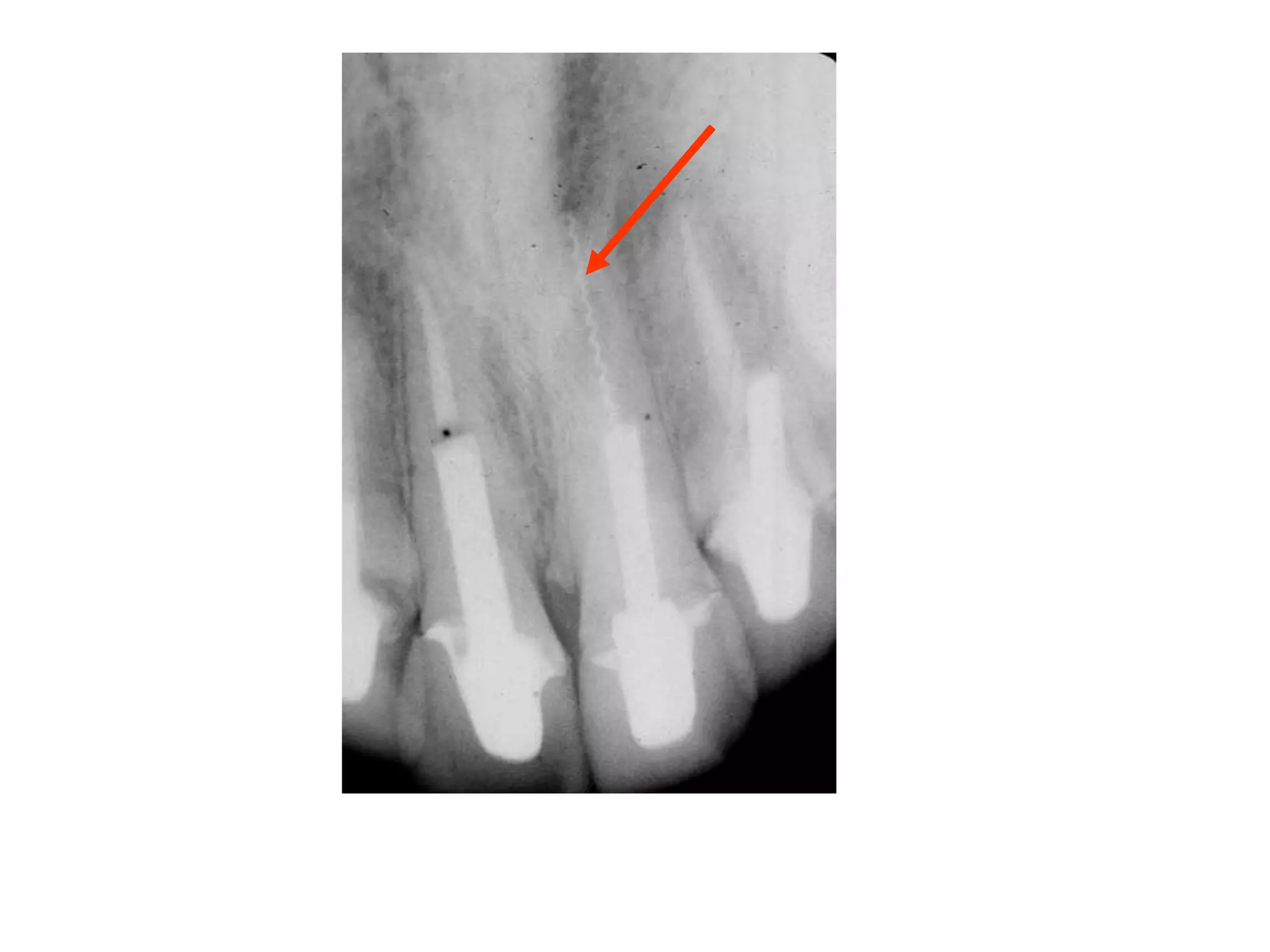
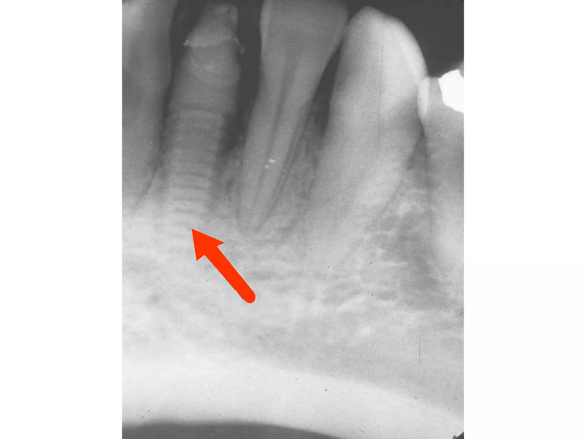
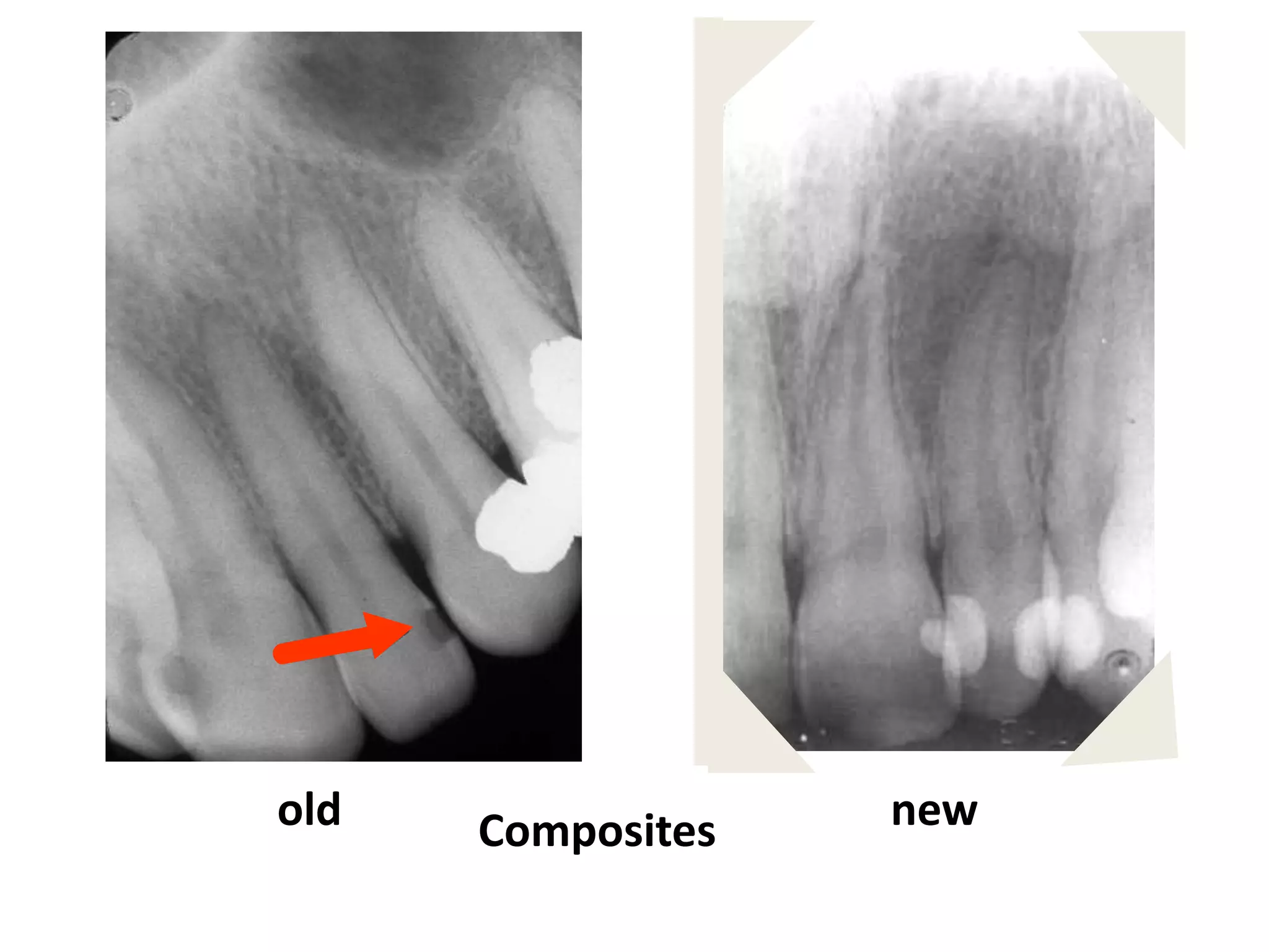
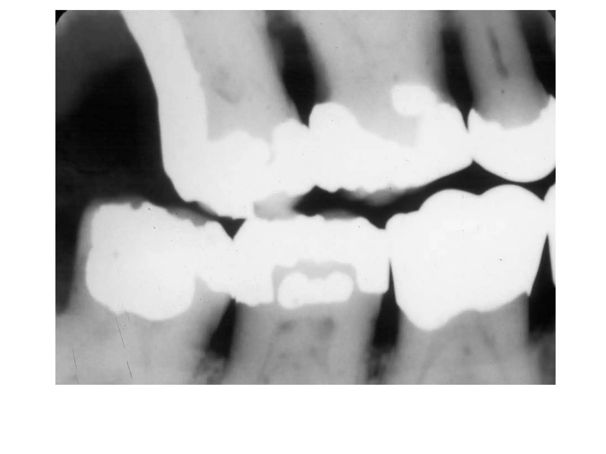

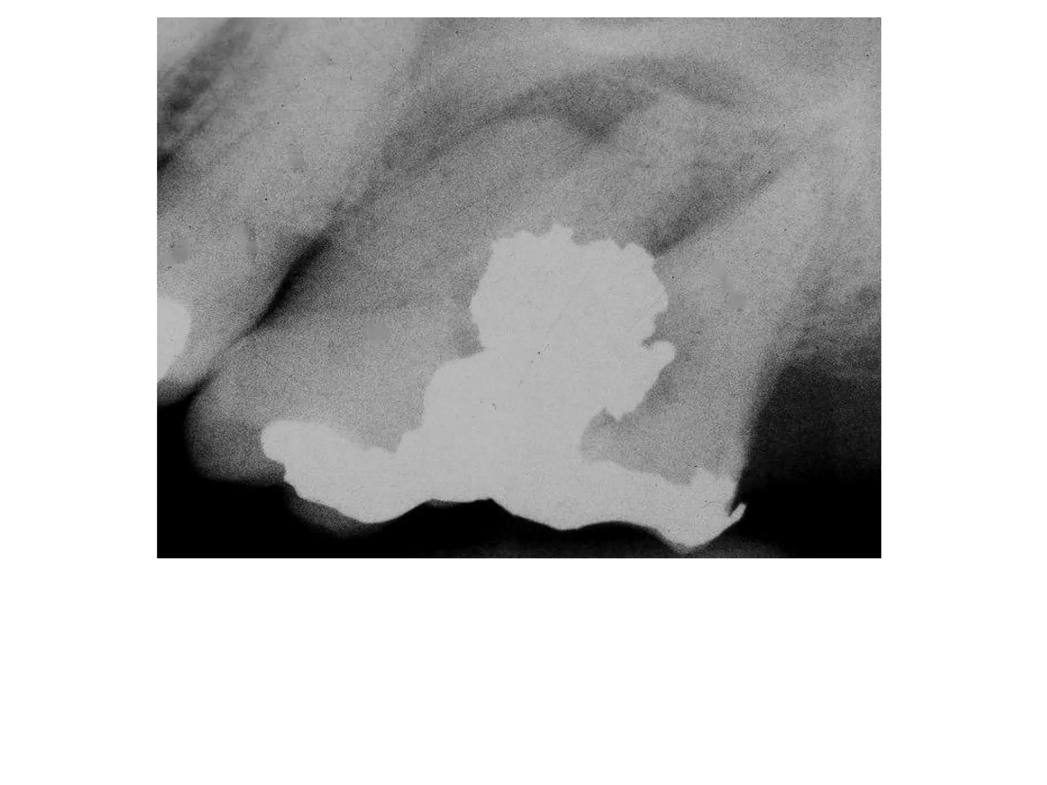


Periodontal disease involves the loss of periodontal ligament attachment and alveolar bone support around teeth. Radiographs are useful but limited for diagnosing periodontal disease, as they provide only a two-dimensional view and structures can be superimposed. Early signs on radiographs include crestal irregularities, triangulation of the periodontal ligament space, and interdental bone changes. Periodontitis can range from localized to more severe and generalized forms with increased bone loss and mobility. Contributing risk factors include occlusal trauma, open contacts, overhangs, calculus, and systemic health issues.


