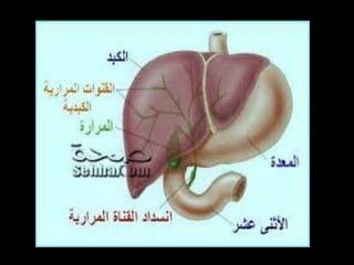
‫د‬.‫حجازى‬ ‫مصطفى‬
 Common
• Common bile duct stones
• Carcinoma of the head of pancreas
• Malignant porta hepatis lymph nodes
 Infrequent
• Ampullary carcinoma
• Pancreatitis
• Liver secondaries
 Rare
• Benign strictures - iatrogenic, trauma
• Recurrent cholangitis
• Mirrizi's syndrome
• Sclerosing cholangitis
• Cholangiocarcinoma
• Biliary atresia
• Choledochal cysts
Common bile duct stones
 Choledocholithiasis may be identified in 5–10% of
patients undergoing elective cholecystectomy.
 The presence of choledocholithiasis correlates with
patient age and is: 5%, 15%, and 35% in patients <60,
60–79, and >80 years of age, respectively.
 Common duct stones can be classified into two types:
primary and secondary.
 Primary stones form de novo in the common duct as a
result of biliary infection or stasis.
 Secondary stones are identical in composition to
gallbladder stones and presumably migrate from the
gallbladder.
 Clinical features of bile duct stones range from an
incidental finding discovered during cholecystectomy
to acute suppurative cholangitis.
 An increased serum alkaline phosphatase activity,
jaundice, and a dilated common bile duct are
characteristic signs of common duct stones, but are
not specific.
 Magnetic resonance cholangiopancreatography
(MRCP) is an effective, noninvasive method for
detecting choledocholithiasis when suspected.
 Endoscopic retrograde cholangiopancreatography
(ERCP) with sphincterotomy and stone retrieval is a
safe and effective treatment of common duct stones.
 When technically possible, laparoscopic, transcystic
common bile duct exploration with stone retrieval is a
safe and effective treatment option in experienced
centers.
 Overall, 5–15% of patients undergoing
cholecystectomy for cholelithiasis have concomitant
bile duct stones, and a small percentage of patients
after cholecystectomy will develop common duct
stones.
 Thus, bile duct stones and their management
constitute an important clinical problem.
 Traditionally, laparotomy with common duct
exploration was the primary therapy since the
performance of the first successful biliary lithotomy by
Courvoisier in 1890.
 The introduction of endoscopic sphincterotomy by
Classen and Kawai in 1974 initiated a change in the
management of retained and recurrent bile duct
stones.
 Advances in endoscopic equipment, techniques, and
the availability of skilled endoscopists have solidified
the success of this minimally invasive treatment
option.
 Today, open common duct exploration is rarely
necessary in most centers.
 Laparoscopic common duct exploration has also been
described and is gaining popularity in several centers.
 Furthermore, modern imaging modalities, including
MRI, multi-detector CT, and endoscopic
ultrasonography (EUS), have allowed improved
detection of choledocholithiasis.
 At present, it would be difficult to set a universally
accepted algorithm for the diagnosis and treatment of
common bile duct stone due to a wide variety of highly
accurate imaging modalities and varying expertise of
biliary endoscopists and laparoscopic surgeons.
 Common duct stones are defined as calculi in the
common duct regardless of the coexistence of stones
in the gallbladder.
 When stones are present proximal to the confluence of
the right and left hepatic ducts, they are termed
intrahepatic.
 Bile duct stones can be classified into two types,
depending on theirsite of origin:
 Primary stones form de novo in the bile duct (after
cholecystectomy), whereas
 Secondary stones are presumed to have migrated from
the gallbladder into the biliary tree.
 Primary common duct stones are composed
predominantly of calcium bilirubinate monomers with
variable, but minor quantities of cholesterol, and
calcium soaps of fatty acids such as calcium palmitate.
 These stones are often described as ‘‘earthy,’’ brownish
yellow, and are usually very soft and friable.
 Their pathogenesis is attributed to two main factors:
bacterial infection and biliary stasis.
 The predominant organisms isolated from bile in the
presence of primary bile duct stones include
gramnegative enteric organisms such as Escherichia
coli and Klebsiella species, and anaerobes, such as
Bacteroides and Clostridium
 These bacteria secrete enzymes such as b-
glucuronidase that catalyze the conjugation of
bilirubin and the lysis of phospholipids, leading to
deposition of calcium bilirubinate and calcium
palmitate.
 Bacterial digestion of bile salts and lecithin further
promotes lithogenesis and adds cholesterol to the
primary duct stones.
 The association of biliary strictures and severe ductal
dilation with the formation of brown pigment stones
supports the contributory role of biliary stasis in the
pathogenesis of primary duct stones.
 Finally, foreign objects, such as nonabsorbable suture
material and hemostatic clips, have also been
associated with primary duct stone formation,
presumably by providing a nidus for bacterial
colonization and subsequent stone growth.
 Secondary stones are identical in composition to
stones in the gallbladder (predominantly cholesterol
in 80% or black pigment in 20%) and are presumed to
form in the gallbladder with subsequent migration
into the biliary tree.
 The events predisposing to this migration remain
unknown; however, the diameter of the cystic duct
appears to be important.
 In a prospective evaluation of 331 patients undergoing
cholecystectomy, stones present in the gallbladder
could be advanced manually through the cystic duct in
60% of patients with concomitant common bile duct
stones, in 67% with gallstone pancreatitis, but in only
3% of those with gallstones alone.
 None of the gallstones with diameters greater than the
diameter of the cystic duct could be forced through it.
 Rarely, large gallstones (unable to traverse the cystic
duct) can enter the bile duct through a fistula.
 The prevalence of common duct stones is reportedly
5–15% of patients undergoing cholecystectomy for
cholelithiasis.
 Several series have demonstrated a correlation
between age and the presence of common duct stones.
For patients aged <60, 60–79, and >75 years
undergoing cholecystectomy for cholecystolithiasis,
concomitant choledocholithiasis has been identified
in 5%, 15%, and 35%, respectively.
 One theory for this relationship with age is the time at
risk for choledocholithiasis in patients with
cholelithiasis.
 Stones discovered in the bile ducts after
cholecystectomy are defined as retained, residual, or
recurrent.
 When choledocholithiasis is identified by
cholangiography shortly after cholecystectomy, they
are termed ‘‘retained,’’ because these stones are missed
during the operation.
 Stones that are found later (<2 years after
cholecystectomy) and contain the same composition
(black pigment or cholesterol) as the gallbladder
stones are termed ‘‘residual’’ stones.
 Stones that form within the bile duct (primary duct
stones, brown stones) or those discovered >2 years
after the cholecystectomy are termed ‘‘recurrent’’
stones and are assumed to be primary common duct
stones.
 Analysis of composition of 56 sets of common duct
stones revealed that most stones (75%) found at, or
shortly after, cholecystectomy were of the cholesterol
or black pigment type.
 In contrast, the majority discovered 21 months or more
after cholecystectomy were brown pigment stones.
 The prevalence of either retained stones or residual
stones is estimated to be 1–7% and 2–5%, respectively.
 With the assumption that some patients with
choledocholithiasis may remain asymptomatic, the
actual prevalence of retained common bile duct stones
may be greater.
 The spectrum of clinical features of
choledocholithiasis ranges from an incidental finding
during cholecystectomy to acute suppurative
cholangitis.
 Whereas the overall prevalence of common duct
stones discovered at the time of cholecystectomy is
about 10%, the proportion that is unsuspected (normal
laboratory data and normal bile duct by
ultrasonography) is probably l–3%.
 The patient with choledocholithiasis commonly has
severe discomfort in the epigastrium or right upper
quadrant that often radiates to the back and may be
associated with nausea and vomiting.
 The pain presents as discrete attacks lasting 30 min to
several hours and is indistinguishable from pain
elicited by gallstone impaction in the cystic duct,
except for its often epigastric location.
 These symptoms may occur at any time and are not
necessarily related to food ingestion.
 Ductal stones can cause obstruction to bile flow, pain,
obstructive jaundice, and infection/cholangitis
 The classic triad of right upper quadrant pain, fever
and chills, and jaundice, termed ‘‘Charcot’s triad,’’ is
present in 50–75% of patients with acutecholangitis.
 When patients exhibit hypotension and mental
confusion in addition to Charcot’s triad, it is termed
Reynald’s pentad and portends a high mortality of 30–
50%.
 Gallstone pancreatitis is a potential serious sequela of
choledocholithiasis.
 While most common bile duct stones that cause
pancreatitis pass spontaneously, the severity of the
illness is related generally to the severity of the
pancreatitis, ranging from mild and self-limited to
severe and necrotizing, with the complications of
infection, peripancreatic fluid collections, and death.
 Acute cholangitis may accompany the pancreatitis and
worsens the prognosis.
 Ascending cholangitis
• Charcot's triad is classical clinical picture
Intermittent pain, jaundice and fever
• Cholangitis can lead to hepatic abscesses
• Need parenteral antibiotics and biliary decompression
•Operative mortality in elderly of up to 20%
 Clotting disorders
• Vitamin K required for gamma-carboxylation of Factors
II, VII, IX, XI
• Vitamin K is fat soluble. No absorbed.
• Needs to be given parenterally
• Urgent correction will need Fresh Frozen Plasma
• Also endotoxin activation of complement system
 Hepato-renal syndrome
• Poorly understood
• Renal failure post intervention
• Due to gram negative endotoxinaemia from gut
• Preoperative lactulose may improve outcome
• Improves altered systemic and renal haemodynamics
 Drug Metabolism
• Half life of some drugs prolonged. e.g. morphine
 Impaired wound healing
Investigation will differentiate hepatocellular and
obstructive jaundice in 90% cases
 Blood results
• Conjugated bilirubin more than 35 mmol/l
• Increase in ALP / GGT more than AST / ALT
• Albumin may be reduced
• Prolonged PTT
 Several serum laboratory tests are helpful in the
evaluation of patients with suspected
choledocholithiasis.
 These tests reflect the degree of biliary tract
obstruction; however, they may be normal in patients
with non-obstructing stones.
 Urinalysis findings in jaundice
 Haemolysis Obstruction Hepatocellular
Conjugated bilirubin normal increased normal
 Urobilinoge increased nil normal
 The alkaline phosphatase activity is probably the most
sensitive measure of obstruction and is usually
increased twofold to fivefold in choledocholithiasis.
 Higher levels may be suggestive of a more chronic,
malignant biliary obstruction. Increased serum
bilirubinconcentration adds specificity to the
laboratory diagnosis of bile duct obstruction and is
present in 50–75% of symptomatic patients with
choledocholithiasis.
 Of the patients with acute cholecystitis but without
choledocholithiasis, 30% will have hyperbilirubinemia
(usually <3 mg/dl).
 A serum bilirubin concentration of >7 mg/dl in
patients with acute cholecystitis usually indicates
concomitant bile duct stones.
 Marked increase in serum bilirubin (>10 mg/dl) in the
absence of suppurative cholangitis suggests malignant
obstruction or concomitant parenchymal disease of
the liver.
 Only rarely does choledocholithiasis cause a complete
and progressive biliary obstruction.
 Fluctuation of the serum bilirubin level may also
suggest choledocholithiasis.
 The ability of aspartate aminotransferase activity to
differentiate biliary obstruction due to calculi from
that due to malignant neoplasm has been suggested.
 With choledocholithiasis, the serum level often
increases acutely to 3–5 times normal and may
increase into the thousands (hepatitis range), but
typically returns rapidly toward normal.
 This phenomenon is not seen with malignant
obstruction.
 When superimposed biliary infection occurs, the
white cell count is increased usually to 15,000–18,000
mm–3 but can also be decreased markedly to 2,000–
3,000 mm–3 with overwhelming sepsis.
 Patients who are asymptomatic or who present with
biliary colic frequently have a normal white cell count.
Ultrasonography
 The sensitivity of transcutaneous ultrasonography in
detecting choledocholithiasis is low, but is fairly
specific.
 In patients with a dilated common bile duct,
choledocholithiasis or other causes of obstruction are
inferred; however, 30% of patients with
choledocholithiasis have no evidence of ductal dilation
on ultrasonography.
 Although able to assess accurately biliary dilation,
ultrasonography is able to detect common bile duct
stones in only 15% of patients.
 This sensitivity increases to 33% in patients who are
jaundiced, but this overall low sensitivity requires
other imaging modalities in the majority of patients
with suspected choledocholithiasis
 Ultrasound
• Normal CBD less than 8 mm diameter
• CBD diameter increase with age and after previous
biliary surgery
• For obstructive jaundice ultrasound has a sensitivity 70
- 95% and specificity 80 - 100%
• In future endoscopic ultrasound may become more
widely available
Intravenous cholangiography
 Intravenous cholangiography was used historically in
the detection of choledocholithiasis, but fell into
disfavor because of its low sensitivity and potential for
adverse reactions.
 In a recent study, however, preoperative intravenous
cholangiograms were obtained in l00 consecutive
patients undergoing laparoscopic cholecystectomy; all
patients had subsequent operative cholangiography.
 Intravenous cholangiography identified
choledocholithiasis in eight patients with only one
false-positive result.
 Operative cholangiography discovered only one
additional patient with choledocholithiasis.
 Only two patients had mild reactions to the contrast
agent.
 If the results of this study are verified, intravenous
cholangiography may be of value in the detection of
common duct stones before laparoscopic surgery.
 CT Scanning
• Sensitivity and specificity similar to good quality
ultrasound
• Useful in obese or excessive bowel gas
• Better at imaging lower end of common bile duct
• Stages and assesses operability of tumours
 Radionuclide scanning
• 99 technetium iminodiacetic acid (HIDA)
• Taken up by hepatocytes and actively excreted into bile
• Allows imaging of biliary tree
• Failure to fill gallbladder = acute cholecytitis
• Delay of flow into duodenum = biliary obstruction
 MR imaging has seen a rapid technologic development
in recent years, with dramatic improvement in
noninvasive techniques and diagnostic accuracy for
both abdominal imaging and MRCP.
 For MRCP, the combination of T1- and T2-weighted
thin section scans in coronal oblique planes together
with thick slab radial cholangiographic T2-weighted
images to delineate anatomy are obtained.
 MR has proven to be an excellent, noninvasive,
accurate imaging technique without the need for
ionizing radiation or, in many cases, the need for any
type of oral or intravenously infused contrast
administration.
 Moreover, MR imaging is relatively operator-
independent and has no or negligiblemorbidity.
 The most commonly used contrast agent is a
gadolinium chelate, which has excellent patient safety
and tolerability.
 In one study, MRCP was performed in 202 patients
with a suspicion of choledocholithiasis based on
clinical presentation and/or cholestatic liver function
tests.
 ERCP was performed subsequently in all patients in
whom MRCP indicated choledocholithiasis, as well as
in those patients with a strong clinical suspicion of
choledocholithiasis despite a negative MRCP.
 In 25 patients, MRCP suggested choledocholithiasis,
which was proven subsequently with ERCP in 24
patients.
 Despite a negative MRCP, 27 additional patients had a
subsequent ERCP, none of whom had evidence of
stones.
 In the entire group, MRCP resulted in a 100%
sensitivity and a 96% specificity in detecting stones in
the common bile duct.
 In obstructive jaundice, MRCP has an overall accuracy
of 96–100% for demonstrating the anatomic level of
obstruction and a 90% accuracy for the cause of
obstruction.
 MRCP may, therefore, be able to replace diagnostic
ERCP in patients with suspected choledocholithiasis,
while the more invasive ERCP may be reserved for
patients requiring endoscopic intervention.
 The limitations of MR imaging are due predominantly
to the difficulty in imaging certain groups of patients,
such as those with claustrophobia or those with
cardiac pacemakers.
 MR imaging is also not suitable when therapeutic
intervention is planned.

 ERCP
• Allows biopsy or brush cytology
• Stone extraction or stenting
 PTC
• Rarely required today
• Performed with 22G Chiba Needle
• Also allows biliary drainage and stenting
 ERC and percutaneous transhepatic cholangiograms
Percutaneous transhepatic cholangiography (PTC)
provides direct imaging of the biliary tree and remains
the gold standard in depicting subtle changes within
the bile ducts and in the detection of small calculi. If
opacification of the biliary system is obtained,
cholangiography has a sensitivity approaching 100% in
the detection of obstruction.
 ERCP is now performed more often than PTC.
 Both approaches of direct ductal imaging are invasive
investigations which are operator-dependent and have
a relatively high morbidity of 1–7% for ERCP and 3–5%
for PTC.
 ERCP has a sensitivity of 90–96% and specificity of
98% in detecting choledocholithiasis, but in 3–10% of
patients, cannulation of the bile duct is unsuccessful.
 PTC provides excellent imaging with a success rate of
up to 99%, although this level of success is highly
dependent on the presence of biliary dilation.
 As the diagnostic role of MRCP evolves, the main
advantages of ERCP and PTC are primarily in their
therapeutic use with the ability to extract stones,
perform tissue biopsy, and replace stents
Endoscopic ultrasonography
 EUS offers a diagnostic accuracy comparable to that of
MRCP; however, the invasive nature, operator
dependence, and restricted availability have limited its
widespread use in detection of choledocholithiasis.
 Preoperative biliary decompression improves
postoperative morbidity
 Broad spectrum antibiotic prophylaxis
 Parenteral vitamin K +/- fresh frozen plasma
 IVI and catheter
 Pre operative fluid expansion
 Need careful post operative fluid balance to correct
depleted ECF compartment
 Consider 250 ml 10% mannitol. No proven benefit in
RCT
 Endoscopic Intervention
 Long-Term Endobiliary Stenting
 T-Tube Extraction
 Transhepatic Approach
 Medical Management
 ERCP and endoscopic sphincterotomy is investigation
of choice
o Stones extracted with balloons or Dormia basket
o 90% successful
o Complication rate approximately 8%
o Mortality less than1%
 If fails to clear stones will require:
o Open cholecystectomy + exploration of CBD
o Laparoscopic exploration of CBD
o Mechanical lithotripsy
 80% successful after failure of ERCP
o Extra-corporeal shockwave lithotripsy
o Chemical dissolution with cholesterol solvents
Methyl terbutyl ether or mono-octanoin
Administered via T Tube or nasobiliary catheter
25% complete response and 30% partial response
 If retained stones after CBD exploration need to
consider:
o Early ERCP
o Exploration via T tube tract at 6 weeks
Medical Management
 Oral bile acids have been used for years to dissolve
common duct stones.
 The success rates, however, are variable, ranging from
10% to 44%. In one randomized, double-blind,
placebo-controlled study, 28 patients with
uncomplicated, non-obstructing common duct stones
were treated with ursodeoxycholic acid (12 mg/kg/d
for up to 2 years); the bile duct stones disappeared in 7
of 14 patients in the treatment group and 0 of 14 in the
placebo group.
 Four patients (14%) required operative intervention,
including
 one from the treated and three from the placebo
groups.
 Currently, optimal patient selection, duration of
treatment, and optimal dosing has not been
determined with these medical treatments of
choledocholithiasis.
 The area in which oral bile acid therapy may play a role
is the treatment of asymptomatic patients with small
cholesterol duct stones discovered during laparoscopic
cholecystectomy.
 The composition of bile ductal stones may be inferred
if cholesterol stones were present in the gallbladder,
and small duct stones might dissolve relatively rapidly.
 Because duct exploration during laparoscopic
cholecystectomy might be demanding technically,
especially with a small-diameter cystic and/or
common bile duct, this dissolution therapy is a
reasonable therapeutic alternative; however, this
approach needs to be evaluated in clinical trials.
Thank You

Obstructive jaundice hegazy

  • 1.
  • 3.
     Common • Commonbile duct stones • Carcinoma of the head of pancreas • Malignant porta hepatis lymph nodes  Infrequent • Ampullary carcinoma • Pancreatitis • Liver secondaries
  • 4.
     Rare • Benignstrictures - iatrogenic, trauma • Recurrent cholangitis • Mirrizi's syndrome • Sclerosing cholangitis • Cholangiocarcinoma • Biliary atresia • Choledochal cysts
  • 5.
  • 7.
     Choledocholithiasis maybe identified in 5–10% of patients undergoing elective cholecystectomy.  The presence of choledocholithiasis correlates with patient age and is: 5%, 15%, and 35% in patients <60, 60–79, and >80 years of age, respectively.  Common duct stones can be classified into two types: primary and secondary.
  • 8.
     Primary stonesform de novo in the common duct as a result of biliary infection or stasis.  Secondary stones are identical in composition to gallbladder stones and presumably migrate from the gallbladder.  Clinical features of bile duct stones range from an incidental finding discovered during cholecystectomy to acute suppurative cholangitis.
  • 9.
     An increasedserum alkaline phosphatase activity, jaundice, and a dilated common bile duct are characteristic signs of common duct stones, but are not specific.  Magnetic resonance cholangiopancreatography (MRCP) is an effective, noninvasive method for detecting choledocholithiasis when suspected.
  • 10.
     Endoscopic retrogradecholangiopancreatography (ERCP) with sphincterotomy and stone retrieval is a safe and effective treatment of common duct stones.  When technically possible, laparoscopic, transcystic common bile duct exploration with stone retrieval is a safe and effective treatment option in experienced centers.
  • 11.
     Overall, 5–15%of patients undergoing cholecystectomy for cholelithiasis have concomitant bile duct stones, and a small percentage of patients after cholecystectomy will develop common duct stones.  Thus, bile duct stones and their management constitute an important clinical problem.
  • 12.
     Traditionally, laparotomywith common duct exploration was the primary therapy since the performance of the first successful biliary lithotomy by Courvoisier in 1890.
  • 13.
     The introductionof endoscopic sphincterotomy by Classen and Kawai in 1974 initiated a change in the management of retained and recurrent bile duct stones.  Advances in endoscopic equipment, techniques, and the availability of skilled endoscopists have solidified the success of this minimally invasive treatment option.
  • 14.
     Today, opencommon duct exploration is rarely necessary in most centers.  Laparoscopic common duct exploration has also been described and is gaining popularity in several centers.  Furthermore, modern imaging modalities, including MRI, multi-detector CT, and endoscopic ultrasonography (EUS), have allowed improved detection of choledocholithiasis.
  • 15.
     At present,it would be difficult to set a universally accepted algorithm for the diagnosis and treatment of common bile duct stone due to a wide variety of highly accurate imaging modalities and varying expertise of biliary endoscopists and laparoscopic surgeons.
  • 16.
     Common ductstones are defined as calculi in the common duct regardless of the coexistence of stones in the gallbladder.  When stones are present proximal to the confluence of the right and left hepatic ducts, they are termed intrahepatic.
  • 17.
     Bile ductstones can be classified into two types, depending on theirsite of origin:  Primary stones form de novo in the bile duct (after cholecystectomy), whereas  Secondary stones are presumed to have migrated from the gallbladder into the biliary tree.
  • 18.
     Primary commonduct stones are composed predominantly of calcium bilirubinate monomers with variable, but minor quantities of cholesterol, and calcium soaps of fatty acids such as calcium palmitate.
  • 19.
     These stonesare often described as ‘‘earthy,’’ brownish yellow, and are usually very soft and friable.  Their pathogenesis is attributed to two main factors: bacterial infection and biliary stasis.  The predominant organisms isolated from bile in the presence of primary bile duct stones include gramnegative enteric organisms such as Escherichia coli and Klebsiella species, and anaerobes, such as Bacteroides and Clostridium
  • 20.
     These bacteriasecrete enzymes such as b- glucuronidase that catalyze the conjugation of bilirubin and the lysis of phospholipids, leading to deposition of calcium bilirubinate and calcium palmitate.  Bacterial digestion of bile salts and lecithin further promotes lithogenesis and adds cholesterol to the primary duct stones.
  • 21.
     The associationof biliary strictures and severe ductal dilation with the formation of brown pigment stones supports the contributory role of biliary stasis in the pathogenesis of primary duct stones.  Finally, foreign objects, such as nonabsorbable suture material and hemostatic clips, have also been associated with primary duct stone formation, presumably by providing a nidus for bacterial colonization and subsequent stone growth.
  • 22.
     Secondary stonesare identical in composition to stones in the gallbladder (predominantly cholesterol in 80% or black pigment in 20%) and are presumed to form in the gallbladder with subsequent migration into the biliary tree.
  • 23.
     The eventspredisposing to this migration remain unknown; however, the diameter of the cystic duct appears to be important.  In a prospective evaluation of 331 patients undergoing cholecystectomy, stones present in the gallbladder could be advanced manually through the cystic duct in 60% of patients with concomitant common bile duct stones, in 67% with gallstone pancreatitis, but in only 3% of those with gallstones alone.
  • 24.
     None ofthe gallstones with diameters greater than the diameter of the cystic duct could be forced through it.  Rarely, large gallstones (unable to traverse the cystic duct) can enter the bile duct through a fistula.
  • 25.
     The prevalenceof common duct stones is reportedly 5–15% of patients undergoing cholecystectomy for cholelithiasis.  Several series have demonstrated a correlation between age and the presence of common duct stones. For patients aged <60, 60–79, and >75 years undergoing cholecystectomy for cholecystolithiasis, concomitant choledocholithiasis has been identified in 5%, 15%, and 35%, respectively.
  • 26.
     One theoryfor this relationship with age is the time at risk for choledocholithiasis in patients with cholelithiasis.
  • 27.
     Stones discoveredin the bile ducts after cholecystectomy are defined as retained, residual, or recurrent.  When choledocholithiasis is identified by cholangiography shortly after cholecystectomy, they are termed ‘‘retained,’’ because these stones are missed during the operation.
  • 28.
     Stones thatare found later (<2 years after cholecystectomy) and contain the same composition (black pigment or cholesterol) as the gallbladder stones are termed ‘‘residual’’ stones.  Stones that form within the bile duct (primary duct stones, brown stones) or those discovered >2 years after the cholecystectomy are termed ‘‘recurrent’’ stones and are assumed to be primary common duct stones.
  • 29.
     Analysis ofcomposition of 56 sets of common duct stones revealed that most stones (75%) found at, or shortly after, cholecystectomy were of the cholesterol or black pigment type.  In contrast, the majority discovered 21 months or more after cholecystectomy were brown pigment stones.
  • 30.
     The prevalenceof either retained stones or residual stones is estimated to be 1–7% and 2–5%, respectively.  With the assumption that some patients with choledocholithiasis may remain asymptomatic, the actual prevalence of retained common bile duct stones may be greater.
  • 31.
     The spectrumof clinical features of choledocholithiasis ranges from an incidental finding during cholecystectomy to acute suppurative cholangitis.  Whereas the overall prevalence of common duct stones discovered at the time of cholecystectomy is about 10%, the proportion that is unsuspected (normal laboratory data and normal bile duct by ultrasonography) is probably l–3%.
  • 32.
     The patientwith choledocholithiasis commonly has severe discomfort in the epigastrium or right upper quadrant that often radiates to the back and may be associated with nausea and vomiting.  The pain presents as discrete attacks lasting 30 min to several hours and is indistinguishable from pain elicited by gallstone impaction in the cystic duct, except for its often epigastric location.
  • 33.
     These symptomsmay occur at any time and are not necessarily related to food ingestion.  Ductal stones can cause obstruction to bile flow, pain, obstructive jaundice, and infection/cholangitis
  • 34.
     The classictriad of right upper quadrant pain, fever and chills, and jaundice, termed ‘‘Charcot’s triad,’’ is present in 50–75% of patients with acutecholangitis.  When patients exhibit hypotension and mental confusion in addition to Charcot’s triad, it is termed Reynald’s pentad and portends a high mortality of 30– 50%.
  • 35.
     Gallstone pancreatitisis a potential serious sequela of choledocholithiasis.  While most common bile duct stones that cause pancreatitis pass spontaneously, the severity of the illness is related generally to the severity of the pancreatitis, ranging from mild and self-limited to severe and necrotizing, with the complications of infection, peripancreatic fluid collections, and death.  Acute cholangitis may accompany the pancreatitis and worsens the prognosis.
  • 36.
     Ascending cholangitis •Charcot's triad is classical clinical picture Intermittent pain, jaundice and fever • Cholangitis can lead to hepatic abscesses • Need parenteral antibiotics and biliary decompression •Operative mortality in elderly of up to 20%
  • 37.
     Clotting disorders •Vitamin K required for gamma-carboxylation of Factors II, VII, IX, XI • Vitamin K is fat soluble. No absorbed. • Needs to be given parenterally • Urgent correction will need Fresh Frozen Plasma • Also endotoxin activation of complement system
  • 38.
     Hepato-renal syndrome •Poorly understood • Renal failure post intervention • Due to gram negative endotoxinaemia from gut • Preoperative lactulose may improve outcome • Improves altered systemic and renal haemodynamics
  • 39.
     Drug Metabolism •Half life of some drugs prolonged. e.g. morphine  Impaired wound healing
  • 41.
    Investigation will differentiatehepatocellular and obstructive jaundice in 90% cases  Blood results • Conjugated bilirubin more than 35 mmol/l • Increase in ALP / GGT more than AST / ALT • Albumin may be reduced • Prolonged PTT
  • 42.
     Several serumlaboratory tests are helpful in the evaluation of patients with suspected choledocholithiasis.  These tests reflect the degree of biliary tract obstruction; however, they may be normal in patients with non-obstructing stones.
  • 43.
     Urinalysis findingsin jaundice  Haemolysis Obstruction Hepatocellular Conjugated bilirubin normal increased normal  Urobilinoge increased nil normal
  • 44.
     The alkalinephosphatase activity is probably the most sensitive measure of obstruction and is usually increased twofold to fivefold in choledocholithiasis.  Higher levels may be suggestive of a more chronic, malignant biliary obstruction. Increased serum bilirubinconcentration adds specificity to the laboratory diagnosis of bile duct obstruction and is present in 50–75% of symptomatic patients with choledocholithiasis.
  • 45.
     Of thepatients with acute cholecystitis but without choledocholithiasis, 30% will have hyperbilirubinemia (usually <3 mg/dl).  A serum bilirubin concentration of >7 mg/dl in patients with acute cholecystitis usually indicates concomitant bile duct stones.
  • 46.
     Marked increasein serum bilirubin (>10 mg/dl) in the absence of suppurative cholangitis suggests malignant obstruction or concomitant parenchymal disease of the liver.  Only rarely does choledocholithiasis cause a complete and progressive biliary obstruction.
  • 47.
     Fluctuation ofthe serum bilirubin level may also suggest choledocholithiasis.  The ability of aspartate aminotransferase activity to differentiate biliary obstruction due to calculi from that due to malignant neoplasm has been suggested.  With choledocholithiasis, the serum level often increases acutely to 3–5 times normal and may increase into the thousands (hepatitis range), but typically returns rapidly toward normal.  This phenomenon is not seen with malignant obstruction.
  • 48.
     When superimposedbiliary infection occurs, the white cell count is increased usually to 15,000–18,000 mm–3 but can also be decreased markedly to 2,000– 3,000 mm–3 with overwhelming sepsis.  Patients who are asymptomatic or who present with biliary colic frequently have a normal white cell count.
  • 49.
    Ultrasonography  The sensitivityof transcutaneous ultrasonography in detecting choledocholithiasis is low, but is fairly specific.  In patients with a dilated common bile duct, choledocholithiasis or other causes of obstruction are inferred; however, 30% of patients with choledocholithiasis have no evidence of ductal dilation on ultrasonography.
  • 50.
     Although ableto assess accurately biliary dilation, ultrasonography is able to detect common bile duct stones in only 15% of patients.  This sensitivity increases to 33% in patients who are jaundiced, but this overall low sensitivity requires other imaging modalities in the majority of patients with suspected choledocholithiasis
  • 51.
     Ultrasound • NormalCBD less than 8 mm diameter • CBD diameter increase with age and after previous biliary surgery • For obstructive jaundice ultrasound has a sensitivity 70 - 95% and specificity 80 - 100% • In future endoscopic ultrasound may become more widely available
  • 52.
    Intravenous cholangiography  Intravenouscholangiography was used historically in the detection of choledocholithiasis, but fell into disfavor because of its low sensitivity and potential for adverse reactions.  In a recent study, however, preoperative intravenous cholangiograms were obtained in l00 consecutive patients undergoing laparoscopic cholecystectomy; all patients had subsequent operative cholangiography.
  • 53.
     Intravenous cholangiographyidentified choledocholithiasis in eight patients with only one false-positive result.  Operative cholangiography discovered only one additional patient with choledocholithiasis.  Only two patients had mild reactions to the contrast agent.  If the results of this study are verified, intravenous cholangiography may be of value in the detection of common duct stones before laparoscopic surgery.
  • 54.
     CT Scanning •Sensitivity and specificity similar to good quality ultrasound • Useful in obese or excessive bowel gas • Better at imaging lower end of common bile duct • Stages and assesses operability of tumours
  • 55.
     Radionuclide scanning •99 technetium iminodiacetic acid (HIDA) • Taken up by hepatocytes and actively excreted into bile • Allows imaging of biliary tree • Failure to fill gallbladder = acute cholecytitis • Delay of flow into duodenum = biliary obstruction
  • 56.
     MR imaginghas seen a rapid technologic development in recent years, with dramatic improvement in noninvasive techniques and diagnostic accuracy for both abdominal imaging and MRCP.
  • 57.
     For MRCP,the combination of T1- and T2-weighted thin section scans in coronal oblique planes together with thick slab radial cholangiographic T2-weighted images to delineate anatomy are obtained.  MR has proven to be an excellent, noninvasive, accurate imaging technique without the need for ionizing radiation or, in many cases, the need for any type of oral or intravenously infused contrast administration.
  • 58.
     Moreover, MRimaging is relatively operator- independent and has no or negligiblemorbidity.  The most commonly used contrast agent is a gadolinium chelate, which has excellent patient safety and tolerability.
  • 59.
     In onestudy, MRCP was performed in 202 patients with a suspicion of choledocholithiasis based on clinical presentation and/or cholestatic liver function tests.  ERCP was performed subsequently in all patients in whom MRCP indicated choledocholithiasis, as well as in those patients with a strong clinical suspicion of choledocholithiasis despite a negative MRCP.
  • 60.
     In 25patients, MRCP suggested choledocholithiasis, which was proven subsequently with ERCP in 24 patients.  Despite a negative MRCP, 27 additional patients had a subsequent ERCP, none of whom had evidence of stones.  In the entire group, MRCP resulted in a 100% sensitivity and a 96% specificity in detecting stones in the common bile duct.
  • 61.
     In obstructivejaundice, MRCP has an overall accuracy of 96–100% for demonstrating the anatomic level of obstruction and a 90% accuracy for the cause of obstruction.  MRCP may, therefore, be able to replace diagnostic ERCP in patients with suspected choledocholithiasis, while the more invasive ERCP may be reserved for patients requiring endoscopic intervention.
  • 62.
     The limitationsof MR imaging are due predominantly to the difficulty in imaging certain groups of patients, such as those with claustrophobia or those with cardiac pacemakers.  MR imaging is also not suitable when therapeutic intervention is planned. 
  • 63.
     ERCP • Allowsbiopsy or brush cytology • Stone extraction or stenting  PTC • Rarely required today • Performed with 22G Chiba Needle • Also allows biliary drainage and stenting
  • 64.
     ERC andpercutaneous transhepatic cholangiograms Percutaneous transhepatic cholangiography (PTC) provides direct imaging of the biliary tree and remains the gold standard in depicting subtle changes within the bile ducts and in the detection of small calculi. If opacification of the biliary system is obtained, cholangiography has a sensitivity approaching 100% in the detection of obstruction.
  • 65.
     ERCP isnow performed more often than PTC.  Both approaches of direct ductal imaging are invasive investigations which are operator-dependent and have a relatively high morbidity of 1–7% for ERCP and 3–5% for PTC.  ERCP has a sensitivity of 90–96% and specificity of 98% in detecting choledocholithiasis, but in 3–10% of patients, cannulation of the bile duct is unsuccessful.
  • 66.
     PTC providesexcellent imaging with a success rate of up to 99%, although this level of success is highly dependent on the presence of biliary dilation.  As the diagnostic role of MRCP evolves, the main advantages of ERCP and PTC are primarily in their therapeutic use with the ability to extract stones, perform tissue biopsy, and replace stents
  • 67.
    Endoscopic ultrasonography  EUSoffers a diagnostic accuracy comparable to that of MRCP; however, the invasive nature, operator dependence, and restricted availability have limited its widespread use in detection of choledocholithiasis.
  • 68.
     Preoperative biliarydecompression improves postoperative morbidity  Broad spectrum antibiotic prophylaxis  Parenteral vitamin K +/- fresh frozen plasma  IVI and catheter  Pre operative fluid expansion  Need careful post operative fluid balance to correct depleted ECF compartment  Consider 250 ml 10% mannitol. No proven benefit in RCT
  • 69.
     Endoscopic Intervention Long-Term Endobiliary Stenting  T-Tube Extraction  Transhepatic Approach  Medical Management
  • 70.
     ERCP andendoscopic sphincterotomy is investigation of choice o Stones extracted with balloons or Dormia basket o 90% successful o Complication rate approximately 8% o Mortality less than1%  If fails to clear stones will require: o Open cholecystectomy + exploration of CBD
  • 71.
    o Laparoscopic explorationof CBD o Mechanical lithotripsy  80% successful after failure of ERCP o Extra-corporeal shockwave lithotripsy o Chemical dissolution with cholesterol solvents Methyl terbutyl ether or mono-octanoin Administered via T Tube or nasobiliary catheter 25% complete response and 30% partial response
  • 72.
     If retainedstones after CBD exploration need to consider: o Early ERCP o Exploration via T tube tract at 6 weeks
  • 73.
  • 74.
     Oral bileacids have been used for years to dissolve common duct stones.  The success rates, however, are variable, ranging from 10% to 44%. In one randomized, double-blind, placebo-controlled study, 28 patients with uncomplicated, non-obstructing common duct stones were treated with ursodeoxycholic acid (12 mg/kg/d for up to 2 years); the bile duct stones disappeared in 7 of 14 patients in the treatment group and 0 of 14 in the placebo group.
  • 75.
     Four patients(14%) required operative intervention, including  one from the treated and three from the placebo groups.
  • 76.
     Currently, optimalpatient selection, duration of treatment, and optimal dosing has not been determined with these medical treatments of choledocholithiasis.  The area in which oral bile acid therapy may play a role is the treatment of asymptomatic patients with small cholesterol duct stones discovered during laparoscopic cholecystectomy.
  • 77.
     The compositionof bile ductal stones may be inferred if cholesterol stones were present in the gallbladder, and small duct stones might dissolve relatively rapidly.  Because duct exploration during laparoscopic cholecystectomy might be demanding technically, especially with a small-diameter cystic and/or common bile duct, this dissolution therapy is a reasonable therapeutic alternative; however, this approach needs to be evaluated in clinical trials.
  • 78.