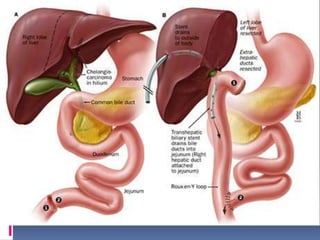
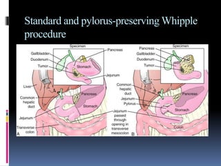
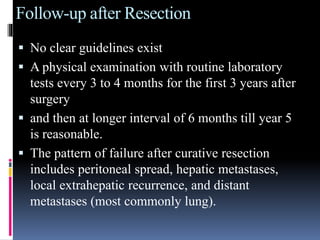
CHOLANGIOCARCINOMA
Dr.Atul Jain
 Denote cancers arising in the intrahepatic or
extrahepatic biliary tree, exclusive of the ampulla
of Vater and gallbladder.
 Incidence of cholangiocarcinoma in the United
States is 1 or 2 cases per 100,000 population
 Majority of patients are 65 years and older
Risk Factors
 Primary sclerosing cholangitis
 Liver flukes infestation (Opisthorchis viverrini
and Clonorchis sinensis)
 Choledochal cysts
 Caroli's disease
 Hepatolithiasis
 Chemicals (e.g., Thorotrast and Dioxin)
 Hepatitis C
 Lynch syndrome II
 Bile duct adenoma
 Multiple biliary papillomatosis
 HIV
 Cirrhosis
 Biliary-enteric anastomosis
 Drugs: methyldopa, isoniazide, OCP.
 Smoking
 Obesity
Pathological classification
 Adenocarcinomas (90%)
 squamous cell carcinomas,
 sarcomas,
 small cell cancer, and
 lymphomas.
Sclerosing (most frequent)
 An intense desmoplastic reaction, and is seen as
diffuse thickening of the ducts without a defined
mass.
 Hilar and extrahepatic cholangiocarcinomas are
more likely to be sclerosing.
 This form is the most difficult to treat.
Nodular (mass-forming)
 Tends to result in a mass lesion, and usually arises
within the liver.
 Intrahepatic forms are more likely to be nodular
and mass-forming
Papillary (rare)
 It represents a low-grade adenocarcinoma that is
represented by a polypoid mass filling the lumen
of the bile duct, with minimal invasion and no
desmoplastic reaction.
 It is associated with a favorable outcome.
Anatomical Location
 Classified into three groups according to their
anatomic location:
(1) intrahepatic or peripheral (10%),
(2) perihilar (65%), and
(3) distal (25%).
 Tumors involving the hepatic duct bifurcation are
known as Klatskin tumors.
Bismuth classification of perihilar
cholangiocarcinomas
 Type i-involvement of common hepatic duct.
 Type ii-bifurcation involved without involvement of
secondary intrahepatic duct.
 Type iiia-extends into the right secondary intrahepatic
duct.
 Type iiib-extends into the left secondary intrahepatic
duct.
 Type iv- secondary intrahepatic ducts involved on both
sides.
AJCC Staging of Intrahepatic
Cholangiocarcinoma
T1 Solitary tumor without vascular invasion
T2 Solitary tumor with vascular invasion or multiple tumors none
more than 5 cm
T3 Multiple tumors more than 5 cm or tumor involving a major
branch of the portal or hepatic veins
T4 Tumor(s) with direct invasion of adjacent organs other than
the gallbladder or with perforation of visceral peritoneum
N1 Nodal metastases to the hepatoduodenal ligament
M1 Any distant metastases
Stage I T1 N0 M0
Stage II T2 N0 M0
Stage IIIA T3 N0 M0
Stage IIIB T4 N0 M0
Stage IIIC AnyT N1 M0
Stage IV AnyT Any N M1
AJCC Staging for Extrahepatic
Cholangiocarcinoma
T1 Tumor confined to the bile duct
T2 Tumor invades beyond the wall of the bile duct
T3 Tumor invades the liver, gallbladder, pancreas, and/or
unilateral branches of the portal vein (right or left) or
hepatic artery (right or left)
T4 Tumor invades any of the following: main portal vein or its
branches bilaterally, common hepatic artery, or other
adjacent structures, such as the colon, stomach,
duodenum, or abdominal wall
N1 Regional lymph node metastasis
M1 Distant metastasis
Stage IA T1 N0 M0
Stage IB T2 N0 M0
Stage IIA T3 N0 M0
Stage IIB T1-3 N1 M0
Stage III T4 Any N M0
Stage IV AnyT Any N M1
Frequency of Mutations in Cholangiocarcinoma
Oncogenes
K- ras 10%-54%
BRAF 22%
EGFR 5%-20%
PIK3CA 9%
ERBB2 5%
Supressor genes
P16Ink4A 55%-88%
TP53 33%-37%
SMAD4 13%-55%
STK11 6%
Clinical Presentation
 Early-stage tumors remain asymptomatic.
 Painless jaundice ( 70% to 90% )
 Pruritis (66%)
 Abdominal pain
 Weight loss (30% to 50%)
 Fever (20%)
 Palpable liver
 Palpable gallbladder
 cholangitis
Laboratory Tests
Liver function tests show elevated
 Bilirubin levels
 alkaline phosphatase, and
 Y-glutamyltransferase.
 Serum aminotransferase levels may be normal
or mildly elevated
Tumor markers
 Serum carcinoembryonic antigen (CEA)
 CA 19-9
 CA 242,
 CA 72-4,
 CA 50,
 CA 125,
 RCAS1, and
 serum MUC5AC
Radiologic Evaluation
 Ultrasound
 MRI/ MRCP
 ERCP
 Percutaneous transhepatic cholangiography (PTC)
 Computed tomography (CT)
 Three-dimensional helical CT cholangiography
 Endoscopic ultrasound may be useful for distal
common bile duct cancers for defining a mass or
abnormal thickening, which can direct biopsies
 Positron emission tomography (PET) scans
Biopsy and Cytology
 Bile can be collected for cytology
 brushings of the bile duct
 Endoscopic ultrasound-directed fine-needle
aspiration
Treatment for bile duct cancer include:
 · Surgery
 · Radiation therapy
 · Chemotherapy
 · Palliative therapy
Surgery for Cholangiocarcinoma
 contraindications include
 (1) major comorbidities precluding safe surgery,
including cirrhosis,
 (2) metastatic disease,
 (3) invasion of the main portal vein or hepatic artery
proximal to their bifurcations,
 (4) bilateral invasion of portal vein and/or hepatic
artery branches,
 (5) bilateral hepatic duct involvement (up to
secondary radicles bilaterally), and
 (6) unilateral duct and/or vessel involvement with
contralateral liver lobe atrophy.
Preoperative Stenting of the Bile Duct
 major advantage is the rapid palliation of
symptoms
 disadvantage is the subsequent bacterial
colonization of bile that leads to cholangitis
Intrahepatic Cholangiocarcinoma
 Resectable intrahepatic cholangiocarcinomas are
treated using standard liver resections
 Usually this requires a lobectomy
 If both lobes of the liver are involved with
metastases, curative resection is unlikely
Perihilar cholangiocarcinomas
 Exploratory laparoscopy to rule out the presence of
disseminated disease
 Proceed with laparotomy through either an upper
midline or a right subcostal incision
 Enlarged regional lymph nodes are biopsied
 The presence of metastasis in lymph nodes beyond
the regional ones (N1) is a contraindication to radical
resection.
 Transect the CBD at proximal to where it assumes a
retroduodenal location
 The resection is completed by transecting the biliary
duct(s) proximal to the tumor
 The bile duct and surrounding lymph node–bearing
soft tissues should be cleared en bloc from the portal
vein and the hepatic artery.
 Reconstruction following resection of Klatskin's
tumors consists of bilateral hepaticojejunostomies
 Hepatic lobectomy, caudate lobectomy
 Pancreaticoduodenectomy may be required
Distal Cholangiocarcinoma
 Requires a pancreaticoduodenectomy with
resection of the extrahepatic bile duct to the level
of the confluens for complete clearance of
disease.
 A pancreaticoduodenectomy has a reasonable
chance of providing a margin-negative resection
for tumors of the distal bile duct.
 If resection is not possible owing to vascular
encasement, cholecystectomy, Roux-en-Y
hepaticojejunostomy proximal to the tumor, and a
gastrojejunostomy to prevent gastric outlet
obstruction should be performed.
Standard and pylorus-preserving Whipple
procedure
Follow-up after Resection
 No clear guidelines exist
 A physical examination with routine laboratory
tests every 3 to 4 months for the first 3 years after
surgery
 and then at longer interval of 6 months till year 5
is reasonable.
 The pattern of failure after curative resection
includes peritoneal spread, hepatic metastases,
local extrahepatic recurrence, and distant
metastases (most commonly lung).
Adjuvant Therapy
 Adjuvant chemotherapy, radiotherapy, or
chemoradiotherapy is offered commonly
 Postoperative adjuvant radiotherapy for biliary
tract cancer can be administered either by
external-beam radiotherapy (EBRT),
brachytherapy, intraoperative radiotherapy
(IORT), or a combination of radiotherapy
modalities.
Palliation
 The major goal of palliation is relief of symptoms
of biliary obstruction.
 Endoscopic or percutaneous biliary stenting
 Metal stents tend to provide more durable
palliation than plastic (polyethylene) stents
 The palliative options for patients with
unresectable perihilar cholangiocarcinoma include
(1) Tumor debulking with Roux-en-Y
hepaticojejunostomy and intraoperative
placement of Silastic transhepatic catheters
(2) Roux-en-Y hepaticojejunostomy using the
segment 3 or 4 duct
 External-beam radiation and transcatheter
brachytherapy may contribute to pain relief and
biliary decompression
 Palliative chemotherapy regimens include agents
such as 5-FU/leucovorin alone or with
gemcitabine, gemcitabine plus cisplatin, and
gemcitabine alone.
 Photodynamic therapy (PDT), in which
endoscopic application of light activates a
photosensitizer, leading to local cell death, has
been associated with promising results.
Transplantation for Cholangiocarcinoma
 Transplantation as a primary treatment modality
for hilar and intrahepatic cholangiocarcinoma is a
viable option.
 Extrahepatic nodal disease or metastases are a
contraindication to transplant.
Thank you

Cholangiocarcinoma

  • 1.
  • 2.
     Denote cancersarising in the intrahepatic or extrahepatic biliary tree, exclusive of the ampulla of Vater and gallbladder.  Incidence of cholangiocarcinoma in the United States is 1 or 2 cases per 100,000 population  Majority of patients are 65 years and older
  • 3.
    Risk Factors  Primarysclerosing cholangitis  Liver flukes infestation (Opisthorchis viverrini and Clonorchis sinensis)  Choledochal cysts  Caroli's disease  Hepatolithiasis  Chemicals (e.g., Thorotrast and Dioxin)  Hepatitis C  Lynch syndrome II  Bile duct adenoma
  • 4.
     Multiple biliarypapillomatosis  HIV  Cirrhosis  Biliary-enteric anastomosis  Drugs: methyldopa, isoniazide, OCP.  Smoking  Obesity
  • 5.
    Pathological classification  Adenocarcinomas(90%)  squamous cell carcinomas,  sarcomas,  small cell cancer, and  lymphomas.
  • 6.
    Sclerosing (most frequent) An intense desmoplastic reaction, and is seen as diffuse thickening of the ducts without a defined mass.  Hilar and extrahepatic cholangiocarcinomas are more likely to be sclerosing.  This form is the most difficult to treat.
  • 7.
    Nodular (mass-forming)  Tendsto result in a mass lesion, and usually arises within the liver.  Intrahepatic forms are more likely to be nodular and mass-forming
  • 8.
    Papillary (rare)  Itrepresents a low-grade adenocarcinoma that is represented by a polypoid mass filling the lumen of the bile duct, with minimal invasion and no desmoplastic reaction.  It is associated with a favorable outcome.
  • 9.
  • 10.
     Classified intothree groups according to their anatomic location: (1) intrahepatic or peripheral (10%), (2) perihilar (65%), and (3) distal (25%).  Tumors involving the hepatic duct bifurcation are known as Klatskin tumors.
  • 11.
    Bismuth classification ofperihilar cholangiocarcinomas  Type i-involvement of common hepatic duct.  Type ii-bifurcation involved without involvement of secondary intrahepatic duct.  Type iiia-extends into the right secondary intrahepatic duct.  Type iiib-extends into the left secondary intrahepatic duct.  Type iv- secondary intrahepatic ducts involved on both sides.
  • 13.
    AJCC Staging ofIntrahepatic Cholangiocarcinoma T1 Solitary tumor without vascular invasion T2 Solitary tumor with vascular invasion or multiple tumors none more than 5 cm T3 Multiple tumors more than 5 cm or tumor involving a major branch of the portal or hepatic veins T4 Tumor(s) with direct invasion of adjacent organs other than the gallbladder or with perforation of visceral peritoneum N1 Nodal metastases to the hepatoduodenal ligament M1 Any distant metastases
  • 14.
    Stage I T1N0 M0 Stage II T2 N0 M0 Stage IIIA T3 N0 M0 Stage IIIB T4 N0 M0 Stage IIIC AnyT N1 M0 Stage IV AnyT Any N M1
  • 15.
    AJCC Staging forExtrahepatic Cholangiocarcinoma T1 Tumor confined to the bile duct T2 Tumor invades beyond the wall of the bile duct T3 Tumor invades the liver, gallbladder, pancreas, and/or unilateral branches of the portal vein (right or left) or hepatic artery (right or left) T4 Tumor invades any of the following: main portal vein or its branches bilaterally, common hepatic artery, or other adjacent structures, such as the colon, stomach, duodenum, or abdominal wall N1 Regional lymph node metastasis M1 Distant metastasis
  • 16.
    Stage IA T1N0 M0 Stage IB T2 N0 M0 Stage IIA T3 N0 M0 Stage IIB T1-3 N1 M0 Stage III T4 Any N M0 Stage IV AnyT Any N M1
  • 17.
    Frequency of Mutationsin Cholangiocarcinoma Oncogenes K- ras 10%-54% BRAF 22% EGFR 5%-20% PIK3CA 9% ERBB2 5% Supressor genes P16Ink4A 55%-88% TP53 33%-37% SMAD4 13%-55% STK11 6%
  • 18.
    Clinical Presentation  Early-stagetumors remain asymptomatic.  Painless jaundice ( 70% to 90% )  Pruritis (66%)  Abdominal pain  Weight loss (30% to 50%)  Fever (20%)  Palpable liver  Palpable gallbladder  cholangitis
  • 19.
    Laboratory Tests Liver functiontests show elevated  Bilirubin levels  alkaline phosphatase, and  Y-glutamyltransferase.  Serum aminotransferase levels may be normal or mildly elevated
  • 20.
    Tumor markers  Serumcarcinoembryonic antigen (CEA)  CA 19-9  CA 242,  CA 72-4,  CA 50,  CA 125,  RCAS1, and  serum MUC5AC
  • 21.
    Radiologic Evaluation  Ultrasound MRI/ MRCP  ERCP  Percutaneous transhepatic cholangiography (PTC)  Computed tomography (CT)  Three-dimensional helical CT cholangiography
  • 22.
     Endoscopic ultrasoundmay be useful for distal common bile duct cancers for defining a mass or abnormal thickening, which can direct biopsies  Positron emission tomography (PET) scans
  • 23.
    Biopsy and Cytology Bile can be collected for cytology  brushings of the bile duct  Endoscopic ultrasound-directed fine-needle aspiration
  • 24.
    Treatment for bileduct cancer include:  · Surgery  · Radiation therapy  · Chemotherapy  · Palliative therapy
  • 25.
    Surgery for Cholangiocarcinoma contraindications include  (1) major comorbidities precluding safe surgery, including cirrhosis,  (2) metastatic disease,  (3) invasion of the main portal vein or hepatic artery proximal to their bifurcations,  (4) bilateral invasion of portal vein and/or hepatic artery branches,  (5) bilateral hepatic duct involvement (up to secondary radicles bilaterally), and  (6) unilateral duct and/or vessel involvement with contralateral liver lobe atrophy.
  • 26.
    Preoperative Stenting ofthe Bile Duct  major advantage is the rapid palliation of symptoms  disadvantage is the subsequent bacterial colonization of bile that leads to cholangitis
  • 27.
    Intrahepatic Cholangiocarcinoma  Resectableintrahepatic cholangiocarcinomas are treated using standard liver resections  Usually this requires a lobectomy  If both lobes of the liver are involved with metastases, curative resection is unlikely
  • 28.
    Perihilar cholangiocarcinomas  Exploratorylaparoscopy to rule out the presence of disseminated disease  Proceed with laparotomy through either an upper midline or a right subcostal incision  Enlarged regional lymph nodes are biopsied  The presence of metastasis in lymph nodes beyond the regional ones (N1) is a contraindication to radical resection.  Transect the CBD at proximal to where it assumes a retroduodenal location  The resection is completed by transecting the biliary duct(s) proximal to the tumor
  • 29.
     The bileduct and surrounding lymph node–bearing soft tissues should be cleared en bloc from the portal vein and the hepatic artery.  Reconstruction following resection of Klatskin's tumors consists of bilateral hepaticojejunostomies  Hepatic lobectomy, caudate lobectomy  Pancreaticoduodenectomy may be required
  • 32.
    Distal Cholangiocarcinoma  Requiresa pancreaticoduodenectomy with resection of the extrahepatic bile duct to the level of the confluens for complete clearance of disease.  A pancreaticoduodenectomy has a reasonable chance of providing a margin-negative resection for tumors of the distal bile duct.  If resection is not possible owing to vascular encasement, cholecystectomy, Roux-en-Y hepaticojejunostomy proximal to the tumor, and a gastrojejunostomy to prevent gastric outlet obstruction should be performed.
  • 33.
  • 34.
    Follow-up after Resection No clear guidelines exist  A physical examination with routine laboratory tests every 3 to 4 months for the first 3 years after surgery  and then at longer interval of 6 months till year 5 is reasonable.  The pattern of failure after curative resection includes peritoneal spread, hepatic metastases, local extrahepatic recurrence, and distant metastases (most commonly lung).
  • 35.
    Adjuvant Therapy  Adjuvantchemotherapy, radiotherapy, or chemoradiotherapy is offered commonly  Postoperative adjuvant radiotherapy for biliary tract cancer can be administered either by external-beam radiotherapy (EBRT), brachytherapy, intraoperative radiotherapy (IORT), or a combination of radiotherapy modalities.
  • 36.
    Palliation  The majorgoal of palliation is relief of symptoms of biliary obstruction.  Endoscopic or percutaneous biliary stenting  Metal stents tend to provide more durable palliation than plastic (polyethylene) stents  The palliative options for patients with unresectable perihilar cholangiocarcinoma include (1) Tumor debulking with Roux-en-Y hepaticojejunostomy and intraoperative placement of Silastic transhepatic catheters (2) Roux-en-Y hepaticojejunostomy using the segment 3 or 4 duct
  • 37.
     External-beam radiationand transcatheter brachytherapy may contribute to pain relief and biliary decompression  Palliative chemotherapy regimens include agents such as 5-FU/leucovorin alone or with gemcitabine, gemcitabine plus cisplatin, and gemcitabine alone.  Photodynamic therapy (PDT), in which endoscopic application of light activates a photosensitizer, leading to local cell death, has been associated with promising results.
  • 38.
    Transplantation for Cholangiocarcinoma Transplantation as a primary treatment modality for hilar and intrahepatic cholangiocarcinoma is a viable option.  Extrahepatic nodal disease or metastases are a contraindication to transplant.
  • 40.