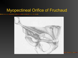
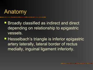
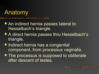
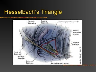
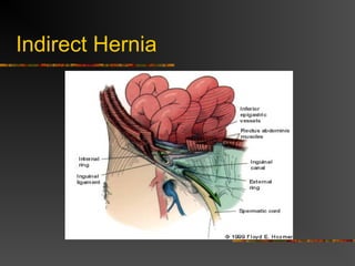
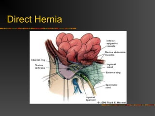
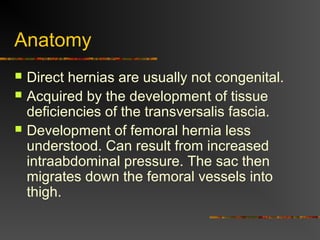
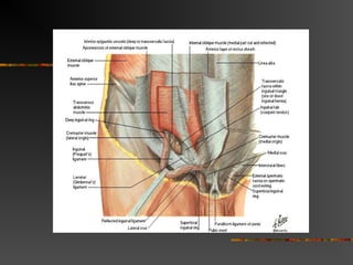
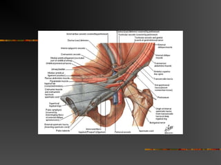
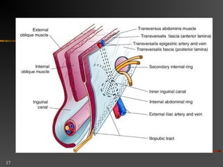
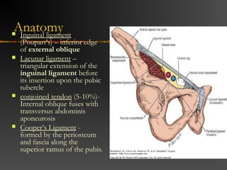
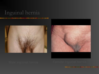
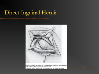
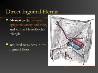
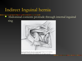
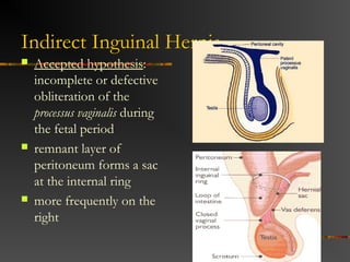
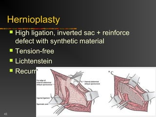
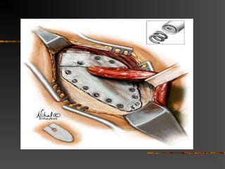
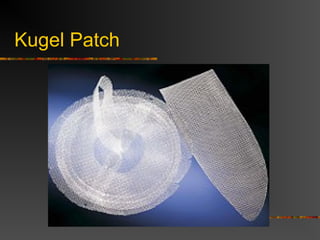
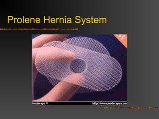
Groin Hernias
Sameh Shehata
Definition
 Abnormal protrusion of a peritoneal lined
sac thru the musculoaponeurotic covering
of the abdomen
Introduction
 In US 96% are inguinal, 4% femoral
 20% bilateral
 Most common in both sexes indirect.
 Femoral hernias more common in elderly
females
 Male to female ratio in 9:1 for inguinal
hernias, 1:3 for femoral hernias
Myopectineal Orifice of Fruchaud
The MPO is bordered:
 Above by the arching fibers of the internal
oblique and transversus abdominus Muscles,
 Medially (towards the center or to the right) by
the Rectus Abdominus Muscle and its fascial
Rectus Sheath
 Inferiorly by Coopers Ligament, and
 Laterally by the Ileopsoas Muscle
 Running diagonally thru the MPO is the
inguinal ligament
Myopectineal Orifice of Fruchaud
Anatomy
 4cm in length
 2-4 cm cephalad to inguinal ligament
 Extends between superficial and deep
rings
 Contains spermatic cord or round
ligament
Anatomy
 Bounded superficially by external oblique
 Cephalad by conjoint tendon
 Inferior border is inguinal ligament
 Floor is transversalis fascia
Layers
 Skin, subcutaneous, campers, scarpa,
external spermatic fascia, cremaster,, int
spermatic fascia , preperitoneal tissues,
peritoneum
Anatomy
 Broadly classified as indirect and direct
depending on relationship to epigastric
vessels.
 Hesselbach’s triangle is inferior epigastric
artery laterally, lateral border of rectus
medially, inguinal ligament inferiorly.
Anatomy
 An indirect hernia passes lateral to
Hesselbach’s triangle.
 A direct hernia passes thru Hesselbach’s
triangle.
 Indirect hernia has a congenital
component, from processus vaginalis.
 The processus is supposed to obliterate
after descent of testes.
Hesselbach’s Triangle
Indirect Inguinal hernia
 Abdominal contents protrude through internal inguinal
ring
Indirect Hernia
Indirect Inguinal Hernia
 Accepted hypothesis:
incomplete or defective
obliteration of the
processus vaginalis during
the fetal period
 remnant layer of
peritoneum forms a sac
at the internal ring
 more frequently on the
right
15
Direct Hernia
Direct Inguinal Hernia
 Medial to the inferior
epigastric artery and vein,
and within Hesselbach's
triangle
 acquired weakness in the
inguinal floor
Anatomy
 Direct hernias are usually not congenital.
 Acquired by the development of tissue
deficiencies of the transversalis fascia.
 Development of femoral hernia less
understood. Can result from increased
intraabdominal pressure. The sac then
migrates down the femoral vessels into
thigh.
Anatomy Inguinal ligament
(Poupart’s) – inferior edge
of external oblique
 Lacunar ligament –
triangular extension of the
inguinal ligament before
its insertion upon the pubic
tubercle
 conjoined tendon (5-10%)-
Internal oblique fuses with
transversus abdominis
aponeurosis
 Cooper’s Ligament -
formed by the periosteum
and fascia along the
superior ramus of the pubis.
Inguinal hernia
Male inguinal hernia Female inguinal hernia
Nyhus Classification
 I indirect, internal ring normal (kids)
 II indirect, dilated internal ring
 III posterior wall defects, direct inguinal
hernia, dilated internal ring, massive
scrotal, sliding, femoral hernia
 IV recurrent hernia
Terminology
 Reducible – can be replaced within
surrounding musculature
 Incarcerated – cannot be reduced
 Strangulated – compromised blood supply to
its contents
24
Hernia complications
Bowel obstruction: usually not partial,
look at groin
Irreducibility .
Strangulation: serious, life-threatening
 Inflammation.
Epidemiology
 Prevelance of hernias increases with age
 Most serious complication – strangulation
 1 to 3% of groin hernias
 Femoral – highest rate of complications 15% to
20%
 recommended all be repaired at time of discovery
Treatment
27
28
History
 1st
century: Surgical treatment
 15th
century: Castration with wound cauterization or
hernia sac debridement with secondary healing
 Early 18th
century: Sir Astley Cooper:
recommended truss > surgery, only in
strangulation
History
 1881: French surgeon, Lucas-
Championni re: high ligation of indirectѐ
inguinal hernia
 1844-1924: Edoardo Bassini (father of
modern inguinal hernia surgery): high
ligation and reconstruction of inguinal floor
Father of Modern Inguinal Hernia Repair
EDUARDO BASSINI
Surgical Techniques
 Open anterior repair (Bassini, McVay,
Shouldice).
 Tension-free repair with
mesh(Liechtenstein, Rutkow)
 Posterior repair
 Open : Nyhus
 Laparoscopic
Open anterior repair
 Herniotomy
 Herniorrhaphy (repair)
 Hernioplasty .
34
Herniotomy
 Patent processus vaginalis ligated at
origin at internal ring (high ligation)
 Nyhus type I
 Children
35
Herniorrhaphy
 Nyhus type II and III
 High ligation + reinforced area of
weakness with patient’s own tissue
 Bassini, Shouldice, McVay
36
Bassini repair
 Transversus abdominis aponeurosis +
transversalis fascia  inguinal
ligament with nonabsorbable
interrupted sutures
37
Shouldice repair
 4 rows of suture
38
McVay repair
 Inguinal and femoral hernias,
Transversus abdominis aponeurosis +
transversalis fascia  Cooper’s
ligament + iliopubic tract
39
Hernioplasty
 High ligation, inverted sac + reinforce
defect with synthetic material
 Tension-free
 Lichtenstein
 Recurrent rate 0.1%
Tension-Free Repair
 Same initial approach as anterior repair
 Instead of sewing fascial layers together
to repair defect, a prosthetic mesh onlay
used
 Simple to learn, easy to perform, suited for
local anesthesia, excellent results with
recurrence less than 4%.
Techniques
 Coined by Liechtenstein in 1989
 Central feature is polypropylene mesh
over unrepaired floor.
 Gilbert repair uses a cone shaped plug
placed thru deep ring.
 Slit placed in mesh for cord structures
Techniques
 Suturing the mesh to the inguinal ligament
is not important.
 Fixing the mesh to the rectus sheath 1-
1.5cm medial and superior to the pubic
tubercle is very important.
 Should have a surplus of mesh over
inguinal ligament, the medial suture
ensures surplus mesh inferiorly
Open Posterior Repair
 Divide the layers of the abdominal wall
superior to the internal ring, enter
preperitoneal space. Dissection continues
behind and deep to the entire inguinal
region.
 Suture tension problems.
Laparoscopic Procedures
 Increasingly popular, controversial
 Early in the development, hernias were
repaired by placing very large mesh over
entire inguinal region on top of the
peritoneum. Was abandoned because of
contact with bowel.
 Today, most performed TEP or TAPP
Laparoscopic Mesh Repair
Note:
Viewed from inside the
pelvis toward the direct
and indirect sites. A
broad portion of mesh is
stapled to span both
hernia defects. Staples
are not used in
proximity to
neurovascular
structures.
Laparoscopic Procedures
 The argued advantage of these
procedures was less pain and disability,
faster return to work.
 Great for bilateral hernia, with no increase
in morbidity.
 For recurrent hernia
 Disadvantages are cost, time.
Recurrence
Type of repair Recurrence
McVay 9%
Shouldice 7-11%
Liechtenstein 0-4%
Laparoscopic 0-1%

Groin hernia 4th year

  • 1.
  • 2.
    Definition  Abnormal protrusionof a peritoneal lined sac thru the musculoaponeurotic covering of the abdomen
  • 3.
    Introduction  In US96% are inguinal, 4% femoral  20% bilateral  Most common in both sexes indirect.  Femoral hernias more common in elderly females  Male to female ratio in 9:1 for inguinal hernias, 1:3 for femoral hernias
  • 4.
    Myopectineal Orifice ofFruchaud The MPO is bordered:  Above by the arching fibers of the internal oblique and transversus abdominus Muscles,  Medially (towards the center or to the right) by the Rectus Abdominus Muscle and its fascial Rectus Sheath  Inferiorly by Coopers Ligament, and  Laterally by the Ileopsoas Muscle  Running diagonally thru the MPO is the inguinal ligament
  • 5.
  • 6.
    Anatomy  4cm inlength  2-4 cm cephalad to inguinal ligament  Extends between superficial and deep rings  Contains spermatic cord or round ligament
  • 7.
    Anatomy  Bounded superficiallyby external oblique  Cephalad by conjoint tendon  Inferior border is inguinal ligament  Floor is transversalis fascia
  • 8.
    Layers  Skin, subcutaneous,campers, scarpa, external spermatic fascia, cremaster,, int spermatic fascia , preperitoneal tissues, peritoneum
  • 9.
    Anatomy  Broadly classifiedas indirect and direct depending on relationship to epigastric vessels.  Hesselbach’s triangle is inferior epigastric artery laterally, lateral border of rectus medially, inguinal ligament inferiorly.
  • 10.
    Anatomy  An indirecthernia passes lateral to Hesselbach’s triangle.  A direct hernia passes thru Hesselbach’s triangle.  Indirect hernia has a congenital component, from processus vaginalis.  The processus is supposed to obliterate after descent of testes.
  • 11.
  • 12.
    Indirect Inguinal hernia Abdominal contents protrude through internal inguinal ring
  • 13.
  • 14.
    Indirect Inguinal Hernia Accepted hypothesis: incomplete or defective obliteration of the processus vaginalis during the fetal period  remnant layer of peritoneum forms a sac at the internal ring  more frequently on the right
  • 15.
  • 16.
  • 17.
  • 18.
    Direct Inguinal Hernia Medial to the inferior epigastric artery and vein, and within Hesselbach's triangle  acquired weakness in the inguinal floor
  • 19.
    Anatomy  Direct herniasare usually not congenital.  Acquired by the development of tissue deficiencies of the transversalis fascia.  Development of femoral hernia less understood. Can result from increased intraabdominal pressure. The sac then migrates down the femoral vessels into thigh.
  • 20.
    Anatomy Inguinal ligament (Poupart’s)– inferior edge of external oblique  Lacunar ligament – triangular extension of the inguinal ligament before its insertion upon the pubic tubercle  conjoined tendon (5-10%)- Internal oblique fuses with transversus abdominis aponeurosis  Cooper’s Ligament - formed by the periosteum and fascia along the superior ramus of the pubis.
  • 21.
    Inguinal hernia Male inguinalhernia Female inguinal hernia
  • 22.
    Nyhus Classification  Iindirect, internal ring normal (kids)  II indirect, dilated internal ring  III posterior wall defects, direct inguinal hernia, dilated internal ring, massive scrotal, sliding, femoral hernia  IV recurrent hernia
  • 23.
    Terminology  Reducible –can be replaced within surrounding musculature  Incarcerated – cannot be reduced  Strangulated – compromised blood supply to its contents
  • 24.
    24 Hernia complications Bowel obstruction:usually not partial, look at groin Irreducibility . Strangulation: serious, life-threatening  Inflammation.
  • 25.
    Epidemiology  Prevelance ofhernias increases with age  Most serious complication – strangulation  1 to 3% of groin hernias  Femoral – highest rate of complications 15% to 20%  recommended all be repaired at time of discovery
  • 26.
  • 27.
  • 28.
    28 History  1st century: Surgicaltreatment  15th century: Castration with wound cauterization or hernia sac debridement with secondary healing  Early 18th century: Sir Astley Cooper: recommended truss > surgery, only in strangulation
  • 29.
    History  1881: Frenchsurgeon, Lucas- Championni re: high ligation of indirectѐ inguinal hernia  1844-1924: Edoardo Bassini (father of modern inguinal hernia surgery): high ligation and reconstruction of inguinal floor
  • 30.
    Father of ModernInguinal Hernia Repair EDUARDO BASSINI
  • 31.
    Surgical Techniques  Openanterior repair (Bassini, McVay, Shouldice).  Tension-free repair with mesh(Liechtenstein, Rutkow)
  • 32.
     Posterior repair Open : Nyhus  Laparoscopic
  • 33.
    Open anterior repair Herniotomy  Herniorrhaphy (repair)  Hernioplasty .
  • 34.
    34 Herniotomy  Patent processusvaginalis ligated at origin at internal ring (high ligation)  Nyhus type I  Children
  • 35.
    35 Herniorrhaphy  Nyhus typeII and III  High ligation + reinforced area of weakness with patient’s own tissue  Bassini, Shouldice, McVay
  • 36.
    36 Bassini repair  Transversusabdominis aponeurosis + transversalis fascia  inguinal ligament with nonabsorbable interrupted sutures
  • 37.
  • 38.
    38 McVay repair  Inguinaland femoral hernias, Transversus abdominis aponeurosis + transversalis fascia  Cooper’s ligament + iliopubic tract
  • 39.
    39 Hernioplasty  High ligation,inverted sac + reinforce defect with synthetic material  Tension-free  Lichtenstein  Recurrent rate 0.1%
  • 40.
    Tension-Free Repair  Sameinitial approach as anterior repair  Instead of sewing fascial layers together to repair defect, a prosthetic mesh onlay used  Simple to learn, easy to perform, suited for local anesthesia, excellent results with recurrence less than 4%.
  • 43.
    Techniques  Coined byLiechtenstein in 1989  Central feature is polypropylene mesh over unrepaired floor.  Gilbert repair uses a cone shaped plug placed thru deep ring.  Slit placed in mesh for cord structures
  • 44.
    Techniques  Suturing themesh to the inguinal ligament is not important.  Fixing the mesh to the rectus sheath 1- 1.5cm medial and superior to the pubic tubercle is very important.  Should have a surplus of mesh over inguinal ligament, the medial suture ensures surplus mesh inferiorly
  • 45.
    Open Posterior Repair Divide the layers of the abdominal wall superior to the internal ring, enter preperitoneal space. Dissection continues behind and deep to the entire inguinal region.  Suture tension problems.
  • 46.
    Laparoscopic Procedures  Increasinglypopular, controversial  Early in the development, hernias were repaired by placing very large mesh over entire inguinal region on top of the peritoneum. Was abandoned because of contact with bowel.  Today, most performed TEP or TAPP
  • 48.
    Laparoscopic Mesh Repair Note: Viewedfrom inside the pelvis toward the direct and indirect sites. A broad portion of mesh is stapled to span both hernia defects. Staples are not used in proximity to neurovascular structures.
  • 49.
    Laparoscopic Procedures  Theargued advantage of these procedures was less pain and disability, faster return to work.  Great for bilateral hernia, with no increase in morbidity.  For recurrent hernia  Disadvantages are cost, time.
  • 50.
    Recurrence Type of repairRecurrence McVay 9% Shouldice 7-11% Liechtenstein 0-4% Laparoscopic 0-1%

Editor's Notes

  • #15 -through which intra-abdominal contents may herniate -where descent of the gonads occurs later during fetal development.)
  • #21 This structure is posterior to the iliopubic tract and forms the posterior border of the femoral canal
  • #24 (more in large hernias that have small necks obstructing arterial flow and/or venous drainage)
  • #26 (increases with age)
  • #31 Bassini revolutionized the surgical repair of the groin hernia with his novel anatomical dissection and low recurrence rates – first operation in 1884