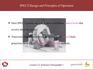
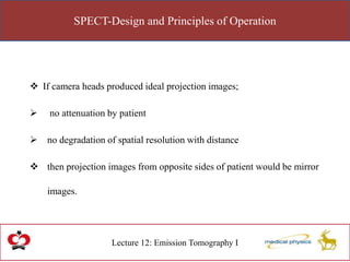
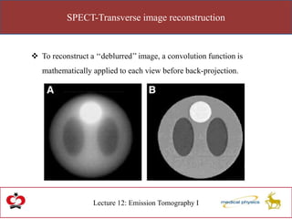
Lecture 12: Emission Tomography I
Shahid Younas
NUCLEAR IMAGING
Emission Tomography I
Single Photon Emission Computed Tomography (SPECT)
Introduction
Lecture 12: Emission Tomography I
 Nuclear Medicine projection image depicts a two-dimensional projection
of the three-dimensional activity distribution.
 Contribution to the image from structures at different depths overlap.
 Hindering the ability to discern the image of a structure at a particular
depth.
Introduction
Lecture 12: Emission Tomography I
 Tomographic imaging attempts to depict the
activity distribution in a single cross section
of the patient.
Types of Tomography
Lecture 12: Emission Tomography I
 Conventional Tomography
 Computed Tomography (CT)
Types of Tomography
Lecture 12: Emission Tomography I
 Conventional Tomography also called Geometric or Focal Plane.
 Structures out of a focal plane are not removed from the resultant
image.
 They are blurred by an amount proportional to their distance from
the local plane.
Types of Tomography
Lecture 12: Emission Tomography I
 Computed Tomography uses mathematical methods to remove
overlying structures completely.
 CT requires the acquisition of a set of projection image from at least
a 180-degree arc about the patient.
Types of Tomography
Lecture 12: Emission Tomography I
CT uses mathematical methods; do you know what instrument was
used in nuclear medicine to carry on conventional tomography?
Focused or seven pin-hole collimators
SPECT
Lecture 12: Emission Tomography I
Three Rivals of SPECT
 Attenuation of photons in the patient
 Compton scattered photons in the image
 Degradation of spatial resolution with distance from collimator.
SPECT-Design and Principles of Operation
Lecture 12: Emission Tomography I
 Single photon emission computed tomography (SPECT) generates transverse
images depicting the distribution of x- or gamma ray emitting nuclides in
patients.
 Standard planar projection images are acquired from an arc of 180 degrees
(most cardiac SPECT) or 360 degrees (most non-cardiac SPECT) about the
patient.
SPECT-Design and Principles of Operation
Lecture 12: Emission Tomography I
 Most SPECT systems use one or more scintillation camera heads that
revolve about the patient.
 Transverse images are reconstructed using either filtered back-
projection (as in CT) or iterative reconstruction methods.
SPECT-Design and Principles of Operation
Lecture 12: Emission Tomography I
 If camera heads produced ideal projection images;
 no attenuation by patient
 no degradation of spatial resolution with distance
 then projection images from opposite sides of patient would be mirror
images.
SPECT
Lecture 12: Emission Tomography I
 Attenuation greatly reduces number of photons from activity in the half
of patient opposite camera head; this information is blurred by distance.
SPECT-Image acquisition
Lecture 12: Emission Tomography I
 SPECT projection images usually acquired in either a 64 x 64 (60 or 64
projections) or a 128 x 128 (120 or 128 projections) pixel format.
 Using too small a pixel format reduces spatial resolution of the projection
images and of the resultant reconstructed transverse images.
 Using too few projections creates radial streak artifacts in the reconstructed
transverse images.
SPECT-Image acquisition
Lecture 12: Emission Tomography I
Brain SPECT are acquired over 360o whereas Cardiac SPECT are
acquired at 180o? What is reduced and what is enhanced?
Attenuation is reduced whereas contrast and resolution is enhanced
SPECT-Image acquisition
Lecture 12: Emission Tomography I
 Camera heads on older SPECT systems used circular orbits
around the patient while acquiring images,
 Satisfactory for imaging of the brain.
 Loss of spatial resolution in body imaging because of distance
from surface.
SPECT-Image acquisition
Lecture 12: Emission Tomography I
Newer systems provide noncircular orbits that keep camera heads in close
proximity to surface of body throughout the orbit.
SPECT-Image acquisition
Lecture 12: Emission Tomography I
What is PSD? What is its role in SPECT imaging?
 Pressure Sensitive Device.
 Detect nearest position w.r.t.
patient body without touching
SPECT-Transverse image reconstruction
Lecture 12: Emission Tomography I
 The goal of SPECT image reconstruction methods is to estimate
the true radioactivity distribution in vivo from the measured
projection data.
 After projection images are acquired, they are usually corrected
for axis-of-rotation misalignments.
SPECT-Transverse image reconstruction
Lecture 12: Emission Tomography I
 The substantial effects of attenuation, scatter , collimator and
detector response are ignored.
 The Following these corrections, transverse image reconstruction
is performed using either filtered back-projection or iterative
methods.
SPECT-Transverse image reconstruction
Lecture 12: Emission Tomography I
Suppose that a simple test object containing 3 objects with different attenuation
values is scanned and views (attenuation measurements) are obtained at 3
angles.
SPECT-Transverse image reconstruction
Lecture 12: Emission Tomography I
The attenuation measurements of each view are simply divided evenly along the
path of the ray.
SPECT-Transverse image reconstruction
Lecture 12: Emission Tomography I
after back projection of only 4 views, an image of the test object is beginning to
appear in Figure 5C.
SPECT-Transverse image reconstruction
Lecture 12: Emission Tomography I
 Back-projection is efficient each measurement is processed just
once and involves relatively simple calculations but has a serious
flaw,
The resulting images are blurry- poor spatial resolution
SPECT-Transverse image reconstruction
Lecture 12: Emission Tomography I
Why resulting images have poor spatial resolution?
The substantial effects of attenuation, scatter , collimator and detector
response are ignored.
SPECT-Transverse image reconstruction
Lecture 12: Emission Tomography I
 The blurring can be reversed by a mathematic process known as
“Convolution”.
 Consider a scan of a phantom containing a single cylinder with an
attenuation higher than that of its surroundings.
SPECT-Transverse image reconstruction
Lecture 12: Emission Tomography I
 The attenuation of the cylinder is highest through its center
(where it is thickest) and decreases toward its edges.
 Back-projection builds a cylinder image whose intensity
decreases from the maximum at the center toward the edges.
SPECT-Transverse image reconstruction
Lecture 12: Emission Tomography I
 To reconstruct a ‘‘deblurred’’ image, a convolution function is
mathematically applied to each view before back-projection.
SPECT-Transverse image reconstruction
Lecture 12: Emission Tomography I
The mathematic operation is called convolution. Do you know how
the process is referred?
Filtering
SPECT-Transverse image reconstruction
Lecture 12: Emission Tomography I
 Choice of filter kernel for a particular type of study is determined by
the amount of statistical noise in the projection images
 Mainly determined by injected activity, collimator, and acquisition
time per image
SPECT-Transverse image reconstruction
Lecture 12: Emission Tomography I
 Their spatial resolution is,
 Determined by collimator and the typical distances from the camera
head(s) from the organ being imaged
SPECT-Transverse image reconstruction
Lecture 12: Emission Tomography I
 Their spatial resolution is,
 Determined by collimator and the typical distances from the camera
head(s) from the organ being imaged

Nuclear imaging, PET CT MEDICAL PHYSICS

  • 1.
    Lecture 12: EmissionTomography I Shahid Younas NUCLEAR IMAGING Emission Tomography I Single Photon Emission Computed Tomography (SPECT)
  • 2.
    Introduction Lecture 12: EmissionTomography I  Nuclear Medicine projection image depicts a two-dimensional projection of the three-dimensional activity distribution.  Contribution to the image from structures at different depths overlap.  Hindering the ability to discern the image of a structure at a particular depth.
  • 3.
    Introduction Lecture 12: EmissionTomography I  Tomographic imaging attempts to depict the activity distribution in a single cross section of the patient.
  • 4.
    Types of Tomography Lecture12: Emission Tomography I  Conventional Tomography  Computed Tomography (CT)
  • 5.
    Types of Tomography Lecture12: Emission Tomography I  Conventional Tomography also called Geometric or Focal Plane.  Structures out of a focal plane are not removed from the resultant image.  They are blurred by an amount proportional to their distance from the local plane.
  • 6.
    Types of Tomography Lecture12: Emission Tomography I  Computed Tomography uses mathematical methods to remove overlying structures completely.  CT requires the acquisition of a set of projection image from at least a 180-degree arc about the patient.
  • 7.
    Types of Tomography Lecture12: Emission Tomography I CT uses mathematical methods; do you know what instrument was used in nuclear medicine to carry on conventional tomography? Focused or seven pin-hole collimators
  • 8.
    SPECT Lecture 12: EmissionTomography I Three Rivals of SPECT  Attenuation of photons in the patient  Compton scattered photons in the image  Degradation of spatial resolution with distance from collimator.
  • 9.
    SPECT-Design and Principlesof Operation Lecture 12: Emission Tomography I  Single photon emission computed tomography (SPECT) generates transverse images depicting the distribution of x- or gamma ray emitting nuclides in patients.  Standard planar projection images are acquired from an arc of 180 degrees (most cardiac SPECT) or 360 degrees (most non-cardiac SPECT) about the patient.
  • 10.
    SPECT-Design and Principlesof Operation Lecture 12: Emission Tomography I  Most SPECT systems use one or more scintillation camera heads that revolve about the patient.  Transverse images are reconstructed using either filtered back- projection (as in CT) or iterative reconstruction methods.
  • 11.
    SPECT-Design and Principlesof Operation Lecture 12: Emission Tomography I  If camera heads produced ideal projection images;  no attenuation by patient  no degradation of spatial resolution with distance  then projection images from opposite sides of patient would be mirror images.
  • 12.
    SPECT Lecture 12: EmissionTomography I  Attenuation greatly reduces number of photons from activity in the half of patient opposite camera head; this information is blurred by distance.
  • 13.
    SPECT-Image acquisition Lecture 12:Emission Tomography I  SPECT projection images usually acquired in either a 64 x 64 (60 or 64 projections) or a 128 x 128 (120 or 128 projections) pixel format.  Using too small a pixel format reduces spatial resolution of the projection images and of the resultant reconstructed transverse images.  Using too few projections creates radial streak artifacts in the reconstructed transverse images.
  • 14.
    SPECT-Image acquisition Lecture 12:Emission Tomography I Brain SPECT are acquired over 360o whereas Cardiac SPECT are acquired at 180o? What is reduced and what is enhanced? Attenuation is reduced whereas contrast and resolution is enhanced
  • 15.
    SPECT-Image acquisition Lecture 12:Emission Tomography I  Camera heads on older SPECT systems used circular orbits around the patient while acquiring images,  Satisfactory for imaging of the brain.  Loss of spatial resolution in body imaging because of distance from surface.
  • 16.
    SPECT-Image acquisition Lecture 12:Emission Tomography I Newer systems provide noncircular orbits that keep camera heads in close proximity to surface of body throughout the orbit.
  • 17.
    SPECT-Image acquisition Lecture 12:Emission Tomography I What is PSD? What is its role in SPECT imaging?  Pressure Sensitive Device.  Detect nearest position w.r.t. patient body without touching
  • 18.
    SPECT-Transverse image reconstruction Lecture12: Emission Tomography I  The goal of SPECT image reconstruction methods is to estimate the true radioactivity distribution in vivo from the measured projection data.  After projection images are acquired, they are usually corrected for axis-of-rotation misalignments.
  • 19.
    SPECT-Transverse image reconstruction Lecture12: Emission Tomography I  The substantial effects of attenuation, scatter , collimator and detector response are ignored.  The Following these corrections, transverse image reconstruction is performed using either filtered back-projection or iterative methods.
  • 20.
    SPECT-Transverse image reconstruction Lecture12: Emission Tomography I Suppose that a simple test object containing 3 objects with different attenuation values is scanned and views (attenuation measurements) are obtained at 3 angles.
  • 21.
    SPECT-Transverse image reconstruction Lecture12: Emission Tomography I The attenuation measurements of each view are simply divided evenly along the path of the ray.
  • 22.
    SPECT-Transverse image reconstruction Lecture12: Emission Tomography I after back projection of only 4 views, an image of the test object is beginning to appear in Figure 5C.
  • 23.
    SPECT-Transverse image reconstruction Lecture12: Emission Tomography I  Back-projection is efficient each measurement is processed just once and involves relatively simple calculations but has a serious flaw, The resulting images are blurry- poor spatial resolution
  • 24.
    SPECT-Transverse image reconstruction Lecture12: Emission Tomography I Why resulting images have poor spatial resolution? The substantial effects of attenuation, scatter , collimator and detector response are ignored.
  • 25.
    SPECT-Transverse image reconstruction Lecture12: Emission Tomography I  The blurring can be reversed by a mathematic process known as “Convolution”.  Consider a scan of a phantom containing a single cylinder with an attenuation higher than that of its surroundings.
  • 26.
    SPECT-Transverse image reconstruction Lecture12: Emission Tomography I  The attenuation of the cylinder is highest through its center (where it is thickest) and decreases toward its edges.  Back-projection builds a cylinder image whose intensity decreases from the maximum at the center toward the edges.
  • 27.
    SPECT-Transverse image reconstruction Lecture12: Emission Tomography I  To reconstruct a ‘‘deblurred’’ image, a convolution function is mathematically applied to each view before back-projection.
  • 28.
    SPECT-Transverse image reconstruction Lecture12: Emission Tomography I The mathematic operation is called convolution. Do you know how the process is referred? Filtering
  • 29.
    SPECT-Transverse image reconstruction Lecture12: Emission Tomography I  Choice of filter kernel for a particular type of study is determined by the amount of statistical noise in the projection images  Mainly determined by injected activity, collimator, and acquisition time per image
  • 30.
    SPECT-Transverse image reconstruction Lecture12: Emission Tomography I  Their spatial resolution is,  Determined by collimator and the typical distances from the camera head(s) from the organ being imaged
  • 31.
    SPECT-Transverse image reconstruction Lecture12: Emission Tomography I  Their spatial resolution is,  Determined by collimator and the typical distances from the camera head(s) from the organ being imaged