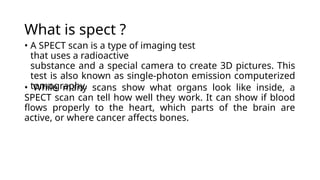
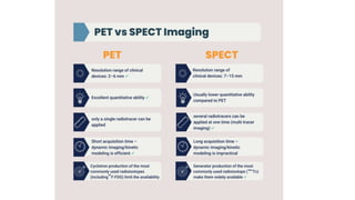
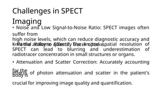
Current advancements
in the field of SPECT
(advancement in SPECT
imaging
algorithms)
Group
members
Ali
hassan
2021-BME-
128
Muhammad
saad Sarmad
mukhtar
2021-BME-
134 2021-
BME-135



What is spect ?
• A SPECT scan is a type of imaging test
that uses a radioactive
substance and a special camera to create 3D pictures. This
test is also known as single-photon emission computerized
tomography.
• While many scans show what organs look like inside, a
SPECT scan can tell how well they work. It can show if blood
flows properly to the heart, which parts of the brain are
active, or where cancer affects bones.
Why it's
done?
Brain Disorders:

 


SPECT scanning creates a detailed 3D map of blood flow activity in the brain. It helps diagnose
vascular brain disorders like Moyamoya disease by identifying issues with blood flow. SPECT is
useful in diagnosing and treating seizure disorders such as epilepsy by pinpointing areas of
seizure
activity.
In rare cases, a specific SPECT scan called DaTscan can help confirm a diagnosis of Parkinson's
disease. Some medical institutions use SPECT scanning to investigate other brain conditions
like dementia or head
trauma.
Heart
Problems:


SPECT scans can detect clogged coronary arteries, which can lead to heart muscle damage
or death. They assess the pumping action of the heart chambers during contractions,
indicating the heart's efficiency.
Bone
Disorders:

 
SPECT scans highlight areas of bone healing and are used to diagnose hidden
bone fractures. They aid in diagnosing and tracking cancer that has
metastasized to the bones. SPECT can identify sites for bone biopsy procedures.
How imiging can be done in
Spect ?
• Radioactive Tracer Injection: A small amount of radioactive tracer is injected into the
bloodstream. This tracer is typically a compound that emits gamma rays, allowing it to be
detected by the SPECT scanner.
•
TracerDistribution:Thetracercirculatesthroughthebodyandaccumulatesintissuesororgansofinte
rest,
such as the brain, heart, or bones, depending on the purpose
of the scan.
• Gamma Camera Detection: The patient lies down on a bed or table that can move through
the SPECT
scanner. The gamma camera rotates around the patient, capturing multiple images from
different angles.
• Data Acquisition: As the gamma camera rotates, it detects the gamma rays emitted by the
tracer in the
body. Each angle provides a different perspective, capturing information about the distribution
of the tracer
in three dimensions.
• Image Reconstruction: The raw data collected by the gamma camera are processed by a
computer to
reconstruct a 3D image of the distribution of the tracer within the body. This image represents
the areas of
interest, such as blood flow in the brain or uptake of the tracer in the heart.
• Image Interpretation: The reconstructed SPECT image is then interpreted by a radiologist or
nuclear
medicine physician. They analyze the image to assess blood flow, detect abnormalities, or
evaluate the
function of organs or tissues, depending on the clinical indication for the scan.
Overall, SPECT imaging provides valuable information about the physiological processes
occurring within the
body, aiding in the diagnosis, treatment planning, and monitoring of various medical
conditions.
Challenges in SPECT
Imaging
• Noise and Low Signal-to-Noise Ratio: SPECT images often
suffer from
high noise levels, which can reduce diagnostic accuracy and
limit the ability to quantify tracer uptake.
• Partial Volume Effects: The limited spatial resolution of
SPECT can lead to blurring and underestimation of
radiotracer concentration in small structures or organs.
• Attenuation and Scatter Correction: Accurately accounting
for the
effects of photon attenuation and scatter in the patient's
body is
crucial for improving image quality and quantification.
Working of gamma
camera
detailed picture showing where the tracer is in
the body.
• A special tracer is injected into the body and goes to specific
areas. • The tracer emits gamma rays in all directions. • These
gamma rays pass through a filter called a collimator that only
lets through the ones going
straight.
• The filtered gamma rays hit a layer that turns them into
visible light.
• The visible light is then converted into electrical signals by
tubes
called PhotoMultiplier Tubes
(PMTs).
• These signals are sent to a computer, which uses them to
create a
Working of Gamma
camera
Noise Reduction Techniques
• Filtering Techniques: Employ low-pass filters to reduce high-
frequency noise while preserving important image
features.
• Wavelet-based Methods: Leverage wavelet transform
s
to
selective noise
reconstruction
algorithms that model and compensate for noise during the
image reconstruction process.
separate noise
suppression.
from the signal, enabling
•
Iterative
Reconstruction
:
Utilize iterative
Optimization of Collimator
Design
The collimator is a critical component in SPECT imaging, responsible
for filtering and directing the detected photons. Optimizing the
collimator design can significantly enhance image quality by
improving spatial resolution, sensitivity, and contrast.
techniques have enabled
the development of innovative collimator designs tailored to specific
clinical applications. These optimizations help minimize image
distortions, reduce radiation exposure, and provide higher-
qualitydiagnosticinformation.
Advancement
s
in collimator materials, geometr
y,
and
manufacturing
Advances in Detector
Technology
Cutting-edge SPECT detector technology has and diagnostic capabilities. Novel
materials, such as cadmium zinc telluride (CZT), offer enhanced
energy resolution and sensitivity, enabling more
accurate quantification of radiotracer uptake.
dramaticall
y
improve
d
image quality
Additionally, pixelated solid-state detectors
have
crystals,
and
reducing image distortions caused by scatter
and penetration effects.
replaced
providin
g
traditiona
l
scintillatio
n
superio
r
spatial resolutio
n
Motion Correction in SPECT
Imaging
Respiratory Gating
By synchronizing data acquisition with the patient's breathing cycle, respiratory
motion can be mitigated, reducing image blurring and enhancing the visualization
of thoracic
and abdominal structures.
Patient Motion Tracking
Advances in motion tracking technologies, such as optical markers and image-
based techniques, enable real-time monitoring of patient movements during
SPECT scans.
Motion Compensated Reconstruction
Sophisticated algorithms incorporate the detected motion information to correct
for patient movement, improving the spatial fidelity and quantitative accuracy of
SPECT im ag es.
Cardiac SPECT scan with different
motion correction methods
applied.
Breakdown of what each row shows: Top
row: No motion correction applied, so the
image is blurry. Middle row: Conventional
motion correction method applied,
resulting in a clearer image. Bottom row:
A new method proposed by the
researchers for motion correction,
resulting in a clear image.
Attenuation Correction
Techniques
Photon
Absorption
As photons emitted
from radioactive
tracers traverse the
body, they can be
absorbed or
scattered by
surrounding
tissues, distorting
the final image.
Attenuation
Mapping
Using CT or MRI
data, SPECT
imaging systems
can create a 3D
attenuation map
of
the patient's
anatomy,
quantifying the
amount of photon
absorption in
each region.
Correction Algo
rit hm s
Advanced
mathematical
algorithms then use
this attenuation
map
to compensate for
the loss of photons,
improving image
contrast and
quantitative
accuracy.
Hybrid
Imaging
anatomical
information,
enabling more
robust
attenuation
correction.
Combining SPECT
with CT or MRI in a
single hybrid
system provides
both functional
and
Attenuation correction of lung SPECT images (a) No corrected image, (b) Attenuation
correction after tomographic reconstruction (Chang's method) (c) 3D reconstruction slice by
slice
Scatter Correction
Techniques
Compton
Scatter
Modeling
Advanced
algorithms model
the Compton
scattering of
gamma rays
through the
patient's body,
allowing for
accurate
compensation of
scattered photons
in the SPECT image
reconstruction
process.
Energy-based
Scatter C
orrection
By analyzing the
energy spectra of
detected photons,
sophisticated
scatter correction
methods can
identify and
remove the
contribution of
scattered events,
improving image
contrast and
quantitative
accuracy.
Dual-Energy
Window
Techniques
Acquiring SPECT
data at multiple
energy windows
enables the
separation of
primary and
scattered photons,
enabling effective
scatter subtraction
without the need
for complex
analytical models.
Monte Carlo S
imulation
Detailed Monte
Carlo simulations
of
photon transport
through the patient
anatomy can
provide highly
accurate scatter
correction, though
at the cost of
increased
computational
complexity.
Explanation of
Iterative
Reconstruction
Techniques
with an initial estimate of the radiotracer distribution, then iteratively
updates and improves the image by comparing the estimated
projections to the actual measured data. With each iteration, the
image quality and quantitative accuracy are enhanced, delivering
superior spatial resolution and more precise uptake measurements
compared to traditional filtered back-projection methods.
Iterative reconstruction in SPECT imaging involves repeatedly refining
the image data through complex mathematical algorithms. This
process starts
Benefits of Iterative
Reconstruction in SPECT Imaging
Enhanced Image Quality
Iterative reconstruction techniques
improve spatial resolution, reduce
noise, and deliver higher-quality SPECT
images for more accurate diagnosis
and treatment monitoring.
Reduced Radiation
Exposure
Iterative methods can achieve
comparable image quality with lower
radiation doses, minimizing the
health risks for patients undergoing
repeated SPECT scans.
Improved Quantification
The refined image data enables more
precise quantification of radiotracer
uptake, providing valuable insights
into physiological processes and
organ func tion.
Customizable Algorithms
Iterative reconstruction allows for the
optimization of algorithms to account
for specific patient anatomy, imaging
acquisition parameters, and clinical
needs.
Artificial Intelligence in SPECT
Image Analysis
Machine
Learning
AI-driven
algorithms can
automate image
segmentation,
lesion detection,
and quantification,
improving workflow
efficiency and
diagnostic accuracy.
Deep Learning
Deep learning
models have shown
promising
results in tasks such
as image
reconstruction, noise
reduction, and
image classification.
Computer
Vision
Advanced computer
vision techniques
can
enhance image
interpretation,
facilitating the
identification of
subtle abnormalities
and treatment
response
monitoring.
Data Analytics
Integrating SPECT
data with other
clinical
information can
provide valuable
insights and support
personalized
treatment decisions.
Applications and Benefits of AI
in SPECT Analysis
Automated Disease Detection
AI algorithms can rapidly scan SPECT
images to identify anomalies and
suspicious patterns, aiding early
diagnosis of conditions like heart
disease and neurological disorders.
Adaptive Image
Reconstruction
AI models can optimize iterative
reconstruction techniques, adapting
algorithms to individual patient
anatomy and imaging conditions for
enhanced image quality and reduced
radiation exposure.
Quantitative Evaluation
AI-powered analysis can provide
precise, objective measurements of
radiotracer uptake, organ function, and
other clinically relevant parameters to
support more informed treatment
decisions.
Clinical Workflow Efficiency
AI-driven automation of tedious tasks
like image segmentation and
quantification can streamline SPECT
imaging workflows, allowing clinicians
to focus on higher-level analysis and
patient care.
Enhanced Diagnostic
Capabilities Through Fusion
Imaging
•
•
•
Combining SPECT with other modalities like CT or MRI provides a
comprehensive view of anatomy and physiology.
Multimodal fusion enhances diagnostic accuracy by leveraging the unique
strengths and complementary information of each imaging technique.
Integrated analysis of structural, functional, and molecular data enables
more
precise localization, characterization, and staging of diseases.
Fusion imaging supports personalized treatment planning by identifying
optimal therapeutic targets and monitoring response to interventions.
•
Improved Spatial Resolution in
SPECT Imaging
1 2 3
Pinhole Collimators
Pinhole collimators provide higher spatial
resolution by focusing the photons from a
small region of the body.
Multipinhole Collimators
Multipinhole collimators use multiple
pinholes to further improve spatial
resolution and se n s i tiv ity.
Solid-State Detectors
Solid-state detectors, such as CZT, offer
higher intrinsic resolution compared to
traditional scintillation detectors.
Quantitative SPECT Imaging for
Therapy Monitoring
Tracer
Uptake
Tumor
Volume
Advanced image analysis techniques, including kinetic modeling and radiomics,
enable the extraction of quantitative biomarkers from SPECT data. These
biomarkers can help predict treatment outcomes, identify early signs of disease
progression, and personalize dosing strategies to optimize patient care.
Quantitative SPECT imaging provides a powerful
tool for monitoring the efficacy of cancer
therapies.
By tracking changes in tracer uptake and tumor
volume over time, clinicians can objectively assess a
patient's response to treatment and make informed
decisions about continuing,
modifying or discontinuing the therapeutic regimen.
Future Outlook and
Applications of Advanced
SPECT
Molecular
Imaging
Improved Image
Quality
modalities, such as PET, CT, and MRI,
will provide comprehensive,
multiparametric information for
diagnosis and therapy mo n i tori n g.
genomic and clinical information will
enable more targeted and
individualized patient management.
The development of novel radiotracers will
expand the capabilities of SPECT to
visualize
specific molecular targets and pathways,
facilitating early disease detection and
therapy
guidance.
Continued advancements in reconstruction
algorithms and detector technologies will
enhance the spatial and temporal
resolution of SPECT images.
Multimodal
Imaging
Personalized
Medicine
The integration of SPECT with other
imaging
The combination of SPECT imaging data
with
Conclusi
on
precision
medicine.
Advancements in SPECT imaging, powered by innovative
algorithms,
machine learning, and multimodal integration, are
revolutionizing diagnostic capabilities and personalized
healthcare. As this remarkable technology continues to
evolve, we stand on the cusp of a new era in

Single photon emission computed Tomography

  • 1.
    Current advancements in thefield of SPECT (advancement in SPECT imaging algorithms) Group members Ali hassan 2021-BME- 128 Muhammad saad Sarmad mukhtar 2021-BME- 134 2021- BME-135   
  • 2.
    What is spect? • A SPECT scan is a type of imaging test that uses a radioactive substance and a special camera to create 3D pictures. This test is also known as single-photon emission computerized tomography. • While many scans show what organs look like inside, a SPECT scan can tell how well they work. It can show if blood flows properly to the heart, which parts of the brain are active, or where cancer affects bones.
  • 4.
    Why it's done? Brain Disorders:     SPECT scanning creates a detailed 3D map of blood flow activity in the brain. It helps diagnose vascular brain disorders like Moyamoya disease by identifying issues with blood flow. SPECT is useful in diagnosing and treating seizure disorders such as epilepsy by pinpointing areas of seizure activity. In rare cases, a specific SPECT scan called DaTscan can help confirm a diagnosis of Parkinson's disease. Some medical institutions use SPECT scanning to investigate other brain conditions like dementia or head trauma. Heart Problems:   SPECT scans can detect clogged coronary arteries, which can lead to heart muscle damage or death. They assess the pumping action of the heart chambers during contractions, indicating the heart's efficiency. Bone Disorders:    SPECT scans highlight areas of bone healing and are used to diagnose hidden bone fractures. They aid in diagnosing and tracking cancer that has metastasized to the bones. SPECT can identify sites for bone biopsy procedures.
  • 5.
    How imiging canbe done in Spect ? • Radioactive Tracer Injection: A small amount of radioactive tracer is injected into the bloodstream. This tracer is typically a compound that emits gamma rays, allowing it to be detected by the SPECT scanner. • TracerDistribution:Thetracercirculatesthroughthebodyandaccumulatesintissuesororgansofinte rest, such as the brain, heart, or bones, depending on the purpose of the scan. • Gamma Camera Detection: The patient lies down on a bed or table that can move through the SPECT scanner. The gamma camera rotates around the patient, capturing multiple images from different angles. • Data Acquisition: As the gamma camera rotates, it detects the gamma rays emitted by the tracer in the body. Each angle provides a different perspective, capturing information about the distribution of the tracer in three dimensions. • Image Reconstruction: The raw data collected by the gamma camera are processed by a computer to reconstruct a 3D image of the distribution of the tracer within the body. This image represents the areas of interest, such as blood flow in the brain or uptake of the tracer in the heart. • Image Interpretation: The reconstructed SPECT image is then interpreted by a radiologist or nuclear medicine physician. They analyze the image to assess blood flow, detect abnormalities, or evaluate the function of organs or tissues, depending on the clinical indication for the scan. Overall, SPECT imaging provides valuable information about the physiological processes occurring within the body, aiding in the diagnosis, treatment planning, and monitoring of various medical conditions.
  • 7.
    Challenges in SPECT Imaging •Noise and Low Signal-to-Noise Ratio: SPECT images often suffer from high noise levels, which can reduce diagnostic accuracy and limit the ability to quantify tracer uptake. • Partial Volume Effects: The limited spatial resolution of SPECT can lead to blurring and underestimation of radiotracer concentration in small structures or organs. • Attenuation and Scatter Correction: Accurately accounting for the effects of photon attenuation and scatter in the patient's body is crucial for improving image quality and quantification.
  • 8.
    Working of gamma camera detailedpicture showing where the tracer is in the body. • A special tracer is injected into the body and goes to specific areas. • The tracer emits gamma rays in all directions. • These gamma rays pass through a filter called a collimator that only lets through the ones going straight. • The filtered gamma rays hit a layer that turns them into visible light. • The visible light is then converted into electrical signals by tubes called PhotoMultiplier Tubes (PMTs). • These signals are sent to a computer, which uses them to create a
  • 9.
  • 10.
    Noise Reduction Techniques •Filtering Techniques: Employ low-pass filters to reduce high- frequency noise while preserving important image features. • Wavelet-based Methods: Leverage wavelet transform s to selective noise reconstruction algorithms that model and compensate for noise during the image reconstruction process. separate noise suppression. from the signal, enabling • Iterative Reconstruction : Utilize iterative
  • 11.
    Optimization of Collimator Design Thecollimator is a critical component in SPECT imaging, responsible for filtering and directing the detected photons. Optimizing the collimator design can significantly enhance image quality by improving spatial resolution, sensitivity, and contrast. techniques have enabled the development of innovative collimator designs tailored to specific clinical applications. These optimizations help minimize image distortions, reduce radiation exposure, and provide higher- qualitydiagnosticinformation. Advancement s in collimator materials, geometr y, and manufacturing
  • 12.
    Advances in Detector Technology Cutting-edgeSPECT detector technology has and diagnostic capabilities. Novel materials, such as cadmium zinc telluride (CZT), offer enhanced energy resolution and sensitivity, enabling more accurate quantification of radiotracer uptake. dramaticall y improve d image quality Additionally, pixelated solid-state detectors have crystals, and reducing image distortions caused by scatter and penetration effects. replaced providin g traditiona l scintillatio n superio r spatial resolutio n
  • 13.
    Motion Correction inSPECT Imaging Respiratory Gating By synchronizing data acquisition with the patient's breathing cycle, respiratory motion can be mitigated, reducing image blurring and enhancing the visualization of thoracic and abdominal structures. Patient Motion Tracking Advances in motion tracking technologies, such as optical markers and image- based techniques, enable real-time monitoring of patient movements during SPECT scans. Motion Compensated Reconstruction Sophisticated algorithms incorporate the detected motion information to correct for patient movement, improving the spatial fidelity and quantitative accuracy of SPECT im ag es.
  • 14.
    Cardiac SPECT scanwith different motion correction methods applied. Breakdown of what each row shows: Top row: No motion correction applied, so the image is blurry. Middle row: Conventional motion correction method applied, resulting in a clearer image. Bottom row: A new method proposed by the researchers for motion correction, resulting in a clear image.
  • 15.
    Attenuation Correction Techniques Photon Absorption As photonsemitted from radioactive tracers traverse the body, they can be absorbed or scattered by surrounding tissues, distorting the final image. Attenuation Mapping Using CT or MRI data, SPECT imaging systems can create a 3D attenuation map of the patient's anatomy, quantifying the amount of photon absorption in each region. Correction Algo rit hm s Advanced mathematical algorithms then use this attenuation map to compensate for the loss of photons, improving image contrast and quantitative accuracy. Hybrid Imaging anatomical information, enabling more robust attenuation correction. Combining SPECT with CT or MRI in a single hybrid system provides both functional and
  • 16.
    Attenuation correction oflung SPECT images (a) No corrected image, (b) Attenuation correction after tomographic reconstruction (Chang's method) (c) 3D reconstruction slice by slice
  • 17.
    Scatter Correction Techniques Compton Scatter Modeling Advanced algorithms model theCompton scattering of gamma rays through the patient's body, allowing for accurate compensation of scattered photons in the SPECT image reconstruction process. Energy-based Scatter C orrection By analyzing the energy spectra of detected photons, sophisticated scatter correction methods can identify and remove the contribution of scattered events, improving image contrast and quantitative accuracy. Dual-Energy Window Techniques Acquiring SPECT data at multiple energy windows enables the separation of primary and scattered photons, enabling effective scatter subtraction without the need for complex analytical models. Monte Carlo S imulation Detailed Monte Carlo simulations of photon transport through the patient anatomy can provide highly accurate scatter correction, though at the cost of increased computational complexity.
  • 18.
    Explanation of Iterative Reconstruction Techniques with aninitial estimate of the radiotracer distribution, then iteratively updates and improves the image by comparing the estimated projections to the actual measured data. With each iteration, the image quality and quantitative accuracy are enhanced, delivering superior spatial resolution and more precise uptake measurements compared to traditional filtered back-projection methods. Iterative reconstruction in SPECT imaging involves repeatedly refining the image data through complex mathematical algorithms. This process starts
  • 19.
    Benefits of Iterative Reconstructionin SPECT Imaging Enhanced Image Quality Iterative reconstruction techniques improve spatial resolution, reduce noise, and deliver higher-quality SPECT images for more accurate diagnosis and treatment monitoring. Reduced Radiation Exposure Iterative methods can achieve comparable image quality with lower radiation doses, minimizing the health risks for patients undergoing repeated SPECT scans. Improved Quantification The refined image data enables more precise quantification of radiotracer uptake, providing valuable insights into physiological processes and organ func tion. Customizable Algorithms Iterative reconstruction allows for the optimization of algorithms to account for specific patient anatomy, imaging acquisition parameters, and clinical needs.
  • 20.
    Artificial Intelligence inSPECT Image Analysis Machine Learning AI-driven algorithms can automate image segmentation, lesion detection, and quantification, improving workflow efficiency and diagnostic accuracy. Deep Learning Deep learning models have shown promising results in tasks such as image reconstruction, noise reduction, and image classification. Computer Vision Advanced computer vision techniques can enhance image interpretation, facilitating the identification of subtle abnormalities and treatment response monitoring. Data Analytics Integrating SPECT data with other clinical information can provide valuable insights and support personalized treatment decisions.
  • 21.
    Applications and Benefitsof AI in SPECT Analysis Automated Disease Detection AI algorithms can rapidly scan SPECT images to identify anomalies and suspicious patterns, aiding early diagnosis of conditions like heart disease and neurological disorders. Adaptive Image Reconstruction AI models can optimize iterative reconstruction techniques, adapting algorithms to individual patient anatomy and imaging conditions for enhanced image quality and reduced radiation exposure. Quantitative Evaluation AI-powered analysis can provide precise, objective measurements of radiotracer uptake, organ function, and other clinically relevant parameters to support more informed treatment decisions. Clinical Workflow Efficiency AI-driven automation of tedious tasks like image segmentation and quantification can streamline SPECT imaging workflows, allowing clinicians to focus on higher-level analysis and patient care.
  • 22.
    Enhanced Diagnostic Capabilities ThroughFusion Imaging • • • Combining SPECT with other modalities like CT or MRI provides a comprehensive view of anatomy and physiology. Multimodal fusion enhances diagnostic accuracy by leveraging the unique strengths and complementary information of each imaging technique. Integrated analysis of structural, functional, and molecular data enables more precise localization, characterization, and staging of diseases. Fusion imaging supports personalized treatment planning by identifying optimal therapeutic targets and monitoring response to interventions. •
  • 23.
    Improved Spatial Resolutionin SPECT Imaging 1 2 3 Pinhole Collimators Pinhole collimators provide higher spatial resolution by focusing the photons from a small region of the body. Multipinhole Collimators Multipinhole collimators use multiple pinholes to further improve spatial resolution and se n s i tiv ity. Solid-State Detectors Solid-state detectors, such as CZT, offer higher intrinsic resolution compared to traditional scintillation detectors.
  • 24.
    Quantitative SPECT Imagingfor Therapy Monitoring Tracer Uptake Tumor Volume Advanced image analysis techniques, including kinetic modeling and radiomics, enable the extraction of quantitative biomarkers from SPECT data. These biomarkers can help predict treatment outcomes, identify early signs of disease progression, and personalize dosing strategies to optimize patient care. Quantitative SPECT imaging provides a powerful tool for monitoring the efficacy of cancer therapies. By tracking changes in tracer uptake and tumor volume over time, clinicians can objectively assess a patient's response to treatment and make informed decisions about continuing, modifying or discontinuing the therapeutic regimen.
  • 25.
    Future Outlook and Applicationsof Advanced SPECT Molecular Imaging Improved Image Quality modalities, such as PET, CT, and MRI, will provide comprehensive, multiparametric information for diagnosis and therapy mo n i tori n g. genomic and clinical information will enable more targeted and individualized patient management. The development of novel radiotracers will expand the capabilities of SPECT to visualize specific molecular targets and pathways, facilitating early disease detection and therapy guidance. Continued advancements in reconstruction algorithms and detector technologies will enhance the spatial and temporal resolution of SPECT images. Multimodal Imaging Personalized Medicine The integration of SPECT with other imaging The combination of SPECT imaging data with
  • 26.
    Conclusi on precision medicine. Advancements in SPECTimaging, powered by innovative algorithms, machine learning, and multimodal integration, are revolutionizing diagnostic capabilities and personalized healthcare. As this remarkable technology continues to evolve, we stand on the cusp of a new era in