2. NOTCH SIGNALING PATHWAY
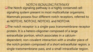
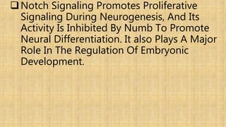
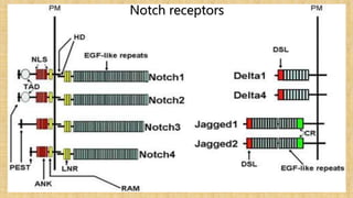
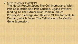
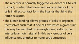
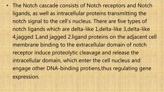
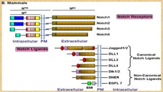
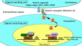
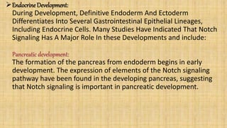
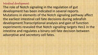
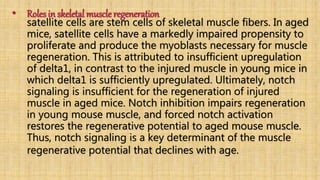
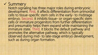
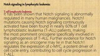
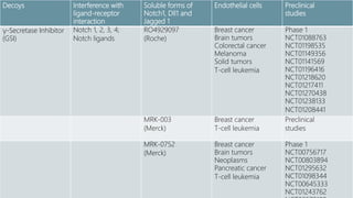
The Notch signaling pathway is a highly conserved cell
signaling system present in most multicellular organisms.
Mammals possess four different notch receptors, referred to
as NOTCH1, NOTCH2, NOTCH3, and NOTCH4.
The notch receptor is a single-pass transmembrane receptor
protein. It is a hetero-oligomer composed of a large
extracellular portion, which associates in a calcium-
dependent, non-covalent interaction with a smaller piece of
the notch protein composed of a short extracellular region, a
single transmembrane-pass, and a small intracellular region.
3. Notch Signaling Promotes Proliferative
Signaling During Neurogenesis, And Its
Activity Is Inhibited By Numb To Promote
Neural Differentiation. It also Plays A Major
Role In The Regulation Of Embryonic
Development.
5. MECHANISM OF ACTION
The Notch Protein Spans The Cell Membrane, With
Part Of It Inside And Part Outside. Ligand Proteins
Binding To The Extracellular Domain Induce
Proteolytic Cleavage And Release Of The Intracellular
Domain, Which Enters The Cell Nucleus To Modify
Gene Expression.
6. •The receptor is normally triggered via direct cell-to-cell
contact, in which the transmembrane proteins of the
cells in direct contact form the ligands that bind the
notch receptor.
•The Notch binding allows groups of cells to organize
themselves such that, if one cell expresses a given trait,
this may be switched off in neighboring cells by the
intercellular notch signal. In this way, groups of cells
influence one another to make large structures.
7. • The Notch cascade consists of Notch receptors and Notch
ligands, as well as intracellular proteins transmitting the
notch signal to the cell's nucleus. There are five types of
notch ligands which are delta-like 1,delta-like 3,delta-like
4,jagged 1,and jagged 2.ligand proteins on the adjacent cell
membrane binding to the extracellular domain of notch
receptor induce proteolytic cleavage and release the
intracellular domain, which enter the cell nucleus and
engage other DNA-binding protiens,thus regulating gene
expression.
8.
9.
10. FUNCTION
The Notch signaling pathway is important for cell-cell
communication, which involves gene regulation
mechanisms that control multiple cell differentiation
processes during embryonic and adult life. Notch
signaling has a role in the following processes:
Embryogenesis:
The Notch signaling pathway plays an important role in cell-cell
communication, and further regulates Embryo polarity,
Somitogenesis,and Epidermal differentiation.
11. Embryo Polarity:
Notch Signaling Is Required In The Regulation Of Polarity. For
Example, Mutation Experiments Have Shown That Loss Of Notch
Signaling Causes Abnormal Anterior-posterior Polarity In
Somites.Also, Notch Signaling Is Required During Left-right
Asymmetry Determination In Vertebrates.
Somitogenesis :
Notch signaling is central to somitogenesis. Notch1 was shown to be
important for coordinating the segmentation of somites in mice.
Further studies identified the role of Notch signaling in the
segmentation clock (clock and wavefront model).
12. These studies hypothesized that the primary function of Notch
signaling does not act on an individual cell, but coordinates cell
clocks and keep them synchronized. This hypothesis explained
the role of Notch signaling in the development of segmentation
and has been supported by experiments in mice and zebrafish.
Experiments with Delta1 mutant mice that show abnormal
somitogenesis with loss of anterior/posterior polarity suggest
that Notch signaling is also necessary for the maintenance of
somite borders.
13. Epidermal differentiation :
Notch signaling is known to occur inside ciliated, differentiating
cells found in the first epidermal layers during early skin
development. Furthermore, it has found that presenilin-2 has an
important role in regulating Notch signaling during this
development through involving in the cleavage of notch receptor.
14. Central nervous systemdevelopment and function:
The Notch signaling pathway plays an important role in Neuron cell
differentiation, Neurite development,Gliogenesis,and Adult brain
function as learning and memory.
Neuron cell differentiation:
The Notch pathway is essential for maintaining NPCs in the
developing brain. Activation of the pathway is sufficient to maintain
NPCs in a proliferating state, whereas loss-of-function mutations in
the critical components of the pathway cause precocious neuronal
differentiation and NPC depletion.
15. Modulators of the Notch signal, e.g., the Numb protein are
able to antagonize Notch effects, resulting in the halting of
cell cycle and differentiation of NPCs.
In adult rodents and in cell culture, Notch3 promotes
neuronal differentiation, having a role opposite to
Notch1/2.This indicates that individual Notch receptors can
have divergent functions, depending on cellular context.
16. Neurite development :
In vitro studies show that Notch can influence neurite
development. In vivo, deletion of the Notch signaling
modulator, Numb, disrupts neuronal maturation in the
developing cerebellum, and also disrupts axonal arborization
in sensory ganglia. Although the mechanism underlying this
phenomenon is not clear, together these findings suggest
Notch signaling might be crucial in neuronal maturation.
17. Gliogenesis :
In gliogenesis, Notch appears to have an instructive role that
can directly promote the differentiation of many glial cell
subtypes. For example, activation of Notch signaling in the
retina favors the generation of Muller glia cells at the expense
of neurons, whereas reduced Notch signaling induces
production of ganglion cells, causing a reduction in the
number of Muller glia.
18. Adult brain function :
Apart from its role in development, evidence shows that Notch
signaling is also involved in neuronal apoptosis, neurite retraction, and
neurodegeneration of ischemic stroke in the brain. In addition to
developmental functions, Notch proteins and ligands are expressed in
cells of the adult nervous system, suggesting a role in CNS plasticity
throughout life.
Several gamma secretase inhibitors that underwent human clinical
trials in Alzheimer's disease and MCI patients resulted in statistically
significant worsening of cognition relative to controls, which is
thought to be due to its incidental effect on Notch signaling.
19. Cardiovascular Development:
The Notch signaling pathway is a critical
of cardiovascular formation and morphogenesis in
both development and disease. It regulates:
Cardiac development
Notch signal pathway plays a crucial role in at least
three cardiac development processes:
Atrioventricular canal development, myocardial
development, and cardiac outflow tract (OFT)
development.
20. Atrioventricular canal development:
AV boundary formation :
Notch signaling can regulate the atrioventricular
boundary formation between the AV canal and the
chamber myocardium.
Studies have revealed that both loss- and gain-of-
function of the Notch pathway results in defects in AV
canal development. In addition, the Notch target genes
HEY1 and HEY2 are involved in restricting the expression
of two critical developmental regulator proteins, BMP2
and Tbx2, to the AV canal.
21. AV epithelial-mesenchymal transition (EMT) :
Notch signaling is also important for the process of
EMT, which is required for AV canal maturation. After
the AV canal boundary formation, a subset of
endocardial cells lining the AV canal are activated by
signals emanating from the myocardium and by
interendocardial signaling pathways to undergo
EMT.Notch1 deficiency results in defective induction of
EMT. Very few migrating cells are seen and these lack
mesenchymal morphology. Notch may regulate this
process by activating matrix metalloproteinase2
(MMP2) expression, or by inhibiting vascular
endothelial (VE)-cadherin expression in the AV canal
22. Ventricular development:
Notch signaling sustains immature cardiomyocyte
proliferation in mammals. The downstream
of Notch signaling, HEY2, was demonstrated to
important in regulating ventricular development
its expression in the interventricular septum and
the endocardial cells of the cardiac cushions.
Cardiomyocyte and smooth muscle cell-specific
deletion of HEY2 results in impaired cardiac
contractility, malformed right ventricle, and
ventricular septal defects.
23. Ventricular outflow tract development:
During development of the aortic arch and the
aortic arch arteries, the Notch receptors, ligands,
and target genes display a unique expression
pattern. When the Notch pathway was blocked, the
induction of vascular smooth muscle cell marker
expression failed to occur, suggesting that Notch is
involved in the differentiation of cardiac neural
crest cells into vascular cells during outflow tract
development.
24. Angiogenesis
Endothelial cells use the Notch signaling pathway to
coordinate cellular behaviors during the blood vessel
sprouting that occurs in angiogenesis. When cells
within a patent vessel are exposed to VEGF signaling,
only a restricted number of them initiate the
angiogenic process. VEGF is able to induce DLL4
expression. In turn, DLL4 expressing cells down-
regulate VEGF receptors in neighboring cells through
activation of Notch, thereby preventing their migration
into the developing sprout. Likewise, during the
sprouting process itself, the migratory behavior of
connector cells must be limited to retain a patent
25. Endocrine Development:
During Development, Definitive Endoderm And Ectoderm
Differentiates Into Several Gastrointestinal Epithelial Lineages,
Including Endocrine Cells. Many Studies Have Indicated That Notch
Signaling Has A Major Role In these Developments and include:
Pancreatic development:
The formation of the pancreas from endoderm begins in early
development. The expression of elements of the Notch signaling
pathway have been found in the developing pancreas, suggesting
that Notch signaling is important in pancreatic development.
26. Intestinal development:
The role of Notch signaling in the regulation of gut
development has been indicated in several reports.
Mutations in elements of the Notch signaling pathway affect
the earliest intestinal cell fate decisions during zebrafish
development.Transcriptional analysis and gain of function
experiments revealed that Notch signaling targets Hes1 in the
intestine and regulates a binary cell fate decision between
adsorptive and secretory cell fates.
27. Bone development:
the Notch signaling pathway functions as down-regulator in
osteoclastogenesis and osteoblastogenesis.Notch1 is expressed in
the mesenchymal condensation area and subsequently in the
hypertrophic chondrocytes during chondrogenesis.Overexpression of
Notch signaling inhibits bone morphogenetic protein2-induced
osteoblast differentiation. Overall, Notch signaling has a major role
in the commitment of mesenchymal cells to the osteoblastic lineage
and provides a possible therapeutic approach to bone regeneration.
Respiratory systemdevelopment:
Notch is implicated in development of alveoli in the lung.
28. • Roles in skeletal muscle regeneration
satellite cells are stem cells of skeletal muscle fibers. In aged
mice, satellite cells have a markedly impaired propensity to
proliferate and produce the myoblasts necessary for muscle
regeneration. This is attributed to insufficient upregulation
of delta1, in contrast to the injured muscle in young mice in
which delta1 is sufficiently upregulated. Ultimately, notch
signaling is insufficient for the regeneration of injured
muscle in aged mice. Notch inhibition impairs regeneration
in young mouse muscle, and forced notch activation
restores the regenerative potential to aged mouse muscle.
Thus, notch signaling is a key determinant of the muscle
regenerative potential that declines with age.
29. Summery
Notch signaling has three major roles during embryonic
development. First, it affects differentiation from primordial
cells to tissue‐specific stem cells in the early‐ to midstage
embryo. Second, it inhibits tissue‐ or organ‐specific stem
cells or immature progenitors from further differentiation
and presumably helps them expand while maintaining the
immature state. Third, it blocks the default pathway and
promotes the alternative pathway, which is typically
observed during mid‐ to late‐stage embryo development,
such as during organ formation.
30. Notch in cancer
A role for notch signaling in cancer was originally
suggested because A chromosomal translocation that
was found in A patient with T cell acute lymphoblastic
leukemia (T-ALL), which opened the door to an ever-
widening understanding of tumor growth controlled or
influenced by notch signaling. Notch has been shown
promote or limit tumor growth, which is highly
dependent on signal dose, notch homolog, and
Accumulating data have demonstrated that notch
signaling is A more complex process than originally
thought. Here we provide A brief overview on the roles
of the notch signaling pathway in the progression of A
31. Notch signaling in lymphocytic leukemia:
T cell lymphocytic leukemia:
It has been shown that Notch signaling is abnormally
regulated in many human malignancies. Notch1
mutations causing Notch signaling continuously
activated have been found in nearly 60% of T cell
lymphoblastic leukemia (T-ALL) patients, making
the most prominent oncogene specifically involved in
the pathogenesis of T-ALL. Further evidence for Notch
signaling as an oncogene may lie in that Notch1
regulates the expression of c-MYC, a potent driver of
cell cycle entry, contributing to cell cycle progression in
T-ALL.
32. • Notch1 directly induces the expression of c-MYC and
that inhibition of Notch1 using small molecule
inhibitors of the γ-secretase complex resulted in cell
cycle arrest and apoptosis and decreased c-MYC
levels.Notch1 also suppressed p53 function in T-ALL
cells, which could promote oncogenesis through
increased cell survival and genomic instability.
Additionally, other Notch signaling and target genes
are also involved in the initiation and progression of
T-ALL. It has been reported that Notch3 and Hes1 are
highly expressed by T-ALL cells, as well as
dramatically reduced or absent in remission.
33. B cell lymphocytic leukemia:
Interestingly, the function of Notch signaling in
leukemogenesis has been shown to be either
or tumor suppressive, and it could be context
dependent. Notch signaling and target genes have
demonstrated to be tumor suppressive rather than
oncogenic in a limited number of leukemia types,
including B-ALL. It has been reported that in contrast
T-ALL, Notch3, Jagged1, Hes2, Hes4 and Hes5 were
frequently hypermethylated in B-ALL, associated with
gene silencing. Furthermore, restoration of Hes5
expression by lentiviral transduction could give rise to
growth arrest and apoptosis in Hes5 negative B-ALL
34. In contrast with B-ALL, Notch signaling could
maintain B cell chronic lymphoblastic leukemia (B-
CLL) cell survival and apoptosis resistance,
undoubtedly indicating an oncogenic role in B-CLL.
Emerging evidence suggests that the Notch signaling
network is frequently deregulated in human B-CLL
with up-regulated expression of Notch1 and Notch2
as well as their ligands Jagged1 and Jagged2.
Moreover, Notch signaling inhibition by the gamma-
secretase inhibitors (GSIs) and the specific Notch2
down-regulation using small interfering RNA (siRNA)
could promote B-CLL cell apoptosis.
35. Inhibitors of Notch signaling and the potential clinical application
The specific and profound involvement of Notch signaling in
various leukemic types makes it an ideal target for
pharmacological intervention. Several strategies have been
proposed to inhibit or modulate this signaling. The most
used drug to globally inhibit Notch signaling is GSIs, which
block the cleavage of Notch at the cell membrane, inhibiting
release of the transcriptionally active Notch intracellular
domain (NICD) subunit. A lot of clinical research or preclinical
testing have focused on testing GSIs in the treatment of
leukemia, but the results were initially disappointing. It has
been reported that RO4929097, one of GSIs, could induce
insignificant differences in event free survival distribution
compared to control in 0 of 8 (0%) of the evaluable ALL
xenografts mice.
36. A phase I clinical trial also showed that MK-0752, another
GSIs, had limited antitumor activity in relapsed T-ALL
patients. What is more, GSIs are nonspecific and can inhibit
Notch signaling in the gut, leading to gastrointestinal
toxicity, which also limit its application. However, in an
attempt to the clinical application of GSIs, dexamethasone
was found to abrogate GSI-induced toxicity in the gut and
as well GSIs treatment could reverse glucocorticoid
resistance in T-ALL patients. Therefore, these results
supported a role for combination therapy with GSIs plus
glucocorticoids in the treatment T-ALL. In another attempt
to remedy this issue, inhibitory antibodies have recently
been synthesized for all Notch receptors.
37. A Notch1-specific antibody significantly induced
cell cycle arrest and reduced cell proliferation in
T-ALL cells. Moreover, in mouse xenograft T-ALL
and colon cancer models, the Notch1-specific
antibody could induce significant tumor
regression and slowing of growth, which would
pave the way for new clinical trials to evaluate
the efficacy of more selective and less toxic
antibody-based therapies.
38. Notchsignaling pathway and breast cancer
After the discovery of its involvement in T-ALL, Notch
was also implicated in breast cancer. The oncogenic potential
of Notch activation in solid tumors was first observed in
mammary cancer, which is induced by the mouse mammary
tumor virus (MMTV).
Breast cancer patients with high levels of Notch1 and Jagged1
showed a poorer prognostic profile and lower survival rates.
Similarly, one study has shown that more than 50% of human
breast tumors express reduced protein levels of Numb, a
negative regulator of Notch signaling, which has been
associated with high-grade breast cancers. Also can be
reversed after treatment with γ-secretase inhibitor.
39. Role of Notch signaling pathway in acute kidney injury, repair, and regeneration
The Notch signaling pathway is induced in mature
organs after injury. Notch-1 and Jagged-1 proteins are
upregulated in the rat liver after partial
hepatectomy,and NICD levels are significantly elevated
in the brain after cerebral ischemia–reperfusion.With
regard to the renal injury, one study reported an
upregulation of Jagged-1 expression in the kidney of
mice with ureteral obstruction. the Delta/Notch/Hes
pathway is activated after ischemic AKI and plays a role
in the proliferation of renal tubules.
40. Notch pathway in the ischemic heart
Myocardial infarction (MI), one of the leading causes
of death worldwide, mainly depends on coronary
artery occlusion and ischemia followed by
reperfusion (I/R), in which blood flow restoration is
accompanied by oxidative stress exacerbating
myocardial damage. Noteworthy, the adult
myocardium can re-express fetal genes as an
adaptive response to injury: in this context,
increased notch1 signaling was demonstrated in
surviving cardiomyocytes of the MI border zone.
41. Several studies have shown that notch signaling
protects the heart from I/R-induced myocardial
injury: activation of notch1 pathway limits the extent
of ischemic damage, promotes coronary neo-
angiogenesis and revascularization of the ischemic
myocardium, reduces myocardial fibrosis and
improves heart function. Conversely, in systemic
notch1 deficient mice, I/R leads to the development
of a larger myocardial infarct area and worsening of
heart function than wild-type controls.
42. The mechanisms underlying Notch-mediated cardio-
protection are complex and involve an interplay
between mature and immature cardiomyocytes,
cardiac progenitors cells (CPCs) and bone marrow
(BM)-derived cells. Notch1 prevents cardiomyocyte
apoptosis by activation of PI3K/AKT pro-survival
signaling and regulation of apoptotic genes.
Moreover, Notch signaling induces cell cycle re-entry
of immature cardiomyocytes, promotes proliferation
and myogenic differentiation of CPCs, decreases
oxidative/nitrosative stress and prevents cardiac
fibrosis.
43.
44. Notch pathway in the I/R of liver
Hepatic ischemia/reperfusion (I/R) injury is initiated by reactive
oxygen species (ROS) accumulated during the early reperfusion
phase after ischemia, but cellular mechanisms controlling ROS
production and scavenging have not been fully understood.
The blocking of Notch signal by knockout of the transcription
factor RBP-J or a pharmacological inhibitor led to aggravated
hepatic I/R injury, as manifested by deteriorated liver function
and increased apoptosis, necrosis, and inflammation, both in
vitro and in vivo. Interruption of Notch signaling resulted in
increased intracellular ROS in hepatocytes, and a ROS
scavenger cured exacerbated hepatic I/R injury after Notch
signaling blockade, suggesting that Notch signal deficiency
aggravated I/R injury through increased ROS levels.
45. Notch signal blockade resulted in down-regulation of
Hes5, leading to reduced formation of the Hes5-
STAT3 complex and hypophosphorylation of STAT3,
which further attenuated manganese superoxide
dismutase (MnSOD) expression and increased ROS
and apoptosis. Indeed, overexpression of a
constitutively active STAT3 rescued MnSOD
expression and I/R injury-induced apoptosis in the
absence of Notch signaling. Finally, forced Notch
activation by ligand stimulation or Hes5
overexpression reduced intracellular ROS and
protected hepatocytes from apoptosis after I/R injury
through the activation of STAT3 and MnSOD
46. Notch as tumor suppressor
Although Notch was originally identified as an
oncogene, studies have also demonstrated that
components of the same pathway may have growth-
suppressive functions in some hematopoietic cells, skin,
and pancreatic epithelium, as well as in hepatocytes,
illustrating the highly context-dependent nature of the
pathway. The first evidence describing Notch signaling
as a factor for suppressing tumors was derived from
Nicolas et al. In their study, mice with Notch1-deficient
epithelia increased and sustained expression of Gli2,
which is a downstream component of the Sonic–
hedgehog (SHH)-signaling pathway, causing the
47. Consistent with this, Thelu et al.reported that
expressions of Notch1, Notch2, and Jagged1
were down-regulated in human basal-cell
carcinomas. These results indicated that a loss
of Notch signaling in human epidermis, as well
as in mouse epithelia, could lead to the
development of basal-cell carcinomas through
suppression of the SHH pathways.
48. • The studies on Notch function in skin lead to an interesting
question: Is the tumor suppressive activity of Notch
manifested in a broader range of tissues? Evidence from
several studies on Notch function in neuroendocrine tumors
(NETs), such as small-cell lung cancer (SCLC), pancreatic
carcinoid, and medullary thyroid cancer (MTC), seem to
support this notion. In non-small cell lung cancer (NSCLC),
Notch shows a growth promoting function, whereas in SCLC
it exerts an inhibitory effect. These apparent but paradoxical
functions clearly indicate that the role of Notch signaling is
dependent on its cellular context. In SCLC, constitutively
active Notch receptors (Notch1, Notch2) have been shown
to cause a profound growth arrest.
49. Development
Phase
ConditionCompoundNotch pathway
Target
Agent
Phase 1
NCT01277146
Solid tumorsOMP-59R5
anti-Notch2/3
mAb
(OncoMed
Pharmaceuticals)
Interference with
ligand-induced
Notch subunit
separation and
Notch ligands.
Specific for
Notch 1, 2, 3;
DLL1, 4
Neutralizing
antibodies
Preclinical and
In vitro studies
Breast cancer
Colon Cancer
Anaplastic carcinoma
T-cell leukemia
T-ALL cell line
NRR1
anti-Notch1 mAb
(Genentech and
Exelixis; Merck)
Preclinical
studies
Breast cancer
Colon Cancer
Anaplastic carcinoma
HEK293T cell line
NRR2
anti-Notch2 mAb
(Genentech and
Exelixis)
Notch inhibitors and their current development stage
50. Preclinical
studies
Endothelial cellsSoluble forms of
Notch1, Dll1 and
Jagged 1
Interference with
ligand-receptor
interaction
Decoys
Phase 1
NCT01088763
NCT01198535
NCT01149356
NCT01141569
NCT01196416
NCT01218620
NCT01217411
NCT01270438
NCT01238133
NCT01208441
Breast cancer
Brain tumors
Colorectal cancer
Melanoma
Solid tumors
T-cell leukemia
RO4929097
(Roche)
Notch 1, 2, 3, 4;
Notch ligands
γ-Secretase Inhibitor
(GSI)
Preclinical
studies
Breast cancer
T-cell leukemia
MRK-003
(Merck)
Phase 1
NCT00756717
NCT00803894
NCT01295632
NCT01098344
NCT00645333
NCT01243762
Breast cancer
Brain tumors
Neoplasms
Pancreatic cancer
T-cell leukemia
MRK-0752
(Merck)
51. Preclinical and
In vitro studies
Pancreatic cancer
Prostate cancer
Thyroid cancer
Carcinoid
T-ALL cells
Glioblastoma cells
Oral cancer cells
Genistein
Sulforaphane
Quercetin
Curcumin
Resveratrol
Downregulation
of Notch activity
and Notch
pathway
Natural compounds