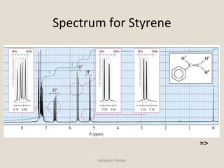
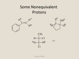
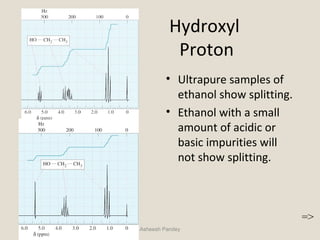
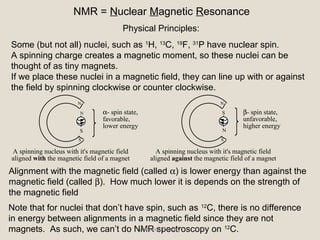
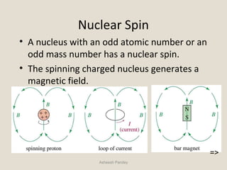
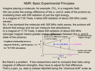
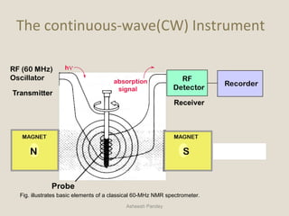
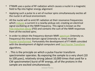
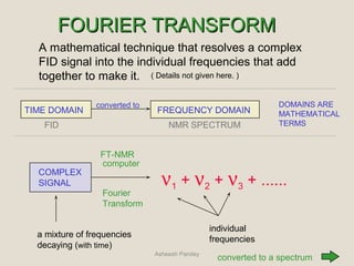
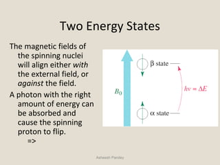
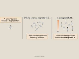
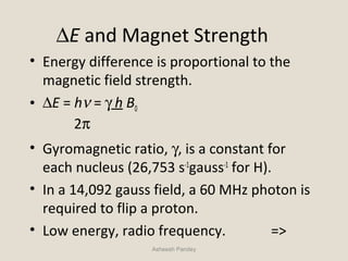
Nuclear Magnetic
Resonance Spectroscopy
Asheesh Pandey
NMR = Nuclear Magnetic Resonance
Some (but not all) nuclei, such as 1
H, 13
C, 19
F, 31
P have nuclear spin.
A spinning charge creates a magnetic moment, so these nuclei can be
thought of as tiny magnets.
If we place these nuclei in a magnetic field, they can line up with or against
the field by spinning clockwise or counter clockwise.
Alignment with the magnetic field (called α) is lower energy than against the
magnetic field (called β). How much lower it is depends on the strength of
the magnetic field
Physical Principles:
Note that for nuclei that don’t have spin, such as 12
C, there is no difference
in energy between alignments in a magnetic field since they are not
magnets. As such, we can’t do NMR spectroscopy on 12
C.
S
A spinning nucleus with it's magnetic field
aligned with the magnetic field of a magnet
α- spin state,
favorable,
lower energy
N
S
N
N
S β- spin state,
unfavorable,
higher energy
A spinning nucleus with it's magnetic field
aligned against the magnetic field of a magnet
S
N
Asheesh Pandey
• NMR is the most powerful tool available for
organic structure determination.
• It is used to study a wide variety of nuclei:
– 1
H
– 13
C
– 15
N
– 19
F
– 31
P
=>
Asheesh Pandey
Nuclear Spin
• A nucleus with an odd atomic number or an
odd mass number has a nuclear spin.
• The spinning charged nucleus generates a
magnetic field.
Asheesh Pandey
=>
External Magnetic Field
When placed in an external field, spinning protons act
like bar magnets.
Asheesh Pandey
=>
NMR: Basic Experimental Principles
Imagine placing a molecule, for example, CH4, in a magnetic field.
We can probe the energy difference of the α- and β- state of the protons by
irradiating them with EM radiation of just the right energy.
In a magnet of 7.05 Tesla, it takes EM radiation of about 300 MHz (radio
waves).
So, if we bombard the molecule with 300 MHz radio waves, the protons will
absorb that energy and we can measure that absorbance.
In a magnet of 11.75 Tesla, it takes EM radiation of about 500 MHz
(stronger magnet means greater energy difference between the α- and β-
state of the protons)
But there’s a problem. If two researchers want to compare their data using
magnets of different strengths, they have to adjust for that difference.
That’s a pain, so, data is instead reported using the “chemical shift” scale as
E
Bo
∆E = h x 300 M Hz ∆E = h x 500 MHz
7.05 T 11.75 T
α proton spin state
(lower energy)
β proton spin state
(higher energy)
Graphical relationship between
magnetic field (B o) and frequency ( ν)
for 1
H NMR absorptions
at no magnetic field,
there is no difference beteen
α- and β- states.
0 T
Asheesh Pandey
Chemical shift δ is usually expressed in parts per million (ppm) by frequency,
because it is calculated from
Asheesh Pandey
Instrumentation:-Instrumentation:-
• Types of NMR high resolution spectra
Continuous wave Fourier transform
NMR NMR
• Imp parts of the NMR spectrometer:-
 Permanent magnet/electromagnet
 RF generator
 RF detector
 Sample holder
 Magnetic coils
Asheesh Pandey
diagram:
Asheesh Pandey
• Principle:- based on
• frequency sweep field sweep
frequency of
RF source frequency is
is varied
constant
Bo is constant Bo is varied
Asheesh Pandey
CONSTRUCTION:-CONSTRUCTION:-
 Magnets:-
• permanent:- constant Bo is generated that is 0.7;1.4;2.1
o adv:-
• construction is simple
• cheaper
• electromagnet:-Bo can be varied which is done by winding the
electromagnetic coil around the magnet
• most expensive components of the nuclear magnetic resonance
spectrometer system
 Shim Coils
• The purpose of shim coils on a spectrometer is to correct minor spatial
inhomogeneities in the Bo magnetic field.
• These inhomogeneities could be caused by the magnet design, materials
in the probe, variations in the thickness of the sample tube, sample
permeability.
Asheesh Pandey
• A shim coil is designed to create a small magnetic field which will oppose
and cancel out an inhomogeneity in the Bo magnetic field.
 Superconducting solenoids:-
• prepared from superconducting niobium-titanium wire and niobium-tin
wire
• operated at lower temp.
• kept in liquid He(mostly preferred) or liquid N2 at temp of 4 K
• liquid N2 should be changed at 10 days while liquid He shouldd be changed
at 80-130 days
• higher Bo can be produced that is upto 21 T.
o Advantage:-
 High stability
 Low operating cost
 High sensitivity
 Small size compared to electromagnets
 simple
Asheesh Pandey
RADIOFREQUNCY TRANSMITTER
• It is a 60 MHz crystal controlled oscillator.
• RF signal is fed into a pair of coils mounted at right angles to the path of
field.
• The coil that transmit RF field is made into 2 halves in order to allow
insertion of sample holder .
• 2 halves are placed in magnetic gap
• For high resolution the transmitted frequency must be highly constant.
• The basic oscillator is crystal controlled followed by a buffer doubler, the
frequency being doubled by tunning the variable
• It is further connected to another buffer doubler tuned to 60 MHz
• Then buffer amplifier is provided to avoid circuit loading.
Asheesh Pandey
Signal amplifier and detector
• Radiofrequency signal is produced by the resonating nuclei is
detected by means of a coil that surrounds the sample holder
• The signal results from the absorption of energy from the
receiver coil, when nuclear transitions are induced and the
voltage across receiver coil drops
• This voltage change is very small and it must be amplified
before it can be displayed.
The display system
• The detected signal is applied to vertical plates of an
oscilloscope to produce NMR spectrum
• Spectrum can also be recorded on a chart recorder.
Asheesh Pandey
 Sample Probe
 The sample probe is the name given to that part of the
spectrometer which accepts the sample, sends RF energy into
the sample, and detects the signal emanating from the
sample.
• It contains the RF coil, sample spinner, temperature
controlling circuitry, and gradient coils.
• It is also provided with an air driven turbine for rotating the
sample tube at several hundred rpm
• This rotation averages out the effects of in homogeneities in
the field and provide better resolution.
Asheesh Pandey
 The spectrum obtained either by CW scan or pulse FT at constant
magnetic field is shown in series of peaks whose areas are proportional to
the number of protons they represent.
 Peak areas are measured by an electronic integrator that traces a series of
steps with heights proportional to the peak areas.
Asheesh Pandey
• A proton count from the integration is useful to
determination or confirm molecular formulas, detect hidden
peaks, determine sample purity, and to quantitative analysis.
• Peak positions are measured in frequency units from a
reference peak.
• A routine sample for proton NMR on a 300MHz instrument
consist of about 2mg of the compound in about 0.4 mL of
solvent in a 5-mm o.d.glass tube.
• Under favorable conditions, it is possible to obtain a spectrum
of 1μg of a compound of a compound of modest molecular
weight in a microtube in a 300-MHz pulsed instrument.
• Microprobe that accept a 2.5mm or 3 mm o.d. tube are
convenient and provide high sensitivity.
Asheesh Pandey
The continuous-wave(CW) Instrument
Asheesh Pandey
Fig. illustrates basic elements of a classical 60-MHz NMR spectrometer.
WORKING:-
• The sample is dissolved in a solvent containing no interfering proton
(usually CCl4 and CDCl3), and a small amount of TMS is added to serve as an
internal reference.
• The sample is a small cylindrical glass tube that is suspended in the gap
between the faces of the pole pieces of the magnet.
• The sample is spun around the axis to ensure that all parts of the solution
experience a relatively uniform magnetic field.
• Also in a magnetic gap is a coil attached to 60-MHz radiofrequency
generator. This coil supplies the electromagnetic energy used to change
the spin orientation of the protons.
• Perpendicular to the RF oscillator coil is a detector coil. When no
absorption of energy is taking place, the detector coil picks up non of the
energy given off by the RF oscillator coil.
Asheesh Pandey
• when sample absorbs energy, however, the reorientation of
the nuclear spins induces a radiofrequency signal in the plane
of the detector coil, and the instrument responds by
recording this as a resonance signal, or peak.
• Rather than changing the frequency of the RF oscillator to
allow each of the protons in a molecule to come into a
resonance, the typical NMR spectrometer uses a constant
frequency RF- signal and varies the magnetic field strength.
• As the magnetic field strength is increased, the precessional
frequency of all the protons increase. When the precesional
frequency of a given type proton reaches 60MHz, it has
resonance.
Asheesh Pandey
• As each chemically distinct type of proton comes into resonance, it is
recorded as a peak on the chart.
• The peak at δ = 0 ppm is due to the internal reference compound TMS.
• IN THE CLASSICAL NMR EXPERIMENT THE INSTRUMENT SCANS FROMIN THE CLASSICAL NMR EXPERIMENT THE INSTRUMENT SCANS FROM
“LOW FIELD” TO “HIGH FIELD”“LOW FIELD” TO “HIGH FIELD”
Asheesh Pandeyscan
increasing Bo
HIGH
FIELD
LOW
FIELD
UPFIELDDOWNFIELD
NMR CHART
Pulsed Fourier Transform Spectroscopy
Asheesh Pandey
• FTNMR uses a pulse of RF radiation which causes a nuclei in a magnetic
field to flip into higher energy alignment
• Applying such a pulse to a set of nuclear spins simultaneously excites all
the nuclei in all local environment.
• All the nuclei will re emit RF radiation at their resonance frequencies
which induces a current in a nearby pickup coil, creating an electrical
signal oscillating at the NMR frequency. This signal is known as the
free induction decay (FID) and contains the sum of the NMR responses
from all the excited spins.
• In order to obtain the frequency-domain NMR spectrum (intensity vs.
frequency) this time-domain signal (intensity vs. time) must be
Fourier transformed. Fortunately the development of FT-NMR coincided
with the development of digital computers and Fast Fourier Transform
algorithms.
• . This is the principle on which a pulse Fourier transform
spectrometer operates. By exposing the sample to a very short (10
to 100 μsec), relatively strong (about 10,000 times that used for a
CW spectrometer) burst of RF energy, all of the protons in the
sample are excited simultaneously.
Asheesh Pandey
FOURIER TRANSFORMFOURIER TRANSFORM
A mathematical technique that resolves a complex
FID signal into the individual frequencies that add
together to make it.
COMPLEX
SIGNAL ν1 + ν2 + ν3 + ......
computer
Fourier
Transform
FT-NMR
individual
frequencies
TIME DOMAIN FREQUENCY DOMAIN
a mixture of frequencies
decaying (with time)
converted to
converted to a spectrum
( Details not given here. )
FID NMR SPECTRUM
DOMAINS ARE
MATHEMATICAL
TERMS
Asheesh Pandey
The Composite FID is Transformed into aThe Composite FID is Transformed into a
classical NMR Spectrum :classical NMR Spectrum :
Asheesh Pandey
CH2 C
O
CH3
“frequency domain” spectrum
• Advantage over continuous wave NMR
 Sample of low conc. can be determined
 Magnetic nuclei with low natural isotopic abundance can be
determined eg 13
C
 Very rapid pulse repetition can be possible
 Entire spectrum can be recorded, computerized and
transformed in a few seconds that is every 2 sec For e.g. in 13
min 400 spectra can be recorded. So thus 20 times signal
enhancement is seen
 Analysis can be possible where magnetogyric ratio is low
Asheesh Pandey
Solvents used in NMRSolvents used in NMR
• Properties:-
 Nonviscous.
 Should dissolve analyte.
 Should not absorb within spectral range of analysis.
 All solvents used in NMR must be aprotic that is they should not possess
proton.
• Chloroform-d (CDCl3) is the most common solvent for NMR
measurements
• other deuterium labeled compounds, such as deuterium oxide
(D2O), benzene-d6 (C6D6), acetone-d6 (CD3COCD3) and DMSO-d6
(CD3SOCD3) are also available for use as NMRsolvents.
• DMF, DMSO, cyclopropane, dimethyl ether can also be used
Asheesh Pandey
iNTRODUCTiON
 Powerful analytical technique used to characterize organic molecules
by identifying carbon-hydrogen frameworks within molecules.
 2 Types :
 The source of energy in NMR is radio waves which have long
wavelengths, and thus low energy and frequency.
 This waves can change the nuclear spins of some elements, including 1
H
and 13
C.
 Frequency in the range of 300Mhz-500Mhz.
Asheesh Pandey
13
C -NMR1
H -NMR
Two Energy States
The magnetic fields of
the spinning nuclei
will align either with
the external field, or
against the field.
A photon with the right
amount of energy can
be absorbed and
cause the spinning
proton to flip.
=>
Asheesh Pandey
Asheesh Pandey
∆E and Magnet Strength
• Energy difference is proportional to the
magnetic field strength.
• ∆E = hν = γ h B0
2π
• Gyromagnetic ratio, γ, is a constant for
each nucleus (26,753 s-1
gauss-1
for H).
• In a 14,092 gauss field, a 60 MHz photon is
required to flip a proton.
• Low energy, radio frequency. =>
Asheesh Pandey
Magnetic Shielding
• If all protons absorbed the same amount of
energy in a given magnetic field, not much
information could be obtained.
• But protons are surrounded by electrons
that shield them from the external field.
• Circulating electrons create an induced
magnetic field that opposes the external
magnetic field. =>
Asheesh Pandey
Shielded Protons
Magnetic field strength must be increased for
a shielded proton to flip at the same
frequency.
Asheesh Pandey
=>
Protons in a Molecule
Depending on their chemical environment,
protons in a molecule are shielded by
different amounts.
Asheesh Pandey
=>
NMR Signals
• The number of signals shows how many
different kinds of protons are present.
• The location of the signals shows how
shielded or deshielded the proton is.
• The intensity of the signal shows the number
of protons of that type.
• Signal splitting shows the number of protons
on adjacent atoms. =>
Asheesh Pandey
The NMR Spectrometer
Asheesh Pandey
=>
The NMR Graph
Asheesh Pandey
=>
Tetramethylsilane
• TMS is added to the sample.
• Since silicon is less electronegative than
carbon, TMS protons are highly shielded.
Signal defined as zero.
• Organic protons absorb downfield (to the
left) of the TMS signal.
=>
Asheesh Pandey
Si
CH3
CH3
CH3
H3C
Chemical Shift
• Measured in parts per million.
• Ratio of shift downfield from TMS (Hz) to total
spectrometer frequency (Hz).
• Same value for 60, 100, or 300 MHz machine.
• Called the delta scale.
=>
Asheesh Pandey
Delta Scale
Asheesh Pandey
=>
Location of Signals
• More electronegative atoms
deshield more and give larger
shift values.
• Effect decreases with
distance.
• Additional electronegative
atoms cause increase in
chemical shift.
=>
Asheesh Pandey
Typical Values
Asheesh Pandey =>
O-H and N-H Signals
• Chemical shift depends on concentration.
• Hydrogen bonding in concentrated solutions
deshield the protons, so signal is around δ3.5
for N-H and δ4.5 for O-H.
• Proton exchanges between the molecules
broaden the peak.
=>
Asheesh Pandey
Carboxylic Acid
Proton, δ10+
Asheesh Pandey
=>
Number of Signals
Equivalent hydrogens have the same
chemical shift.
Asheesh Pandey
=>
Intensity of Signals
• The area under each peak is proportional to
the number of protons.
• Shown by integral trace.
Asheesh Pandey
=>
How Many Hydrogens?
When the molecular formula is known, each
integral rise can be assigned to a particular
number of hydrogens.
Asheesh Pandey
=>
Spin-Spin Splitting
• Nonequivalent protons on adjacent carbons
have magnetic fields that may align with or
oppose the external field.
• This magnetic coupling causes the proton to
absorb slightly downfield when the external
field is reinforced and slightly upfield when the
external field is opposed.
• All possibilities exist, so signal is split. =>
Asheesh Pandey
Calculating SHIFT VALUES:
Added Chemical Shifts
Substituent Type of Hydrogen -Shift -Shift
C C CH3 0.78 ---
CH2 0.75 -0.10
CH --- ---
RC C C
Y
[Y = C or O] CH3 1.08 ---
Aryl- CH3 1.40 0.35
CH2 1.45 0.53
CH 1.33 ---
Cl- CH3 2.43 0.63
CH2 2.30 0.53
CH 2.55 0.03
Br- CH3 1.80 0.83
CH2 2.18 0.60
CH 2.68 0.25
I- CH3 1.28 1.23
CH2 1.95 0.58
CH 2.75 0.00
OH- CH3 2.50 0.33
CH2 2.30 0.13
CH 2.20 ---
RO- (R is saturated) CH3 2.43 0.33
CH2 2.35 0.15
CH 2.00 ---
R–CO
O
or ArO CH3 2.88 0.38
CH2 2.98 0.43
CH 3.43 ---
(ester only)
R–C
O
CH3 1.23 0.18
where R is alkyl, aryl, OH, CH2 1.05 0.31
OR', H, CO, or N CH 1.05 ---
Asheesh Pandey
(Hydrogen under consideration)C C H
H
H
H
H
Cl
β α
Base Chemical Shift = 0.87 ppm
no α substituents = 0.00
one β -Cl (CH3) = 0.63
TOTAL = 1.50 ppm
(Hydrogen under consideration)C C H
H
H
H
H
Cl
βα
Base Chemical Shift = 1.20 ppm
one α -Cl (CH2) = 2.30
no β substituents = 0.00
TOTAL = 3.50 ppm
Asheesh Pandey
1,1,2-Tribromoethane
Asheesh Pandey
Nonequivalent protons on adjacent carbons.
=>
More 1
H - 1
H Coupling
What happens when there is more than one proton splitting a neighboring
proton? We get more lines. Consider the molecule below where we have
two protons on one carbon and one proton on another.
C C
HBHA
HA'
HA + HA' HB
HA and HA ' appear at the same
chemical shift because they are
in identical environments
They are also split into two lines
(called a doublet) because they
feel the magnetic field of HB.
HB is split into three lines
because it feels the magnetic
field of HA and HA '
Note that the signal produced
byHA + HA ' is twice the size
of that produced b y HB
Asheesh Pandey
Why are There Three Lines for HB?
HB feels the splitting of both HA and HA’. So, let’s imagine starting with HB as
a single line, then let’s “turn on” the coupling from HA and HA’ one at a time:
HB
Now, let's "turn on" HB - HA coupling. This splits
the single line into two lines
If uncoupled, H B would appear as a
singlet where the dashed line indicates
the chemical shift of the singlet.
Now, let's "turn on" HB - HA'coupling. This
splits each of the two new lines into two lines,
but notice how the two lines in the middle
overlap. Overall, we then have three lines.
C C
HBHA
HA'
Because the two lines in the middle overlap, that line is twice as big as the
lines on the outside. More neighboring protons leads to more lines as shown
on the next slide.
Asheesh Pandey
no. of neighbors relative intensities pattern
1
1 1
1 2 1
1 3 3 1
1 4 6 4 1
1 5 10 10 5 1
1 6 15 20 15 6 1
0
1
2
3
4
5
6
singlet (s)
doublet (d)
triplet (t)
quartet (q)
pentet
sextet
septet
example
H
C C
H
H
C C
H
H
H
C C
H
H
H
H
C CC
H
H
H
H
H
C CC
H
H
HH
H
H
C CC
H
H
H
H
H
H
Splitting Patterns with Multiple Neighboring
Protons
If a proton has n neighboring protons that are equivalent, that proton will be
split into n+1 lines. So, if we have four equivalent neighbors, we will have
five lines, six equivalent neighbors… well, you can do the math. The lines
will not be of equal intensity, rather their intensity will be given by Pascal’s
triangle as shown below.
We keep emphasizing that this pattern only holds for when the neighboring
protons are equivalent. Why is that? The answer is two slides away.Asheesh Pandey
More About Coupling
Earlier we said that protons couple to each other because they feel the
magnetic field of the neighboring protons. While this is true, the
mechanism by which they feel this field is complicated and is beyond the
scope of this class (they don’t just feel it through space, it’s transmitted
through the electrons in the bonds). It turns out that when two protons
appear at the same chemical shift, they do not split each other. So, in
EtBr, we have a CH3 next to a CH2, and each proton of the CH3 group is
only coupled to the protons of the CH2 group, not the other CH3 protons
because all the CH3 protons come at the same chemical shift.
C C
H
H
H
H
H
Br
The blue protons all come
at the same chemical shift
and do not split each other
The red protons both come
at the same chemical shift
and do not split each other
C C
H
H
H
H
H
Br
C C
H
H
H
H
H
Br
Asheesh Pandey
Not all Couplings are Equal
When protons couple to each other, they do so with a certain intensity. This
is called the “coupling constant.” Coupling constants can vary from 0 Hz
(which means that the protons are not coupled, even though they are
neighbors) to 16 Hz. Typically, they are around 7 Hz, but many molecules
contain coupling constants that vary significantly from that. So, what
happens when a molecule contains a proton which is coupled to two
different protons with different coupling constants? We get a different
pattern as described in the diagram below.
So, if the protons are not equivalent, they can have different coupling
constants and the resulting pattern will not be a triplet, but a “doublet of
doublets.” Sometimes, nonequivalent protons can be on the same carbonAsheesh Pandey
Doublet: 1 Adjacent Proton
Asheesh Pandey
=>
Triplet: 2 Adjacent Protons
Asheesh Pandey
=>
The N + 1 Rule
Asheesh Pandey
If a signal is split by N equivalent protons,
it is split into N + 1 peaks.
=>
Range of Magnetic
Coupling
• Equivalent protons do not split each other.
• Protons bonded to the same carbon will split
each other only if they are not equivalent.
• Protons on adjacent carbons normally will
couple.
• Protons separated by four or more bonds will
not couple.
=>
Asheesh Pandey
Splitting for Ethyl Groups
Asheesh Pandey
=>
Splitting for
Isopropyl Groups
Asheesh Pandey
=>
Coupling Constants
• Distance between the peaks of multiplet
• Measured in Hz
• Not dependent on strength of the external
field
• Multiplets with the same coupling constants
may come from adjacent groups of protons
that split each other.
=>
Asheesh Pandey
Values for
Coupling Constants
Asheesh Pandey
=>
Complex Splitting
• Signals may be split by adjacent protons,
different from each other, with different
coupling constants.
• Example: Ha
of styrene which is split by an
adjacent H trans to it (J = 17 Hz) and an
adjacent H cis to it (J = 11 Hz).
=>
Asheesh Pandey
C C
H
H
H
a
b
c
Splitting Tree
Asheesh Pandey
C C
H
H
H
a
b
c
=>
Spectrum for Styrene
Asheesh Pandey
=>
Some Nonequivalent
Protons
Asheesh Pandey
C C
H
H
H
a
b
c
OH
H
H
H
a
b
c
d
CH3
H Cl
H H
Cl
a b =>
Time Dependence
• Molecules are tumbling relative to the
magnetic field, so NMR is an averaged
spectrum of all the orientations.
• Axial and equatorial protons on cyclohexane
interconvert so rapidly that they give a single
signal.
• Proton transfers for OH and NH may occur so
quickly that the proton is not split by adjacent
protons in the molecule.
=>
Asheesh Pandey
Hydroxyl
Proton
• Ultrapure samples of
ethanol show splitting.
• Ethanol with a small
amount of acidic or
basic impurities will
not show splitting.
Asheesh Pandey
=>
N-H Proton
• Moderate rate of exchange.
• Peak may be broad.
Asheesh Pandey
=>
Identifying the O-H
or N-H Peak
• Chemical shift will depend on
concentration and solvent.
• To verify that a particular peak is due to O-
H or N-H, shake the sample with D2O
• Deuterium will exchange with the O-H or N-
H protons.
• On a second NMR spectrum the peak will
be absent, or much less intense.
=>Asheesh Pandey
Carbon-13
• 12
C has no magnetic spin.
• 13
C has a magnetic spin, but is only 1% of the
carbon in a sample.
• The gyromagnetic ratio of 13
C is one-fourth of
that of 1
H.
• Signals are weak, getting lost in noise.
• Hundreds of spectra are taken, averaged.
=>
Asheesh Pandey
Fourier Transform NMR
• Nuclei in a magnetic field are given a radio-
frequency pulse close to their resonance
frequency.
• The nuclei absorb energy and precess (spin)
like little tops.
• A complex signal is produced, then decays
as the nuclei lose energy.
• Free induction decay is converted to
spectrum. =>
Asheesh Pandey
Hydrogen and Carbon Chemical
Shifts
Asheesh Pandey
=>
Combined 13
C
and 1
H Spectra
Asheesh Pandey
=>
Differences in
13
C Technique
• Resonance frequency is ~ one-fourth, 15.1
MHz instead of 60 MHz.
• Peak areas are not proportional to number
of carbons.
• Carbon atoms with more hydrogens absorb
more strongly.
=>
Asheesh Pandey
Spin-Spin Splitting
• It is unlikely that a 13
C would be adjacent to
another 13
C, so splitting by carbon is negligible.
• 13
C will magnetically couple with attached
protons and adjacent protons.
• These complex splitting patterns are difficult
to interpret.
=>
Asheesh Pandey
Proton Spin Decoupling
• To simplify the spectrum, protons are
continuously irradiated with “noise,” so they
are rapidly flipping.
• The carbon nuclei see an average of all the
possible proton spin states.
• Thus, each different kind of carbon gives a
single, unsplit peak.
=>
Asheesh Pandey
Off-Resonance Decoupling
• 13
C nuclei are split only by the protons
attached directly to them.
• The N + 1 rule applies: a carbon with N
number of protons gives a signal with
N + 1 peaks.
=>
Asheesh Pandey
Interpreting 13
C NMR
• The number of different signals indicates the
number of different kinds of carbon.
• The location (chemical shift) indicates the
type of functional group.
• The peak area indicates the numbers of
carbons (if integrated).
• The splitting pattern of off-resonance
decoupled spectrum indicates the number of
protons attached to the carbon. =>
Asheesh Pandey
Two 13
C NMR Spectra
Asheesh Pandey
=>
MRI
• Magnetic resonance imaging, noninvasive
• “Nuclear” is omitted because of public’s fear
that it would be radioactive.
• Only protons in one plane can be in resonance
at one time.
• Computer puts together “slices” to get 3D.
• Tumors readily detected.
=>
Asheesh Pandey
Asheesh Pandey

NMR by asheesh pandey

  • 1.
  • 2.
    NMR = NuclearMagnetic Resonance Some (but not all) nuclei, such as 1 H, 13 C, 19 F, 31 P have nuclear spin. A spinning charge creates a magnetic moment, so these nuclei can be thought of as tiny magnets. If we place these nuclei in a magnetic field, they can line up with or against the field by spinning clockwise or counter clockwise. Alignment with the magnetic field (called α) is lower energy than against the magnetic field (called β). How much lower it is depends on the strength of the magnetic field Physical Principles: Note that for nuclei that don’t have spin, such as 12 C, there is no difference in energy between alignments in a magnetic field since they are not magnets. As such, we can’t do NMR spectroscopy on 12 C. S A spinning nucleus with it's magnetic field aligned with the magnetic field of a magnet α- spin state, favorable, lower energy N S N N S β- spin state, unfavorable, higher energy A spinning nucleus with it's magnetic field aligned against the magnetic field of a magnet S N Asheesh Pandey
  • 3.
    • NMR isthe most powerful tool available for organic structure determination. • It is used to study a wide variety of nuclei: – 1 H – 13 C – 15 N – 19 F – 31 P => Asheesh Pandey
  • 4.
    Nuclear Spin • Anucleus with an odd atomic number or an odd mass number has a nuclear spin. • The spinning charged nucleus generates a magnetic field. Asheesh Pandey =>
  • 5.
    External Magnetic Field Whenplaced in an external field, spinning protons act like bar magnets. Asheesh Pandey =>
  • 6.
    NMR: Basic ExperimentalPrinciples Imagine placing a molecule, for example, CH4, in a magnetic field. We can probe the energy difference of the α- and β- state of the protons by irradiating them with EM radiation of just the right energy. In a magnet of 7.05 Tesla, it takes EM radiation of about 300 MHz (radio waves). So, if we bombard the molecule with 300 MHz radio waves, the protons will absorb that energy and we can measure that absorbance. In a magnet of 11.75 Tesla, it takes EM radiation of about 500 MHz (stronger magnet means greater energy difference between the α- and β- state of the protons) But there’s a problem. If two researchers want to compare their data using magnets of different strengths, they have to adjust for that difference. That’s a pain, so, data is instead reported using the “chemical shift” scale as E Bo ∆E = h x 300 M Hz ∆E = h x 500 MHz 7.05 T 11.75 T α proton spin state (lower energy) β proton spin state (higher energy) Graphical relationship between magnetic field (B o) and frequency ( ν) for 1 H NMR absorptions at no magnetic field, there is no difference beteen α- and β- states. 0 T Asheesh Pandey
  • 7.
    Chemical shift δis usually expressed in parts per million (ppm) by frequency, because it is calculated from Asheesh Pandey
  • 8.
    Instrumentation:-Instrumentation:- • Types ofNMR high resolution spectra Continuous wave Fourier transform NMR NMR • Imp parts of the NMR spectrometer:-  Permanent magnet/electromagnet  RF generator  RF detector  Sample holder  Magnetic coils Asheesh Pandey
  • 9.
  • 10.
    • Principle:- basedon • frequency sweep field sweep frequency of RF source frequency is is varied constant Bo is constant Bo is varied Asheesh Pandey
  • 11.
    CONSTRUCTION:-CONSTRUCTION:-  Magnets:- • permanent:-constant Bo is generated that is 0.7;1.4;2.1 o adv:- • construction is simple • cheaper • electromagnet:-Bo can be varied which is done by winding the electromagnetic coil around the magnet • most expensive components of the nuclear magnetic resonance spectrometer system  Shim Coils • The purpose of shim coils on a spectrometer is to correct minor spatial inhomogeneities in the Bo magnetic field. • These inhomogeneities could be caused by the magnet design, materials in the probe, variations in the thickness of the sample tube, sample permeability. Asheesh Pandey
  • 12.
    • A shimcoil is designed to create a small magnetic field which will oppose and cancel out an inhomogeneity in the Bo magnetic field.  Superconducting solenoids:- • prepared from superconducting niobium-titanium wire and niobium-tin wire • operated at lower temp. • kept in liquid He(mostly preferred) or liquid N2 at temp of 4 K • liquid N2 should be changed at 10 days while liquid He shouldd be changed at 80-130 days • higher Bo can be produced that is upto 21 T. o Advantage:-  High stability  Low operating cost  High sensitivity  Small size compared to electromagnets  simple Asheesh Pandey
  • 13.
    RADIOFREQUNCY TRANSMITTER • Itis a 60 MHz crystal controlled oscillator. • RF signal is fed into a pair of coils mounted at right angles to the path of field. • The coil that transmit RF field is made into 2 halves in order to allow insertion of sample holder . • 2 halves are placed in magnetic gap • For high resolution the transmitted frequency must be highly constant. • The basic oscillator is crystal controlled followed by a buffer doubler, the frequency being doubled by tunning the variable • It is further connected to another buffer doubler tuned to 60 MHz • Then buffer amplifier is provided to avoid circuit loading. Asheesh Pandey
  • 14.
    Signal amplifier anddetector • Radiofrequency signal is produced by the resonating nuclei is detected by means of a coil that surrounds the sample holder • The signal results from the absorption of energy from the receiver coil, when nuclear transitions are induced and the voltage across receiver coil drops • This voltage change is very small and it must be amplified before it can be displayed. The display system • The detected signal is applied to vertical plates of an oscilloscope to produce NMR spectrum • Spectrum can also be recorded on a chart recorder. Asheesh Pandey
  • 15.
     Sample Probe The sample probe is the name given to that part of the spectrometer which accepts the sample, sends RF energy into the sample, and detects the signal emanating from the sample. • It contains the RF coil, sample spinner, temperature controlling circuitry, and gradient coils. • It is also provided with an air driven turbine for rotating the sample tube at several hundred rpm • This rotation averages out the effects of in homogeneities in the field and provide better resolution. Asheesh Pandey
  • 16.
     The spectrumobtained either by CW scan or pulse FT at constant magnetic field is shown in series of peaks whose areas are proportional to the number of protons they represent.  Peak areas are measured by an electronic integrator that traces a series of steps with heights proportional to the peak areas. Asheesh Pandey
  • 17.
    • A protoncount from the integration is useful to determination or confirm molecular formulas, detect hidden peaks, determine sample purity, and to quantitative analysis. • Peak positions are measured in frequency units from a reference peak. • A routine sample for proton NMR on a 300MHz instrument consist of about 2mg of the compound in about 0.4 mL of solvent in a 5-mm o.d.glass tube. • Under favorable conditions, it is possible to obtain a spectrum of 1μg of a compound of a compound of modest molecular weight in a microtube in a 300-MHz pulsed instrument. • Microprobe that accept a 2.5mm or 3 mm o.d. tube are convenient and provide high sensitivity. Asheesh Pandey
  • 18.
    The continuous-wave(CW) Instrument AsheeshPandey Fig. illustrates basic elements of a classical 60-MHz NMR spectrometer.
  • 19.
    WORKING:- • The sampleis dissolved in a solvent containing no interfering proton (usually CCl4 and CDCl3), and a small amount of TMS is added to serve as an internal reference. • The sample is a small cylindrical glass tube that is suspended in the gap between the faces of the pole pieces of the magnet. • The sample is spun around the axis to ensure that all parts of the solution experience a relatively uniform magnetic field. • Also in a magnetic gap is a coil attached to 60-MHz radiofrequency generator. This coil supplies the electromagnetic energy used to change the spin orientation of the protons. • Perpendicular to the RF oscillator coil is a detector coil. When no absorption of energy is taking place, the detector coil picks up non of the energy given off by the RF oscillator coil. Asheesh Pandey
  • 20.
    • when sampleabsorbs energy, however, the reorientation of the nuclear spins induces a radiofrequency signal in the plane of the detector coil, and the instrument responds by recording this as a resonance signal, or peak. • Rather than changing the frequency of the RF oscillator to allow each of the protons in a molecule to come into a resonance, the typical NMR spectrometer uses a constant frequency RF- signal and varies the magnetic field strength. • As the magnetic field strength is increased, the precessional frequency of all the protons increase. When the precesional frequency of a given type proton reaches 60MHz, it has resonance. Asheesh Pandey
  • 21.
    • As eachchemically distinct type of proton comes into resonance, it is recorded as a peak on the chart. • The peak at δ = 0 ppm is due to the internal reference compound TMS. • IN THE CLASSICAL NMR EXPERIMENT THE INSTRUMENT SCANS FROMIN THE CLASSICAL NMR EXPERIMENT THE INSTRUMENT SCANS FROM “LOW FIELD” TO “HIGH FIELD”“LOW FIELD” TO “HIGH FIELD” Asheesh Pandeyscan increasing Bo HIGH FIELD LOW FIELD UPFIELDDOWNFIELD NMR CHART
  • 22.
    Pulsed Fourier TransformSpectroscopy Asheesh Pandey
  • 23.
    • FTNMR usesa pulse of RF radiation which causes a nuclei in a magnetic field to flip into higher energy alignment • Applying such a pulse to a set of nuclear spins simultaneously excites all the nuclei in all local environment. • All the nuclei will re emit RF radiation at their resonance frequencies which induces a current in a nearby pickup coil, creating an electrical signal oscillating at the NMR frequency. This signal is known as the free induction decay (FID) and contains the sum of the NMR responses from all the excited spins. • In order to obtain the frequency-domain NMR spectrum (intensity vs. frequency) this time-domain signal (intensity vs. time) must be Fourier transformed. Fortunately the development of FT-NMR coincided with the development of digital computers and Fast Fourier Transform algorithms. • . This is the principle on which a pulse Fourier transform spectrometer operates. By exposing the sample to a very short (10 to 100 μsec), relatively strong (about 10,000 times that used for a CW spectrometer) burst of RF energy, all of the protons in the sample are excited simultaneously. Asheesh Pandey
  • 24.
    FOURIER TRANSFORMFOURIER TRANSFORM Amathematical technique that resolves a complex FID signal into the individual frequencies that add together to make it. COMPLEX SIGNAL ν1 + ν2 + ν3 + ...... computer Fourier Transform FT-NMR individual frequencies TIME DOMAIN FREQUENCY DOMAIN a mixture of frequencies decaying (with time) converted to converted to a spectrum ( Details not given here. ) FID NMR SPECTRUM DOMAINS ARE MATHEMATICAL TERMS Asheesh Pandey
  • 25.
    The Composite FIDis Transformed into aThe Composite FID is Transformed into a classical NMR Spectrum :classical NMR Spectrum : Asheesh Pandey CH2 C O CH3 “frequency domain” spectrum
  • 26.
    • Advantage overcontinuous wave NMR  Sample of low conc. can be determined  Magnetic nuclei with low natural isotopic abundance can be determined eg 13 C  Very rapid pulse repetition can be possible  Entire spectrum can be recorded, computerized and transformed in a few seconds that is every 2 sec For e.g. in 13 min 400 spectra can be recorded. So thus 20 times signal enhancement is seen  Analysis can be possible where magnetogyric ratio is low Asheesh Pandey
  • 27.
    Solvents used inNMRSolvents used in NMR • Properties:-  Nonviscous.  Should dissolve analyte.  Should not absorb within spectral range of analysis.  All solvents used in NMR must be aprotic that is they should not possess proton. • Chloroform-d (CDCl3) is the most common solvent for NMR measurements • other deuterium labeled compounds, such as deuterium oxide (D2O), benzene-d6 (C6D6), acetone-d6 (CD3COCD3) and DMSO-d6 (CD3SOCD3) are also available for use as NMRsolvents. • DMF, DMSO, cyclopropane, dimethyl ether can also be used Asheesh Pandey
  • 28.
    iNTRODUCTiON  Powerful analyticaltechnique used to characterize organic molecules by identifying carbon-hydrogen frameworks within molecules.  2 Types :  The source of energy in NMR is radio waves which have long wavelengths, and thus low energy and frequency.  This waves can change the nuclear spins of some elements, including 1 H and 13 C.  Frequency in the range of 300Mhz-500Mhz. Asheesh Pandey 13 C -NMR1 H -NMR
  • 29.
    Two Energy States Themagnetic fields of the spinning nuclei will align either with the external field, or against the field. A photon with the right amount of energy can be absorbed and cause the spinning proton to flip. => Asheesh Pandey
  • 30.
  • 31.
    ∆E and MagnetStrength • Energy difference is proportional to the magnetic field strength. • ∆E = hν = γ h B0 2π • Gyromagnetic ratio, γ, is a constant for each nucleus (26,753 s-1 gauss-1 for H). • In a 14,092 gauss field, a 60 MHz photon is required to flip a proton. • Low energy, radio frequency. => Asheesh Pandey
  • 32.
    Magnetic Shielding • Ifall protons absorbed the same amount of energy in a given magnetic field, not much information could be obtained. • But protons are surrounded by electrons that shield them from the external field. • Circulating electrons create an induced magnetic field that opposes the external magnetic field. => Asheesh Pandey
  • 33.
    Shielded Protons Magnetic fieldstrength must be increased for a shielded proton to flip at the same frequency. Asheesh Pandey =>
  • 34.
    Protons in aMolecule Depending on their chemical environment, protons in a molecule are shielded by different amounts. Asheesh Pandey =>
  • 35.
    NMR Signals • Thenumber of signals shows how many different kinds of protons are present. • The location of the signals shows how shielded or deshielded the proton is. • The intensity of the signal shows the number of protons of that type. • Signal splitting shows the number of protons on adjacent atoms. => Asheesh Pandey
  • 36.
  • 37.
  • 38.
    Tetramethylsilane • TMS isadded to the sample. • Since silicon is less electronegative than carbon, TMS protons are highly shielded. Signal defined as zero. • Organic protons absorb downfield (to the left) of the TMS signal. => Asheesh Pandey Si CH3 CH3 CH3 H3C
  • 39.
    Chemical Shift • Measuredin parts per million. • Ratio of shift downfield from TMS (Hz) to total spectrometer frequency (Hz). • Same value for 60, 100, or 300 MHz machine. • Called the delta scale. => Asheesh Pandey
  • 40.
  • 41.
    Location of Signals •More electronegative atoms deshield more and give larger shift values. • Effect decreases with distance. • Additional electronegative atoms cause increase in chemical shift. => Asheesh Pandey
  • 42.
  • 43.
    O-H and N-HSignals • Chemical shift depends on concentration. • Hydrogen bonding in concentrated solutions deshield the protons, so signal is around δ3.5 for N-H and δ4.5 for O-H. • Proton exchanges between the molecules broaden the peak. => Asheesh Pandey
  • 44.
  • 45.
    Number of Signals Equivalenthydrogens have the same chemical shift. Asheesh Pandey =>
  • 46.
    Intensity of Signals •The area under each peak is proportional to the number of protons. • Shown by integral trace. Asheesh Pandey =>
  • 47.
    How Many Hydrogens? Whenthe molecular formula is known, each integral rise can be assigned to a particular number of hydrogens. Asheesh Pandey =>
  • 48.
    Spin-Spin Splitting • Nonequivalentprotons on adjacent carbons have magnetic fields that may align with or oppose the external field. • This magnetic coupling causes the proton to absorb slightly downfield when the external field is reinforced and slightly upfield when the external field is opposed. • All possibilities exist, so signal is split. => Asheesh Pandey
  • 49.
    Calculating SHIFT VALUES: AddedChemical Shifts Substituent Type of Hydrogen -Shift -Shift C C CH3 0.78 --- CH2 0.75 -0.10 CH --- --- RC C C Y [Y = C or O] CH3 1.08 --- Aryl- CH3 1.40 0.35 CH2 1.45 0.53 CH 1.33 --- Cl- CH3 2.43 0.63 CH2 2.30 0.53 CH 2.55 0.03 Br- CH3 1.80 0.83 CH2 2.18 0.60 CH 2.68 0.25 I- CH3 1.28 1.23 CH2 1.95 0.58 CH 2.75 0.00 OH- CH3 2.50 0.33 CH2 2.30 0.13 CH 2.20 --- RO- (R is saturated) CH3 2.43 0.33 CH2 2.35 0.15 CH 2.00 --- R–CO O or ArO CH3 2.88 0.38 CH2 2.98 0.43 CH 3.43 --- (ester only) R–C O CH3 1.23 0.18 where R is alkyl, aryl, OH, CH2 1.05 0.31 OR', H, CO, or N CH 1.05 --- Asheesh Pandey (Hydrogen under consideration)C C H H H H H Cl β α Base Chemical Shift = 0.87 ppm no α substituents = 0.00 one β -Cl (CH3) = 0.63 TOTAL = 1.50 ppm (Hydrogen under consideration)C C H H H H H Cl βα Base Chemical Shift = 1.20 ppm one α -Cl (CH2) = 2.30 no β substituents = 0.00 TOTAL = 3.50 ppm
  • 50.
  • 51.
  • 52.
    More 1 H -1 H Coupling What happens when there is more than one proton splitting a neighboring proton? We get more lines. Consider the molecule below where we have two protons on one carbon and one proton on another. C C HBHA HA' HA + HA' HB HA and HA ' appear at the same chemical shift because they are in identical environments They are also split into two lines (called a doublet) because they feel the magnetic field of HB. HB is split into three lines because it feels the magnetic field of HA and HA ' Note that the signal produced byHA + HA ' is twice the size of that produced b y HB Asheesh Pandey
  • 53.
    Why are ThereThree Lines for HB? HB feels the splitting of both HA and HA’. So, let’s imagine starting with HB as a single line, then let’s “turn on” the coupling from HA and HA’ one at a time: HB Now, let's "turn on" HB - HA coupling. This splits the single line into two lines If uncoupled, H B would appear as a singlet where the dashed line indicates the chemical shift of the singlet. Now, let's "turn on" HB - HA'coupling. This splits each of the two new lines into two lines, but notice how the two lines in the middle overlap. Overall, we then have three lines. C C HBHA HA' Because the two lines in the middle overlap, that line is twice as big as the lines on the outside. More neighboring protons leads to more lines as shown on the next slide. Asheesh Pandey
  • 54.
    no. of neighborsrelative intensities pattern 1 1 1 1 2 1 1 3 3 1 1 4 6 4 1 1 5 10 10 5 1 1 6 15 20 15 6 1 0 1 2 3 4 5 6 singlet (s) doublet (d) triplet (t) quartet (q) pentet sextet septet example H C C H H C C H H H C C H H H H C CC H H H H H C CC H H HH H H C CC H H H H H H Splitting Patterns with Multiple Neighboring Protons If a proton has n neighboring protons that are equivalent, that proton will be split into n+1 lines. So, if we have four equivalent neighbors, we will have five lines, six equivalent neighbors… well, you can do the math. The lines will not be of equal intensity, rather their intensity will be given by Pascal’s triangle as shown below. We keep emphasizing that this pattern only holds for when the neighboring protons are equivalent. Why is that? The answer is two slides away.Asheesh Pandey
  • 55.
    More About Coupling Earlierwe said that protons couple to each other because they feel the magnetic field of the neighboring protons. While this is true, the mechanism by which they feel this field is complicated and is beyond the scope of this class (they don’t just feel it through space, it’s transmitted through the electrons in the bonds). It turns out that when two protons appear at the same chemical shift, they do not split each other. So, in EtBr, we have a CH3 next to a CH2, and each proton of the CH3 group is only coupled to the protons of the CH2 group, not the other CH3 protons because all the CH3 protons come at the same chemical shift. C C H H H H H Br The blue protons all come at the same chemical shift and do not split each other The red protons both come at the same chemical shift and do not split each other C C H H H H H Br C C H H H H H Br Asheesh Pandey
  • 56.
    Not all Couplingsare Equal When protons couple to each other, they do so with a certain intensity. This is called the “coupling constant.” Coupling constants can vary from 0 Hz (which means that the protons are not coupled, even though they are neighbors) to 16 Hz. Typically, they are around 7 Hz, but many molecules contain coupling constants that vary significantly from that. So, what happens when a molecule contains a proton which is coupled to two different protons with different coupling constants? We get a different pattern as described in the diagram below. So, if the protons are not equivalent, they can have different coupling constants and the resulting pattern will not be a triplet, but a “doublet of doublets.” Sometimes, nonequivalent protons can be on the same carbonAsheesh Pandey
  • 57.
    Doublet: 1 AdjacentProton Asheesh Pandey =>
  • 58.
    Triplet: 2 AdjacentProtons Asheesh Pandey =>
  • 59.
    The N +1 Rule Asheesh Pandey If a signal is split by N equivalent protons, it is split into N + 1 peaks. =>
  • 60.
    Range of Magnetic Coupling •Equivalent protons do not split each other. • Protons bonded to the same carbon will split each other only if they are not equivalent. • Protons on adjacent carbons normally will couple. • Protons separated by four or more bonds will not couple. => Asheesh Pandey
  • 61.
    Splitting for EthylGroups Asheesh Pandey =>
  • 62.
  • 63.
    Coupling Constants • Distancebetween the peaks of multiplet • Measured in Hz • Not dependent on strength of the external field • Multiplets with the same coupling constants may come from adjacent groups of protons that split each other. => Asheesh Pandey
  • 64.
  • 65.
    Complex Splitting • Signalsmay be split by adjacent protons, different from each other, with different coupling constants. • Example: Ha of styrene which is split by an adjacent H trans to it (J = 17 Hz) and an adjacent H cis to it (J = 11 Hz). => Asheesh Pandey C C H H H a b c
  • 66.
  • 67.
  • 68.
    Some Nonequivalent Protons Asheesh Pandey CC H H H a b c OH H H H a b c d CH3 H Cl H H Cl a b =>
  • 69.
    Time Dependence • Moleculesare tumbling relative to the magnetic field, so NMR is an averaged spectrum of all the orientations. • Axial and equatorial protons on cyclohexane interconvert so rapidly that they give a single signal. • Proton transfers for OH and NH may occur so quickly that the proton is not split by adjacent protons in the molecule. => Asheesh Pandey
  • 70.
    Hydroxyl Proton • Ultrapure samplesof ethanol show splitting. • Ethanol with a small amount of acidic or basic impurities will not show splitting. Asheesh Pandey =>
  • 71.
    N-H Proton • Moderaterate of exchange. • Peak may be broad. Asheesh Pandey =>
  • 72.
    Identifying the O-H orN-H Peak • Chemical shift will depend on concentration and solvent. • To verify that a particular peak is due to O- H or N-H, shake the sample with D2O • Deuterium will exchange with the O-H or N- H protons. • On a second NMR spectrum the peak will be absent, or much less intense. =>Asheesh Pandey
  • 73.
    Carbon-13 • 12 C hasno magnetic spin. • 13 C has a magnetic spin, but is only 1% of the carbon in a sample. • The gyromagnetic ratio of 13 C is one-fourth of that of 1 H. • Signals are weak, getting lost in noise. • Hundreds of spectra are taken, averaged. => Asheesh Pandey
  • 74.
    Fourier Transform NMR •Nuclei in a magnetic field are given a radio- frequency pulse close to their resonance frequency. • The nuclei absorb energy and precess (spin) like little tops. • A complex signal is produced, then decays as the nuclei lose energy. • Free induction decay is converted to spectrum. => Asheesh Pandey
  • 75.
    Hydrogen and CarbonChemical Shifts Asheesh Pandey =>
  • 76.
    Combined 13 C and 1 HSpectra Asheesh Pandey =>
  • 77.
    Differences in 13 C Technique •Resonance frequency is ~ one-fourth, 15.1 MHz instead of 60 MHz. • Peak areas are not proportional to number of carbons. • Carbon atoms with more hydrogens absorb more strongly. => Asheesh Pandey
  • 78.
    Spin-Spin Splitting • Itis unlikely that a 13 C would be adjacent to another 13 C, so splitting by carbon is negligible. • 13 C will magnetically couple with attached protons and adjacent protons. • These complex splitting patterns are difficult to interpret. => Asheesh Pandey
  • 79.
    Proton Spin Decoupling •To simplify the spectrum, protons are continuously irradiated with “noise,” so they are rapidly flipping. • The carbon nuclei see an average of all the possible proton spin states. • Thus, each different kind of carbon gives a single, unsplit peak. => Asheesh Pandey
  • 80.
    Off-Resonance Decoupling • 13 Cnuclei are split only by the protons attached directly to them. • The N + 1 rule applies: a carbon with N number of protons gives a signal with N + 1 peaks. => Asheesh Pandey
  • 81.
    Interpreting 13 C NMR •The number of different signals indicates the number of different kinds of carbon. • The location (chemical shift) indicates the type of functional group. • The peak area indicates the numbers of carbons (if integrated). • The splitting pattern of off-resonance decoupled spectrum indicates the number of protons attached to the carbon. => Asheesh Pandey
  • 82.
    Two 13 C NMRSpectra Asheesh Pandey =>
  • 83.
    MRI • Magnetic resonanceimaging, noninvasive • “Nuclear” is omitted because of public’s fear that it would be radioactive. • Only protons in one plane can be in resonance at one time. • Computer puts together “slices” to get 3D. • Tumors readily detected. => Asheesh Pandey
  • 84.