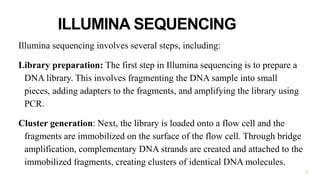
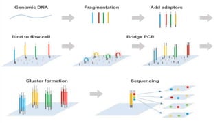
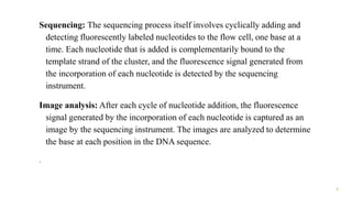
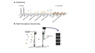
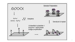
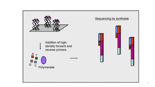
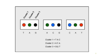
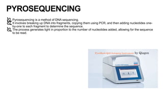
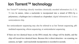
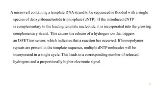
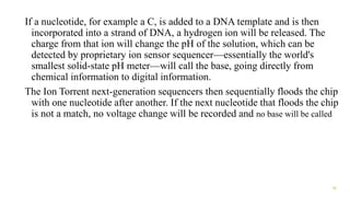
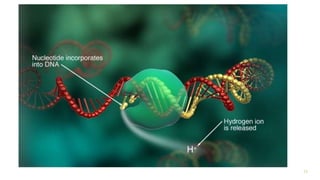
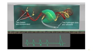
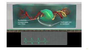
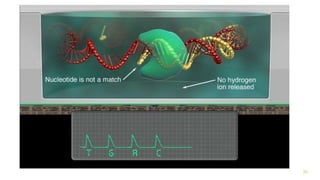
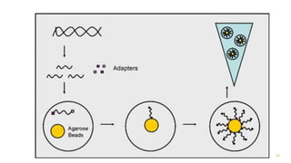
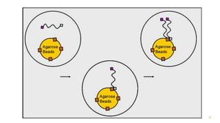
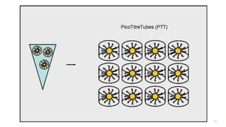
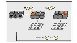
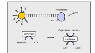
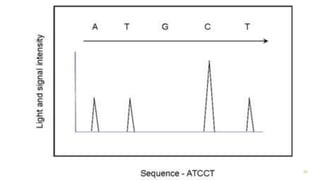
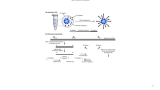
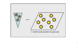
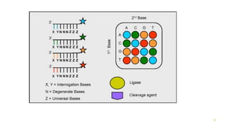
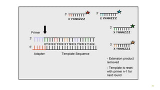
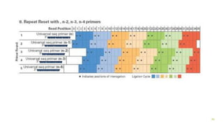
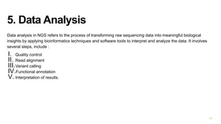
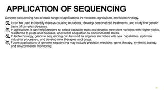
Next generation sequencing
strategies
1
ILLUMINA SEQUENCING
Illumina sequencing involves several steps, including:
Library preparation: The first step in Illumina sequencing is to prepare a
DNA library. This involves fragmenting the DNA sample into small
pieces, adding adapters to the fragments, and amplifying the library using
PCR.
Cluster generation: Next, the library is loaded onto a flow cell and the
fragments are immobilized on the surface of the flow cell. Through bridge
amplification, complementary DNA strands are created and attached to the
immobilized fragments, creating clusters of identical DNA molecules.
2
3
Sequencing: The sequencing process itself involves cyclically adding and
detecting fluorescently labeled nucleotides to the flow cell, one base at a
time. Each nucleotide that is added is complementarily bound to the
template strand of the cluster, and the fluorescence signal generated from
the incorporation of each nucleotide is detected by the sequencing
instrument.
Image analysis: After each cycle of nucleotide addition, the fluorescence
signal generated by the incorporation of each nucleotide is captured as an
image by the sequencing instrument. The images are analyzed to determine
the base at each position in the DNA sequence.
.
4
Data analysis: Finally, the raw sequencing data is processed and analyzed to
generate a final DNA sequence for each fragment in the library. This involves
base calling, alignment to a reference genome or de novo assembly, and
identification of genetic variants, mutations, or other genomic features
5
6
7
8
9
PYROSEQUENCING
Pyrosequencing is a method of DNA sequencing.
It involves breaking up DNA into fragments, copying them using PCR, and then adding nucleotides one-
by-one to each fragment to determine the sequence.
The process generates light in proportion to the number of nucleotides added, allowing for the sequence
to be read.
10
PyroMark Q48 Autoprep Instrument by Qiagen
Pyrosequencing steps:
1. DNA is broken up into fragments of around 100 base pairs of
single-strand DNA.
2. Polymerase chain reaction (PCR) is run to create millions of
identical copies of each DNA fragment, which are split across
thousands of wells, with just one type of DNA fragment per well.
3. DNA fragments are incubated with DNA polymerase, ATP
sulfurylase, apyrase enzymes, and adenosine 5’ phosphosulfate
and luciferin substrates.
4. One of the four types of nucleotides that make up DNA is added
to the wells, which begin to be incorporated onto the single-strand
DNA template by DNA polymerase at the 3’ end, releasing
pyrophosphate.
5. ATP sulfurylase then converts pyrophosphate to adenosine
triphosphate (ATP) in the presence of adenosine 5’
phosphosulfate. 11
6. ATP then takes part in the luciferase-mediated conversion of luciferin to
oxyluciferin, emitting light proportionately to the amount of ATP taking part in
the conversion, which is picked up by a detector.
7. Unused nucleotides and ATP degrade to apyrase, allowing the reaction to
start again with another nucleotide. This process is repeated, adding each
nucleotide one after the other until the synthesis is complete.
8. A detector picks up the intensity of light emitted by the process, which is
then used to infer the number and type of nucleotides added.
12
13
Ion Torrent™ technology
Ion Torrent™ technology directly translates chemically encoded information (A, C, G,
T) into In nature, when a nucleotide is incorporated into a strand of DNA by a
polymerase, a hydrogen ion is released as a byproduct. digital information (0, 1) on a
semiconductor chip.
Ion semiconductor sequencing may also be referred to as Ion Torrent sequencing, pH-
mediated sequencing, silicon sequencing, or semiconductor sequencing.
If there are two identical bases on the DNA strand, the voltage will be double, and the
chip will record two identical bases. Because this is direct detection—no scanning, no
cameras, no light—each nucleotide incorporation is recorded in seconds. 14
A microwell containing a template DNA strand to be sequenced is flooded with a single
species of deoxyribonucleotide triphosphate (dNTP). If the introduced dNTP
is complementary to the leading template nucleotide, it is incorporated into the growing
complementary strand. This causes the release of a hydrogen ion that triggers
an ISFET ion sensor, which indicates that a reaction has occurred. If homopolymer
repeats are present in the template sequence, multiple dNTP molecules will be
incorporated in a single cycle. This leads to a corresponding number of released
hydrogens and a proportionally higher electronic signal.
15
If a nucleotide, for example a C, is added to a DNA template and is then
incorporated into a strand of DNA, a hydrogen ion will be released. The
charge from that ion will change the pH of the solution, which can be
detected by proprietary ion sensor sequencer—essentially the world's
smallest solid-state pH meter—will call the base, going directly from
chemical information to digital information.
The Ion Torrent next-generation sequencers then sequentially floods the chip
with one nucleotide after another. If the next nucleotide that floods the chip
is not a match, no voltage change will be recorded and no base will be called
16
17
18
19
20
454 Sequencing
Essentially, DNA is fragmented, joined to adapters at either end of the
fragmented DNA, amplified in an emulsion PCR (includes 1 μm
agarose bead with complimentary adaptors to fragmented DNA),
PCR amplified allowing up to 1 million identical fragments around
one bead and finally dropped into a PicoTitreTube (PTT). It is here
where the reaction of fluorescence occurs with the addition of
nucleotides. The intensity is read proportional to the number of
homo-polymeric bases added.
21
The DNA is broken up into fragments of around 400 to 600 base pairs using
restriction enzymes that ‘cut’ the DNA at specific points.
Short sequences of DNA called adaptors , are attached to the DNA
fragments.
Tiny resin beads are added to the mix.
DNA sequences on the beads are complementary to sequences on the
adaptors, allowing the DNA fragments to bind directly to the beads,
ideally one fragment to each bead.
22
When the DNA fragments attach to the DNA on the beads the bonds
joining the double-strand together, break and the strands separate,
becoming single-stranded DNA.
The fragments of DNA are then copied numerous times on each bead by
polymerase chain reaction (PCR) . This creates millions of identical
copies of the DNA sequence.
The beads are then filtered to remove any that have either failed to attach to
any DNA or contain more than one type of DNA fragment.
23
Then, the remaining beads are put into wells on a sequencing plate (one bead per well)
along with enzyme beads that contain the DNA polymerase and primer needed for the
sequencing reaction
The polymerase enzyme and primer attach to the DNA fragments on the beads.
Nucleotide bases are added to the wells in waves of one type of base at a time: a wave of
As, followed by a wave of Cs, followed by Gs, followed by Ts.
When each base is incorporated into the DNA, light is given out and this is recorded by a
camera.
The intensity of the light corresponds to the number of nucleotides of the same type that
have been incorporated. For example, if there are three consecutive As in the fragment,
the amount of light generated would be three times that of a single A in the fragment.By
plotting this pattern of light intensity on a graph, the sequence of the original piece of
DNA can be decoded. 24
25
26
27
28
29
30
SOLiD sequencing
SOLiD (Sequencing by Oligonucleotide Ligation and Detection) technology is
a next-generation DNA sequencing method that was developed by Applied
Biosystems (now part of Thermo Fisher Scientific). It is based on a ligation-
based sequencing approach, where short DNA fragments are first ligated to
adaptors that contain a unique barcode sequence, and then amplified by
emulsion PCR. The amplified DNA fragments are then hybridized to a
sequencing slide that contains millions of immobilized oligonucleotides, each
with a specific sequence complementary to one of the four possible
nucleotides (A, C, G, or T).
31
Library preparation: The first step in SOLiD sequencing is the preparation
of a library of DNA fragments. This is typically done by fragmenting the
DNA into small pieces and ligating adaptors that contain a unique barcode
sequence to the ends of the fragments. The barcodes allow multiple
samples to be sequenced simultaneously on the same slide.
Emulsion PCR: The library of DNA fragments is then amplified using
emulsion PCR, where the fragments are partitioned into millions of tiny
water droplets and amplified in parallel. This amplification step generates
a clonal population of DNA fragments, each containing multiple copies of
the same DNA sequence.
32
Bead loading: The amplified DNA fragments are then immobilized onto
small beads and hybridized to a sequencing slide that contains millions of
oligonucleotide probes, each representing a different position in the
genome.
Sequencing-by-ligation: SOLiD sequencing uses a ligation-based
sequencing-by-synthesis approach. The sequencing process starts with
hybridization of the first pair of fluorescently-labeled oligonucleotide
probes to the immobilized bead, which will form a ligation product if the
correct bases are present. The sequence of the first base is read by
detecting the fluorescent signal emitted by the ligated probes.
33
Color detection: After the first base is read, the fluorescent tags are cleaved
off, and the next pair of oligonucleotide probes is hybridized and ligated to
the bead. The fluorescent color emitted by the ligated probes indicates the
identity of the second base in the sequence. This process continues until the
entire sequence of the DNA fragment is determined.
Data analysis: Once the sequencing is complete, the resulting sequence reads
are analyzed to reconstruct the original DNA sequence. This involves
aligning the sequence reads to a reference genome or assembling the reads de
novo to create a new genome or transcriptome assembly.
34
35
36
37
38
39
4. Base calling
Base calling is the process of translating raw sequencing data, which is in the form of fluorescent signals,
into readable DNA sequence data.
During base calling, the fluorescent signals generated by the sequencing reaction are translated into a
sequence of A's, C's, G's, and T's.
Base calling accuracy is crucial to the success of NGS experiments and can be affected by factors such as
sequencing errors, quality of the sequencing data, and the choice of base-calling software
40
5. Data Analysis
Data analysis in NGS refers to the process of transforming raw sequencing data into meaningful biological
insights by applying bioinformatics techniques and software tools to interpret and analyze the data. It involves
several steps, include :
I. Quality control
II. Read alignment
III.Variant calling
IV.Functional annotation
V. Interpretation of results.
41
APPLICATION OF SEQUENCING
Genome sequencing has a broad range of applications in medicine, agriculture, and biotechnology.
It can be used to identify disease-causing mutations, develop personalized treatments, and study the genetic
basis of complex diseases.
In agriculture, it can help breeders to select desirable traits and develop new plant varieties with higher yields,
resistance to pests and diseases, and better adaptation to environmental stress.
In biotechnology, genome sequencing can be used to engineer microbes with new capabilities, optimize
industrial processes, and develop new therapies and drugs.
Future applications of genome sequencing may include precision medicine, gene therapy, synthetic biology,
and environmental monitoring.
42
CONCLUSION
Sequence assembly methods use computational techniques to reconstruct
complete sequences from smaller fragments of DNA, RNA, or protein
through aligning and merging short reads.
NGS involves sample preparation, sequencing, base calling, alignment,
variant calling, and data analysis, resulting in the generation of large
amounts of sequencing data that can be used for a variety of applications
in biological research.
Illumina sequencing is a process of determining the sequence of DNA by
breaking it into small fragments, attaching adaptors, replicating clusters,
adding fluorescent nucleotides, and analyzing the sequence base-by-base
to identify changes in the DNA.
Pyrosequencing is a DNA sequencing method that involves fragmenting
DNA, copying it using PCR, adding nucleotides one-by-one to each
fragment, and generating light to determine the sequence. 43
Reference
Quail, M. A., Swerdlow, H., & Turner, D. J. (2009, July). Improved Protocols for the Illumina Genome
Analyzer Sequencing System. Current Protocols in Human Genetics, 62(1).
https://doi.org/10.1002/0471142905.hg1802s62
Head, S. R., Komori, H. K., LaMere, S. A., Whisenant, T., Van Nieuwerburgh, F., Salomon, D. R., &
Ordoukhanian, P. (2014, February). Library construction for next-generation sequencing: Overviews and
challenges. BioTechniques, 56(2), 61–77. https://doi.org/10.2144/000114133
Syed, F., Grunenwald, H., & Caruccio, N. (2009, October). Optimized library preparation method for next-
generation sequencing. Nature Methods, 6(10), i–ii. https://doi.org/10.1038/nmeth.f.269
Fakruddin, Md, and Abhijit Chowdhury. “Pyrosequencing-An Alternative to Traditional Sanger
Sequencing.” American Journal of Biochemistry and Biotechnology, vol. 8, no. 1, 2012, pp. 14–20.,
doi:10.3844/ajbbsp.2012.14.20.
44

next generation sequencing strategies..pptx

  • 1.
  • 2.
    ILLUMINA SEQUENCING Illumina sequencinginvolves several steps, including: Library preparation: The first step in Illumina sequencing is to prepare a DNA library. This involves fragmenting the DNA sample into small pieces, adding adapters to the fragments, and amplifying the library using PCR. Cluster generation: Next, the library is loaded onto a flow cell and the fragments are immobilized on the surface of the flow cell. Through bridge amplification, complementary DNA strands are created and attached to the immobilized fragments, creating clusters of identical DNA molecules. 2
  • 3.
  • 4.
    Sequencing: The sequencingprocess itself involves cyclically adding and detecting fluorescently labeled nucleotides to the flow cell, one base at a time. Each nucleotide that is added is complementarily bound to the template strand of the cluster, and the fluorescence signal generated from the incorporation of each nucleotide is detected by the sequencing instrument. Image analysis: After each cycle of nucleotide addition, the fluorescence signal generated by the incorporation of each nucleotide is captured as an image by the sequencing instrument. The images are analyzed to determine the base at each position in the DNA sequence. . 4
  • 5.
    Data analysis: Finally,the raw sequencing data is processed and analyzed to generate a final DNA sequence for each fragment in the library. This involves base calling, alignment to a reference genome or de novo assembly, and identification of genetic variants, mutations, or other genomic features 5
  • 6.
  • 7.
  • 8.
  • 9.
  • 10.
    PYROSEQUENCING Pyrosequencing is amethod of DNA sequencing. It involves breaking up DNA into fragments, copying them using PCR, and then adding nucleotides one- by-one to each fragment to determine the sequence. The process generates light in proportion to the number of nucleotides added, allowing for the sequence to be read. 10 PyroMark Q48 Autoprep Instrument by Qiagen
  • 11.
    Pyrosequencing steps: 1. DNAis broken up into fragments of around 100 base pairs of single-strand DNA. 2. Polymerase chain reaction (PCR) is run to create millions of identical copies of each DNA fragment, which are split across thousands of wells, with just one type of DNA fragment per well. 3. DNA fragments are incubated with DNA polymerase, ATP sulfurylase, apyrase enzymes, and adenosine 5’ phosphosulfate and luciferin substrates. 4. One of the four types of nucleotides that make up DNA is added to the wells, which begin to be incorporated onto the single-strand DNA template by DNA polymerase at the 3’ end, releasing pyrophosphate. 5. ATP sulfurylase then converts pyrophosphate to adenosine triphosphate (ATP) in the presence of adenosine 5’ phosphosulfate. 11
  • 12.
    6. ATP thentakes part in the luciferase-mediated conversion of luciferin to oxyluciferin, emitting light proportionately to the amount of ATP taking part in the conversion, which is picked up by a detector. 7. Unused nucleotides and ATP degrade to apyrase, allowing the reaction to start again with another nucleotide. This process is repeated, adding each nucleotide one after the other until the synthesis is complete. 8. A detector picks up the intensity of light emitted by the process, which is then used to infer the number and type of nucleotides added. 12
  • 13.
  • 14.
    Ion Torrent™ technology IonTorrent™ technology directly translates chemically encoded information (A, C, G, T) into In nature, when a nucleotide is incorporated into a strand of DNA by a polymerase, a hydrogen ion is released as a byproduct. digital information (0, 1) on a semiconductor chip. Ion semiconductor sequencing may also be referred to as Ion Torrent sequencing, pH- mediated sequencing, silicon sequencing, or semiconductor sequencing. If there are two identical bases on the DNA strand, the voltage will be double, and the chip will record two identical bases. Because this is direct detection—no scanning, no cameras, no light—each nucleotide incorporation is recorded in seconds. 14
  • 15.
    A microwell containinga template DNA strand to be sequenced is flooded with a single species of deoxyribonucleotide triphosphate (dNTP). If the introduced dNTP is complementary to the leading template nucleotide, it is incorporated into the growing complementary strand. This causes the release of a hydrogen ion that triggers an ISFET ion sensor, which indicates that a reaction has occurred. If homopolymer repeats are present in the template sequence, multiple dNTP molecules will be incorporated in a single cycle. This leads to a corresponding number of released hydrogens and a proportionally higher electronic signal. 15
  • 16.
    If a nucleotide,for example a C, is added to a DNA template and is then incorporated into a strand of DNA, a hydrogen ion will be released. The charge from that ion will change the pH of the solution, which can be detected by proprietary ion sensor sequencer—essentially the world's smallest solid-state pH meter—will call the base, going directly from chemical information to digital information. The Ion Torrent next-generation sequencers then sequentially floods the chip with one nucleotide after another. If the next nucleotide that floods the chip is not a match, no voltage change will be recorded and no base will be called 16
  • 17.
  • 18.
  • 19.
  • 20.
  • 21.
    454 Sequencing Essentially, DNAis fragmented, joined to adapters at either end of the fragmented DNA, amplified in an emulsion PCR (includes 1 μm agarose bead with complimentary adaptors to fragmented DNA), PCR amplified allowing up to 1 million identical fragments around one bead and finally dropped into a PicoTitreTube (PTT). It is here where the reaction of fluorescence occurs with the addition of nucleotides. The intensity is read proportional to the number of homo-polymeric bases added. 21
  • 22.
    The DNA isbroken up into fragments of around 400 to 600 base pairs using restriction enzymes that ‘cut’ the DNA at specific points. Short sequences of DNA called adaptors , are attached to the DNA fragments. Tiny resin beads are added to the mix. DNA sequences on the beads are complementary to sequences on the adaptors, allowing the DNA fragments to bind directly to the beads, ideally one fragment to each bead. 22
  • 23.
    When the DNAfragments attach to the DNA on the beads the bonds joining the double-strand together, break and the strands separate, becoming single-stranded DNA. The fragments of DNA are then copied numerous times on each bead by polymerase chain reaction (PCR) . This creates millions of identical copies of the DNA sequence. The beads are then filtered to remove any that have either failed to attach to any DNA or contain more than one type of DNA fragment. 23
  • 24.
    Then, the remainingbeads are put into wells on a sequencing plate (one bead per well) along with enzyme beads that contain the DNA polymerase and primer needed for the sequencing reaction The polymerase enzyme and primer attach to the DNA fragments on the beads. Nucleotide bases are added to the wells in waves of one type of base at a time: a wave of As, followed by a wave of Cs, followed by Gs, followed by Ts. When each base is incorporated into the DNA, light is given out and this is recorded by a camera. The intensity of the light corresponds to the number of nucleotides of the same type that have been incorporated. For example, if there are three consecutive As in the fragment, the amount of light generated would be three times that of a single A in the fragment.By plotting this pattern of light intensity on a graph, the sequence of the original piece of DNA can be decoded. 24
  • 25.
  • 26.
  • 27.
  • 28.
  • 29.
  • 30.
  • 31.
    SOLiD sequencing SOLiD (Sequencingby Oligonucleotide Ligation and Detection) technology is a next-generation DNA sequencing method that was developed by Applied Biosystems (now part of Thermo Fisher Scientific). It is based on a ligation- based sequencing approach, where short DNA fragments are first ligated to adaptors that contain a unique barcode sequence, and then amplified by emulsion PCR. The amplified DNA fragments are then hybridized to a sequencing slide that contains millions of immobilized oligonucleotides, each with a specific sequence complementary to one of the four possible nucleotides (A, C, G, or T). 31
  • 32.
    Library preparation: Thefirst step in SOLiD sequencing is the preparation of a library of DNA fragments. This is typically done by fragmenting the DNA into small pieces and ligating adaptors that contain a unique barcode sequence to the ends of the fragments. The barcodes allow multiple samples to be sequenced simultaneously on the same slide. Emulsion PCR: The library of DNA fragments is then amplified using emulsion PCR, where the fragments are partitioned into millions of tiny water droplets and amplified in parallel. This amplification step generates a clonal population of DNA fragments, each containing multiple copies of the same DNA sequence. 32
  • 33.
    Bead loading: Theamplified DNA fragments are then immobilized onto small beads and hybridized to a sequencing slide that contains millions of oligonucleotide probes, each representing a different position in the genome. Sequencing-by-ligation: SOLiD sequencing uses a ligation-based sequencing-by-synthesis approach. The sequencing process starts with hybridization of the first pair of fluorescently-labeled oligonucleotide probes to the immobilized bead, which will form a ligation product if the correct bases are present. The sequence of the first base is read by detecting the fluorescent signal emitted by the ligated probes. 33
  • 34.
    Color detection: Afterthe first base is read, the fluorescent tags are cleaved off, and the next pair of oligonucleotide probes is hybridized and ligated to the bead. The fluorescent color emitted by the ligated probes indicates the identity of the second base in the sequence. This process continues until the entire sequence of the DNA fragment is determined. Data analysis: Once the sequencing is complete, the resulting sequence reads are analyzed to reconstruct the original DNA sequence. This involves aligning the sequence reads to a reference genome or assembling the reads de novo to create a new genome or transcriptome assembly. 34
  • 35.
  • 36.
  • 37.
  • 38.
  • 39.
  • 40.
    4. Base calling Basecalling is the process of translating raw sequencing data, which is in the form of fluorescent signals, into readable DNA sequence data. During base calling, the fluorescent signals generated by the sequencing reaction are translated into a sequence of A's, C's, G's, and T's. Base calling accuracy is crucial to the success of NGS experiments and can be affected by factors such as sequencing errors, quality of the sequencing data, and the choice of base-calling software 40
  • 41.
    5. Data Analysis Dataanalysis in NGS refers to the process of transforming raw sequencing data into meaningful biological insights by applying bioinformatics techniques and software tools to interpret and analyze the data. It involves several steps, include : I. Quality control II. Read alignment III.Variant calling IV.Functional annotation V. Interpretation of results. 41
  • 42.
    APPLICATION OF SEQUENCING Genomesequencing has a broad range of applications in medicine, agriculture, and biotechnology. It can be used to identify disease-causing mutations, develop personalized treatments, and study the genetic basis of complex diseases. In agriculture, it can help breeders to select desirable traits and develop new plant varieties with higher yields, resistance to pests and diseases, and better adaptation to environmental stress. In biotechnology, genome sequencing can be used to engineer microbes with new capabilities, optimize industrial processes, and develop new therapies and drugs. Future applications of genome sequencing may include precision medicine, gene therapy, synthetic biology, and environmental monitoring. 42
  • 43.
    CONCLUSION Sequence assembly methodsuse computational techniques to reconstruct complete sequences from smaller fragments of DNA, RNA, or protein through aligning and merging short reads. NGS involves sample preparation, sequencing, base calling, alignment, variant calling, and data analysis, resulting in the generation of large amounts of sequencing data that can be used for a variety of applications in biological research. Illumina sequencing is a process of determining the sequence of DNA by breaking it into small fragments, attaching adaptors, replicating clusters, adding fluorescent nucleotides, and analyzing the sequence base-by-base to identify changes in the DNA. Pyrosequencing is a DNA sequencing method that involves fragmenting DNA, copying it using PCR, adding nucleotides one-by-one to each fragment, and generating light to determine the sequence. 43
  • 44.
    Reference Quail, M. A.,Swerdlow, H., & Turner, D. J. (2009, July). Improved Protocols for the Illumina Genome Analyzer Sequencing System. Current Protocols in Human Genetics, 62(1). https://doi.org/10.1002/0471142905.hg1802s62 Head, S. R., Komori, H. K., LaMere, S. A., Whisenant, T., Van Nieuwerburgh, F., Salomon, D. R., & Ordoukhanian, P. (2014, February). Library construction for next-generation sequencing: Overviews and challenges. BioTechniques, 56(2), 61–77. https://doi.org/10.2144/000114133 Syed, F., Grunenwald, H., & Caruccio, N. (2009, October). Optimized library preparation method for next- generation sequencing. Nature Methods, 6(10), i–ii. https://doi.org/10.1038/nmeth.f.269 Fakruddin, Md, and Abhijit Chowdhury. “Pyrosequencing-An Alternative to Traditional Sanger Sequencing.” American Journal of Biochemistry and Biotechnology, vol. 8, no. 1, 2012, pp. 14–20., doi:10.3844/ajbbsp.2012.14.20. 44