This document discusses several phakomatoses or neurocutaneous syndromes characterized by hamartomas of the skin, eye, central nervous system and other organs. It provides details on the clinical features, genetics, systemic and ophthalmic manifestations as well as management of specific conditions like Neurofibromatosis types 1 and 2, Von Hippel-Lindau disease, Tuberous sclerosis, Sturge-Weber syndrome, and Ataxia telangiectasia. Key aspects like characteristic tumors, genetic mutations, diagnostic criteria and treatment approaches are highlighted for each discussed syndrome.
Progressive multifocal leukoencephalopathy (PML) is a disease of the white matter of the brain, caused by a virus infection that targets cells that make myelin--the material that insulates nerve cells (neurons). Polyomavirus JC (often called JC virus) is carried by a majority of people and is harmless except among those with lowered immune defenses. The disease is rare and occurs in patients undergoing chronic corticosteroid or immunosuppressive therapy for organ transplant, or individuals with cancer (such as Hodgkin’s disease or lymphoma). Individuals with autoimmune conditions such as multiple sclerosis, rheumatoid arthritis, and systemic lupus erythematosus -- some of whom are treated with biological therapies that allow JC virus reactivation -- are at risk for PML as well. PML is most common among individuals with HIV-1 infection / acquired immune deficiency syndrome (AIDS). Currently, the best available therapy is reversal of the immune-deficient state, since there are no effective drugs that block virus infection without toxicity. Reversal may be achieved by using plasma exchange to accelerate the removal of the therapeutic agents that put patients at risk for PML. In the case of HIV-associated PML, immediately beginning anti-retroviral therapy will benefit most individuals. Several new drugs that laboratory tests found effective against infection are being used in PML patients with special permission of the U.S. Food and Drug Administration. Hexadecyloxypropyl-Cidofovir (CMX001) is currently being studied as a treatment option for JVC because of its ability to suppress JVC by inhibiting viral DNA replication.
In general, PML has a mortality rate of 30-50 percent in the first few months following diagnosis but depends on the severity of the underlying disease and treatment received. Those who survive PML can be left with severe neurological disabilities.
Clinically isolated syndromes (CIS) refer to the first clinical episodes of neurological symptoms suggestive of multiple sclerosis. The document discusses CIS in three parts: definition and clinical features of CIS, risk factors for conversion from CIS to multiple sclerosis, and management of CIS. Regarding clinical features, optic neuritis, transverse myelitis, and brainstem syndromes are highlighted as common presentations of CIS. MRI abnormalities, younger age of onset, smoking, and vitamin D deficiency are identified as risk factors for progression to multiple sclerosis. The management section outlines acute treatment with corticosteroids, use of disease-modifying therapies based on MRI findings, and consideration of vitamin D supplementation.
Cerebral microbleeds are small brain hemorrhages detected by MRI that are caused by leakage of blood from damaged small vessel walls. They are increasingly recognized in patients with cerebrovascular disease, Alzheimer's disease, vascular cognitive impairment, and normal elderly populations. Microbleeds in lobar regions may indicate cerebral amyloid angiopathy and link vascular and amyloid neuropathologies, while deep or infratentorial microbleeds often reflect hypertensive vasculopathy. Detection of microbleeds provides insight into cerebral small vessel disease and its relationship to cognitive impairment and dementia.
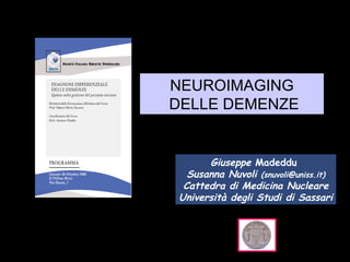
Imaging delle Malattie Neurodegenerative: le DemenzeASMaD
Presentazione a cura del Professor Luigi Mansi - XII° Congresso Nazionale FIMeG 2018 - The Silver Tsunami: l'anziano fra appropriatezza e farmaeconomia
This document provides an overview of approaches to diagnosing leukodystrophies. It begins by defining leukodystrophies and differentiating them from other white matter disorders. Clinical features that suggest a leukodystrophy are described. A 3-step MRI approach is outlined involving identifying symmetric white matter involvement, patterns of involvement, and distinctive features. Common leukodystrophies in adults are discussed in detail including clinical presentation, genetics, imaging findings, and diagnostic testing. The document emphasizes a systematic approach to diagnosis utilizing clinical features, imaging, and ancillary tests.
Cortical dysplasia is a malformation of cortical development caused by abnormal neuronal migration or organization during brain development. It can cause intractable epilepsy and neurodevelopmental disorders like autism. The lecture discusses normal brain development and corticogenesis. It then covers specific malformations including focal cortical dysplasia, describing their histopathology and clinical correlates. Recent research suggests focal disruptions of cortical layering found in children with autism may represent early cortical dysplasia, providing insight into a potential cause of autism.
This document discusses several phakomatoses or neurocutaneous syndromes characterized by hamartomas of the skin, eye, central nervous system and other organs. It provides details on the clinical features, genetics, systemic and ophthalmic manifestations as well as management of specific conditions like Neurofibromatosis types 1 and 2, Von Hippel-Lindau disease, Tuberous sclerosis, Sturge-Weber syndrome, and Ataxia telangiectasia. Key aspects like characteristic tumors, genetic mutations, diagnostic criteria and treatment approaches are highlighted for each discussed syndrome.
Progressive multifocal leukoencephalopathy (PML) is a disease of the white matter of the brain, caused by a virus infection that targets cells that make myelin--the material that insulates nerve cells (neurons). Polyomavirus JC (often called JC virus) is carried by a majority of people and is harmless except among those with lowered immune defenses. The disease is rare and occurs in patients undergoing chronic corticosteroid or immunosuppressive therapy for organ transplant, or individuals with cancer (such as Hodgkin’s disease or lymphoma). Individuals with autoimmune conditions such as multiple sclerosis, rheumatoid arthritis, and systemic lupus erythematosus -- some of whom are treated with biological therapies that allow JC virus reactivation -- are at risk for PML as well. PML is most common among individuals with HIV-1 infection / acquired immune deficiency syndrome (AIDS). Currently, the best available therapy is reversal of the immune-deficient state, since there are no effective drugs that block virus infection without toxicity. Reversal may be achieved by using plasma exchange to accelerate the removal of the therapeutic agents that put patients at risk for PML. In the case of HIV-associated PML, immediately beginning anti-retroviral therapy will benefit most individuals. Several new drugs that laboratory tests found effective against infection are being used in PML patients with special permission of the U.S. Food and Drug Administration. Hexadecyloxypropyl-Cidofovir (CMX001) is currently being studied as a treatment option for JVC because of its ability to suppress JVC by inhibiting viral DNA replication.
In general, PML has a mortality rate of 30-50 percent in the first few months following diagnosis but depends on the severity of the underlying disease and treatment received. Those who survive PML can be left with severe neurological disabilities.
Clinically isolated syndromes (CIS) refer to the first clinical episodes of neurological symptoms suggestive of multiple sclerosis. The document discusses CIS in three parts: definition and clinical features of CIS, risk factors for conversion from CIS to multiple sclerosis, and management of CIS. Regarding clinical features, optic neuritis, transverse myelitis, and brainstem syndromes are highlighted as common presentations of CIS. MRI abnormalities, younger age of onset, smoking, and vitamin D deficiency are identified as risk factors for progression to multiple sclerosis. The management section outlines acute treatment with corticosteroids, use of disease-modifying therapies based on MRI findings, and consideration of vitamin D supplementation.
Cerebral microbleeds are small brain hemorrhages detected by MRI that are caused by leakage of blood from damaged small vessel walls. They are increasingly recognized in patients with cerebrovascular disease, Alzheimer's disease, vascular cognitive impairment, and normal elderly populations. Microbleeds in lobar regions may indicate cerebral amyloid angiopathy and link vascular and amyloid neuropathologies, while deep or infratentorial microbleeds often reflect hypertensive vasculopathy. Detection of microbleeds provides insight into cerebral small vessel disease and its relationship to cognitive impairment and dementia.
Imaging delle Malattie Neurodegenerative: le DemenzeASMaD
Presentazione a cura del Professor Luigi Mansi - XII° Congresso Nazionale FIMeG 2018 - The Silver Tsunami: l'anziano fra appropriatezza e farmaeconomia
This document provides an overview of approaches to diagnosing leukodystrophies. It begins by defining leukodystrophies and differentiating them from other white matter disorders. Clinical features that suggest a leukodystrophy are described. A 3-step MRI approach is outlined involving identifying symmetric white matter involvement, patterns of involvement, and distinctive features. Common leukodystrophies in adults are discussed in detail including clinical presentation, genetics, imaging findings, and diagnostic testing. The document emphasizes a systematic approach to diagnosis utilizing clinical features, imaging, and ancillary tests.
Cortical dysplasia is a malformation of cortical development caused by abnormal neuronal migration or organization during brain development. It can cause intractable epilepsy and neurodevelopmental disorders like autism. The lecture discusses normal brain development and corticogenesis. It then covers specific malformations including focal cortical dysplasia, describing their histopathology and clinical correlates. Recent research suggests focal disruptions of cortical layering found in children with autism may represent early cortical dysplasia, providing insight into a potential cause of autism.
Young onset dementia (YOD) refers to dementia with an onset before age 65. About 5% of all dementias are YOD. Common causes include Alzheimer's disease, vascular dementia, frontotemporal lobar degeneration, and dementia with Lewy bodies. A thorough evaluation includes medical history, physical and neurological exams, imaging like MRI and PET, and may involve genetic testing. Management focuses on treating underlying causes if possible, addressing behavioral and psychiatric symptoms, and providing social support. Prognosis varies by the specific cause but on average YOD results in 10-15 years shorter life expectancy than later onset dementia.
This document provides diagnostic criteria for chronic inflammatory demyelinating polyneuropathy (CIDP), including:
1) Clinical criteria for typical and atypical CIDP with inclusion/exclusion factors.
2) Definite, probable, and possible electrophysiological criteria involving compound muscle action potential tests.
3) Supportive diagnostic criteria including cerebrospinal fluid analysis, MRI findings, nerve conduction studies, and nerve biopsy results.
It also outlines inclusion/exclusion criteria and supportive criteria specifically for diagnosing pure sensory CIDP without motor involvement.
This document discusses highly active multiple sclerosis (MS). It defines several subtypes of aggressive MS including malignant, fulminant, and highly active MS. Predictors of highly active MS are discussed from a 2016 study. The case vignette describes a 34-year-old male physician's MS course from 2006 to 2017, showing recurrent attacks and progression. The timing of therapy is key to preventing disability, with an emphasis on early treatment to preserve brain reserve. Treatment algorithms recommend escalating therapy for aggressive MS to effectively treat within a narrow therapeutic window.
Structural neuroimaging plays an important role in the assessment and diagnosis of different types of dementia. For Alzheimer's disease, MRI typically shows atrophy of the medial temporal lobes including the hippocampus. Vascular dementia is characterized by multiple brain infarcts visible on CT or MRI. Frontotemporal dementia demonstrates frontal and temporal lobe atrophy that can be asymmetric. Dementia with Lewy bodies may show mild generalized atrophy on MRI with occipital hypometabolism on PET. Scales like the MTA scale are used to quantify hippocampal atrophy, while MRS can detect metabolic changes in dementia. Neuroimaging thus aids in distinguishing dementia subtypes and excluding other pathological conditions.
This document discusses various vascular and demyelinating syndromes of the brainstem. It describes several syndromes defined by their anatomical location in the midbrain, pons or specific vascular territories involved. These include Weber's syndrome, Claude syndrome, Benedikt syndrome, and Nothnagel's syndrome in the midbrain as well as Millard-Gubler syndrome, Raymond syndrome, lateral and medial pontine syndromes, and Locked-in syndrome in the pons. Each syndrome is characterized by the neurological deficits caused by lesions to specific brainstem structures. The vascular supply and clinical features of each syndrome are concisely outlined.
This document discusses progressive myoclonus epilepsy (PME), which consists of myoclonic seizures, tonic-clonic seizures, and progressive neurological dysfunction like ataxia and dementia. The main causes of PME include Unverricht-Lundborg disease, myoclonic epilepsy with ragged-red fiber syndrome, Lafora body disease, neuronal ceroid lipofuscinoses, and sialidoses. Each of these disorders is described in detail, outlining their characteristic symptoms, age of onset, genetic basis, diagnostic criteria, management approaches, and other relevant clinical information. Rare causes of PME like dentatorubral-pallidoluysian atrophy and non
Neurofibromatosis (NF) is a genetic disorder that causes tumors to grow on nerves. There are two types, NF1 and NF2, with NF2 generally more severe as it affects the central nervous system. NF is caused by changes in the NF1 or NF2 genes and has autosomal dominant inheritance. Symptoms vary but can include skin spots, nerve tumors, pain, and learning disabilities. While there is no cure, treatments include surgery to remove tumors and chemotherapy if tumors become cancerous. NF affects people differently and can impact daily life through physical problems, pain, or learning disabilities.
This document provides an overview of eye anatomy and physiology as well as clinical assessment of the eye. It discusses the anatomy of the eyelids, muscles, nerves, and retina. It describes types of eye movements including saccades, smooth pursuit, and convergence. Reflexes like the light and accommodation reflex are also covered. The document outlines how to examine the eyes, eyelids, conjunctiva, visual acuity, fields, movements, and pupils. Causes and features of cranial nerve palsies are summarized.
This document provides information on the surgical management of normal pressure hydrocephalus (NPH). It discusses the history and examination findings suggestive of NPH, describes imaging techniques used for diagnosis, and outlines special tests that can aid in evaluation. It also reviews the options for surgical treatment, including ventriculoperitoneal, lumboperitoneal, and endoscopic third ventriculostomy procedures. Factors influencing shunt responsiveness are summarized, and guidelines for proceeding to shunt placement based on diagnostic classification are presented.
This document discusses different types of intradural spinal tumors, including their presentation, diagnosis, and management. The main types discussed are:
- Intradural extramedullary tumors (40%) like meningiomas, schwannomas, and neurofibromas.
- Intramedullary spinal cord tumors (5%) such as astrocytomas and ependymomas.
- Meningiomas are the most common, often occurring in middle-aged women. Diagnosis is typically made using MRI and surgical excision is the main treatment. Prognosis depends on tumor type, with complete resection generally resulting in low recurrence rates.
A 53-year-old female presented with a 4-month history of left periorbital headache and frontal parasthesia. Imaging showed a 1cm enhancing lesion in the cavernous sinus. She was initially treated for tuberculosis but did not improve. Further workup revealed positive Mantoux test but negative CSF studies. Her symptoms improved 90% with a short course of antibiotics and steroids. The differential diagnosis includes cavernous sinus syndrome such as Tolosa-Hunt syndrome, an idiopathic inflammatory condition. A repeat MRI after treatment showed reduction in the size of the lesion, consistent with inflammatory pseudotumor.
Autoimmune encephalitis current conceptsNeurologyKota
1) Autoimmune encephalitis is a debilitating neurological disorder caused by inflammation of the brain. It develops subacutely over weeks and can affect individuals of all ages.
2) It has diverse clinical manifestations and immunological associations. Identification of neural autoantibodies has led to classification of different subtypes.
3) Prominent among these are anti-NMDAR encephalitis commonly seen in young women and children, autoimmune limbic encephalitis, and other syndromes associated with antibodies targeting neuronal cell-surface and intracellular antigens.
Ophthalmodynamometry is a clinical procedure that measures the pressure in the ophthalmic artery to assess patency of the internal carotid artery. It involves applying pressure to the eye until the central retinal artery collapses, noting the diastolic and systolic pressures. This provides information about blood flow through the ophthalmic artery and can detect carotid artery occlusive diseases, helping prevent strokes. The procedure has been used since the early 1900s and modern methods include compression or suction ophthalmodynamometry. Precautions must be taken and it can detect conditions affecting the carotid or cerebral vasculature.
The document defines phakomatoses as multisystem disorders involving the central nervous system, eyes, and skin that cause characteristic lesions and hamartomas. It then describes several common and uncommon phakomatoses syndromes in detail, focusing on their defining clinical features, inheritance patterns, prevalence, and ocular manifestations. The most prominent syndromes discussed are neurofibromatosis types 1 and 2, tuberous sclerosis, Von Hippel-Lindau disease, Sturge-Weber syndrome, and Wyburn-Mason syndrome.
BRAINSTEM LESION INVOLVING 3rd,4th and 6th cranial nerveShivshankar Badole
This document discusses various brainstem lesions that can cause cranial nerve palsies involving the 3rd, 4th, and 6th nerves. It describes the anatomy and functions of these cranial nerves. It then summarizes several clinical syndromes that can result from lesions in specific brainstem locations, including Weber syndrome, Benedikt's syndrome, internuclear ophthalmoplegia, and Parinaud's syndrome. The localization of lesions and characteristic clinical findings for each syndrome are provided.
This document provides information on orbital apex syndrome (OAS) and related conditions. It begins with an overview of applied anatomy of the superior orbital fissure and orbital apex. It then discusses the classification of OAS, cavernous sinus syndrome, and superior orbital fissure syndrome. The clinical presentation, etiology, and management of these conditions is summarized. Common causes include tumors, infections, inflammation, and vascular abnormalities. The document provides details on specific pathologies, treatments, and outcomes.
This document provides information on idiopathic orbital inflammation (IOI), also known as orbital pseudotumor. It is a heterogeneous group of disorders characterized by orbital inflammation without an identifiable cause. The document discusses the classification, symptoms, signs, imaging findings on CT and MRI, and different clinical presentations of IOI including myositis, dacryoadenitis, cellulitis, optic perineuritis, periscleritis, and focal masses. Differential diagnoses are also provided for each clinical presentation.
Horner syndrome results from disruption of the sympathetic nerve supply to the eye. It is characterized by three key signs: ptosis, miosis, and anhidrosis. The syndrome can be classified based on where along the sympathetic pathway the disruption occurs. Testing involves confirming the diagnosis with apraclonidine, which causes reversal of miosis and ptosis in denervated eyes. Investigations depend on classification and may include imaging of the brain, cervical spine, chest, or neck vessels to identify underlying causes.
This document discusses central nervous system vasculitis, including its classification, diagnosis, and treatment. It covers primary angiitis of the CNS and secondary causes. Diagnosis is challenging due to non-specific symptoms and lack of sensitive tests. Evaluation involves clinical assessment, CSF analysis, neuroimaging, and cerebral angiography. Treatment depends on the specific type but often involves immunosuppressants like cyclophosphamide with glucocorticoids. Pathology evaluation can help in difficult cases but has low sensitivity.
Presentation1.pptx, radiological imaging of dementia.Abdellah Nazeer
This document discusses radiological imaging findings in various types of dementia. It provides examples of MRI, FDG-PET, and amyloid PET scans showing characteristic patterns of atrophy and hypometabolism in conditions like Alzheimer's disease, frontotemporal dementia, Creutzfeldt-Jakob disease, and others. Medial temporal lobe atrophy on MRI is highlighted as an important tool for the diagnosis of Alzheimer's, and FDG-PET can help differentiate Alzheimer's from frontotemporal dementia based on patterns of hypometabolism. Magnetic resonance spectroscopy is also discussed as a tool for assessing metabolic changes in Alzheimer's patients.
Webinar | Gentlecare a domicilio per la gestione del malato di Alzheimer: str...Obiettivo Psicologia Srl
La Psicologia dell’Invecchiamento è la disciplina che si occupa delle problematiche di tipo psicologico e neuropsicologico dell’anziano nel corso del processo di invecchiamento.
Sempre più spesso lo psicologo che lavora con gli anziani si trova a contatto con persone affette da demenza, quasi sempre di tipo Alzheimer e con familiari e/o operatori, che necessitano di un aiuto nella gestione dei disturbi comportamentali e dei deficit cognitivi delle persone che assistono. Progettare interventi nel tentativo di ridurre l’impatto che questi disturbi hanno sul quotidiano del malato e di chi se ne occupa significa, innanzitutto, riconoscere fisiologia e patologia della malattia di alzheimer e saperne sfruttare i vari aspetti a vantaggio del mantenimento delle abilità della persona.
Il metodo Gentlecare, ideato da Moyra Jones, si basa su tre punti fermi, persone, programmi e spazio, che contraddistinguono l’esistenza del malato di Alzheimer. Agire su questi tre punti in maniera consapevole significa migliorare di molto la qualità della vita del paziente e di chi lo assiste. Insegnare alle persone come comunicare, cosa dire, come programmare le giornate e in che maniera adattare l’ambiente fisico e psicologico attorno alla persona malata significa fornire una protesi ben più robusta di un deambulatore o di un sollevatore meccanico. A partire da una buona checklist, infatti, è possibile, al domicilio e nelle strutture per anziani, fornire consulenze adeguate sulla costituzione degli ambienti, la disposizione dei mobili, la scelta dei colori e delle suppellettili più utili e protesiche per la persona affetta da demenza.
Capisce una persona malata di alzheimer cosa le sta accadendo?... Idee intorno alla malattia proposte da Letizia Espanoli, formatrice in area sociosanitaria educativa, Centro Studi Internazionale Perusini Alzheimer
Focus sulla Malattia di Alzheimer nell'ottica d'approccio infermieristico.
Sommario.
Demenza Senile:
- Definizione
Letteratura Scientifica:
- Cenni storici
Epidemiologia:
- Diffusione ed incidenza
Patologia:
- Caratteristiche e modalità d'azione
- Fattori di rischio
Quadro clinico:
- Fasi della malattia
- Fase Iniziale
- Fase Intermedia
- Fase Terminale
- Schema di progressione patologica
Diagnosi:
- Anamnesi e segni oggettivi
- Strumentistica diagnostica
- Test valutativi ed istologici
Terapia:
- Trattamenti terapeutici
- Trattamento farmacologico
- Trattamento basato sui training
- Tipologie training
- Trattamenti integrativi
- Ruolo Caregiver
Prevenzione:
- Fattori modificabili
- Teoria della riserva cognitiva
Assistenza Infermieristica:
- Presa in carico del paziente
Slides presentate dal dr Giovanni Brondani, SOS di DPT Radiologia Ugenza ed Emergenza, AOU Santa Maria della Misericordia di Udine, in occasione del corso "Le malattie neuromuscolari", Udine 16 dicembre 2013.
3. Linee Guida sulla diagnosi di demenza e di malattia di Alzheimer * Ufficialmente approvate dalla SIN (2000). SECONDA FASE - FASE DI CONFERMA DIAGNOSTICA E DIAGNOSI DIFFERENZIALE Neuroimaging cerebrale Gli esami di neuroimaging cerebrale dovrebbero essere presi in considerazione in base alle caratteristiche cliniche di presentazione. Sembra comunque ragionevole eseguire un esame CT scan o MRI cerebrale almeno al momento della prima diagnosi. Questo esame è infatti spesso indispensabile per una corretta diagnosi differenziale. Altri esami come la SPECT o la PET, che possono fornire informazioni sullo stato funzionale cerebrale, sono di grande interesse per fini di ricerca e andranno utilizzati all'interno di protocolli di ricerca.
4. Malattie neurodegenerative Ricorso ad un ampio spettro di informazioni che consentano di formulare l’ipotesi diagnostica più probabile e di adottare i protocolli terapeutici più adeguati. Definizione eziopatogenetica “ DEMENZA” Conferma istopatologica
5.
6. dati economici Costo globale complessivo di 315 miliardi di dollari/anno nei paesi avanzati
8. capacità di valutare in vivo le alterazioni morfologiche, funzionali ed ultrastrutturali tipiche di una determinata patologia, costituendo in tal modo un valido e sicuro supporto al clinico…… …… .. RAZIONALE nell’’utilizzo del neuroimaging in fase di diagnosi iniziale FUNZIONALE (medico nucleare) MORFOLOGICO (radiologico)
12. MISURE LINEARI DEL SISTEMA VENTRICOLARE E DEI SOLCHI A =ampiezza del III ventricolo B = somma delle distanze più brevi tra nucleo caudato ed estremità anteriore del setto pellucido C = ampiezza dei ventricoli laterali davanti al forame di Monrow D = diametro trasverso minimo dei ventricoli laterali a livello delle celle medie VS(punteggio ventricolare) = A+ B+ C+ D diametro interparietale IMAGING Morfologico Radiologico VALUTAZIONE IN VIVO DELLA ATROFIA CORTICALE TC
13. ……… Nessuna misura lineare di atrofia globale è risultata utile per la diagnosi di demenza. IMAGING Morfologico Radiologico
14. MISURE LINEARI DI ATROFIA REGIONALE Atrofia delle strutture del lobo temporale mediale (MTL: ippocampo,amigdala,corteccia entorinale): IMAGING Morfologico Radiologico VALUTAZIONE IN VIVO DELLA ATROFIA CORTICALE (TC/RM) 1 =ampiezza del corno temporale 2 =altezza dell’ippocampo 3 =ampiezza della fessura coroidale
15. IMAGING Morfologico Radiologico VALUTAZIONE QUANTITATIVA ISPETTIVA DI ATROFIA DEL MTL SCALA che attribuisce un punteggio da 0 a 4 all’atrofia dell’ippocampo (da Scheltens e al.1992) punti ampiezza della fessura ampiezza del corno altezza dell'ippocampo coroidale temporale 0 N N N 1 + N N 2 ++ + - 3 +++ ++ - - 4 +++ +++ - - - Dilatazione del corno temporale=espressione dell'atrofia dell'ippocampo
16.
17. IMAGING Morfologico Radiologico CT o RM (da preferire) Maggiore accuratezza rispetto alle misure lineari Impiego clinico: richiede ACCURATEZZA e RIPRODUCIBILITA' Limite maggiore di queste tecniche : delimitazione della regione di interesse ( ROI) in cui effettuare la misura. Disegno manuale (TRACHING) della ROI su immagini CT/RM ( tecniche semiautomatiche : sono risultate spesso inadatte per la complessità tridimensionale delle strutture esaminate) - tempo - allenamento degli operatori - conoscenza dettagliata dell'anatomia tridimensionale - variabilità tra operatori diversi e per lo stesso operatore Tecniche impiegate solo nei centri di ricerca. MISURE VOLUMETRICHE DI ATROFIA
18. IMAGING Morfologico Radiologico ATROFIA limite Non e' stato possibile definire una “ SOGLIA DI NORMALITA'” che possa fornire elementi di supporto alla diagnosi differenziale tra invecchiamento fisiologico e demenza.
19. IMAGING Morfologico Radiologico Aree di iperintensità di segnale alla RM (TC: leucoaraiosi) substrato patologico incerto Riscontrata: encefalopatia sottocorticale di Binswanger (sempre) demenza multinfartuale (frequentemente) malattia di Alzheimer (spesso) nell'invecchiamento fisiologico (spesso) Diagnosi differenziale tra lesioni di natura vascolare e non: - vascolari : lesioni “confluenti”, irregolari, spesso a sede periventricolare - non vascolari : lesioni “puntate”, piccole, rotondeggianti (dilatazione degli spazi perivasali di Virchow-Robin, aumento di H2O interstiziale) Alterazioni della Sostanza Bianca (immagini RM T2w)
24. Accertamento non invasivo dei livelli di alcuni metaboliti nel tessuto cerebrale: -NAA (acetilaspartato) marcatore della funzione neuronale (si riduce in corso di patologie cerebrali) - MI (mio-inositolo) marcatore della gliosi (glial marker) - Cho (colina) marcatore della proliferazione cellulare di membrana. - Creatinina (Cr) espressione di funzione energetica - Lattato (Lac) presente solo per alterazioni del metabolismo Possibilità di impiegare i livelli di alcuni metaboliti come strumento diagnostico per differenziare la AD dalle altre demenze: - in AD <NAA e >MI -in DFT <NAA e >MI ma in sede diverse Strumento di ricerca. IMAGING Morfologico Radiologico TECNICHE NON CONVENZIONALI Spettroscopia RM
27. fMRI BOLD activation in the left and right peri-Sylvian areas when listening to dichotic presentations of CV-syllables. The upper row shows activations for the Drug-naive condition, the lower row shows the corresponding activations for the Memantine condition. Bozzali M. Magnetic Resonance Imaging 25 (2007) 969–977
28. Faraci FM, Circ Res. 1993. Yang ST, Neuroreport . 1998. Brenman JE Cell. 1996 Ma….. Condizione irreversibile Regioni ippocampali in AD (atrofia) MRI: Rilievo morfologico di morte neuronale nella sede iniziale di malattia.
31. IMAGING Funzionale Medico-Nucleare Faraci FM, Circ Res. 1993. Yang ST, Neuroreport . 1998. Brenman JE Cell. 1996
32. IMAGING Funzionale Medico- Nucleare PET/SPET : prima di MRI strutturale convenzionale… In quanto consente rilievo precoce di danno funzionale, prima della morte neuronale, sia nella sede iniziale di malattia che e a distanza nelle efferenze sinaptiche…….. monitoraggio della progressione di malattia
33. La PET con 18FDG e la SPECT di perfusione con 99mTcHMPAO consentono lo studio del METABOLISMO NEURONALE BASALE IMAGING Funzionale Medico- Nucleare Brenman JE J Neurosci . 1996. Perea G, J neural Transm. 2005. Zonta M, Nat Neurosci . 2003. fMRI: valuta i picchi di attivazione metabolica ……… Il 95% della Energia viene consumata quando il cervello è teoricamente a riposo ovvero quando i neuroni comunicano continuamente nella c.d. funzione di plasticità sinaptica Solo il 5% viene consumato durante attivazione a seguito di task specifici PET/SPET: prima di MRI funzionale
34. IMAGING Funzionale Medico- Nucleare inoltre rispetto a Neuroimaging strutturale esiste un altro parametro valutabile: Intensità di captazione del radiofarmaco Nelle immagini fisiologiche tomografiche la densità di informazione (intensità di colore o di grigio) è una funzione NOTA del parametro fisiologico in esame: MISURA di concentrazione del radiofarmaco
35. Voxel-Based Correlation between Coregistered Single-Photon Emission Computed Tomography and Dynamic Susceptibility Contrast Magnetic Resonance Imaging in Subjects with Suspected Alzheimer Disease. To compare SPECT and MRI in a cohort of patients examined for suspected dementia, including patients with no objective cognitive impairment (control group), mild cognitive impairment (MCI), and Alzheimer disease (AD). Cavallin L, Acta Radiol. Oct. 2008 SPECT remains superior to DSC-MRI in differentiating normal from pathological perfusion, and DSC-MRI could not replace SPECT in the diagnosis of patients with Alzheimer disease.
36. IMAGING Funzionale Medico- Nucleare inoltre rispetto a Neuroimaging strutturale esiste un altro parametro valutabile: Intensità di captazione del radiofarmaco Nelle immagini fisiologiche tomografiche la densità di informazione (intensità di colore o di grigio) è una funzione NOTA del parametro fisiologico in esame: MISURA di concentrazione del radiofarmaco
39. NEUROIMAGING FUNZIONALE specifico stato funzionale della cellula e.g. metabolismo cerebrale regionale, flusso cerebrale regionale INNOVAZIONE FARMACOLOGICA PET 1. 18F-Deossi Glucosio -> target: metabolismo del glucosio cerebrale SPECT 2. 99m-Tc HMPAO -> target: flusso cerebrale regionale
40. 18FDG-PET (tomografia ad emissione di positroni) Il [18F]-Fluorodesossiglucosio è un analogo marcato del glucosio il quale, in quanto tale, si accumula nelle cellule con elevato metabolismo glucidico, quali appunto le cellule del SNC. Una volta incorporato all’interno delle cellule, l’FDG va incontro ad un processo di fosforilazione da parte di una esochinasi, trasformandosi in FDG-6-fosfato; sotto questa forma il radiofarmaco non rappresenta un substrato per la glicolisi e non viene quindi metabolizzato, rimanendo intrappolato all’interno delle cellule per un tempo sufficientemente lungo da consentire l’acquisizione delle immagini tomografiche.
45. Informazioni: sulla fisiopatologia del sistema di perfusione e di metabolismo conseguenti alle variazioni patologiche della struttura bio-molecolare delle cellule nervose Malattia di Alzheimer Demenza fronto-temporale Demenza cerebro-vascolare ……… Radiofarmaci per l’Imaging Funzionale 18FDG PET e 99mTcHMPAO SPECT
46. specifica funzione/espressione cellulare e.g. recettori, attività di sintesi, espressione genica NEUROIMAGING BIO-MOLECOLARE INNOVAZIONE FARMACOLOGICA PET 1. 18F DOPA ->target: studio della attività della DOPA decarbossilasi SPECT 2. 123I-DaTSCAN (123I IOFLUPANO) -> target: trasportatore della dopamina nelle terminazioni pre-sinaptiche delle fibre nigro-striatali 3. 123I-IBZM -> target: recettori post-sinaptici striatali D2
47. Studio della attività della DOPA decarbossilasi La 18F DOPA dopo la somministrazione viene captata dalle proiezioni dopaminergiche nigrostriatali dove viene successivamente metabolizzata a dopamina e concentrata nelle vescicole presinaptiche. L’accumulo del 18F nei gangli della base rilevato mediante PET, riflette quindi l’attività della decarbossilasi degli aminoacidi aromatici in funzione della densità delle terminazioni sinaptiche striatali contenenti tali enzimi. (Brucke T. et all; J. Neurol. 2000) Sebbene la 18F DOPA non consenta di misurare direttamente la capacità endogena di sintesi della dopamina, correla in maniera estremamente precisa con la concentrazione di dopamina intracellulare come dimostrato da studi postmortem (Snow B 1993; Pate BD 1993) 18F DOPA PET
49. integrità fibre dopaminergiche pre-sinaptiche SPECT 123I-DaTSCAN recettori striatali post-sinaptici D2 SPECT 123I-IBZM Studio SPECT della sinapsi dopaminergica nigrostriatale compromissione delle vie striatonigriche nei pazienti con forme neurodegenerative complesse
59. 2. Presenza ed entità del deficit colinergico [123I]-iodobenzovesamicol ([123I]-IBVM) In Vivo [125I]-Iodobenzovesamicol Binding Reflects Cortical Cholinergic Deficiency Induced by Specific Immunolesion of Rat Basal Forebrain Cholinergic System Dietlind S. Nuclear Medicine & Biology; 2000
62. Analisi delle immagini funzionali concepite come volumi Miglioramento Software (SPECT/PET) Voxel = valore numerico (concentrazione; es. ml/min/gr) del rCBF, metabolismo, captazione recettoriale
63.
64. Analisi Semiquantitativa delle Immagini di Metabolismo e Flusso: Statistical Parametric Mapping ( SPM) Del Sole et a EJNM 2008l Statistical parametric maps appear to be considerably more reliable than simple visual interpretation of 99mTc-HMPAO images Kemp PM. 2005
65. NEUROGAM Analisi Semiquantitativa delle Immagini di Metabolismo e Flusso: …… ..applying the Talaraich technique (NEUROGAM, SEGAMI Corporation) which renders the single brain volume into a normalized one and allows a voxel by voxel comparative analysis with a normal age matched control group; quantitative analysis, expressed as Standard Deviation (SD) below the normal mean for age group…… (Nuvoli S. 2008)
68. Insufficient diagnostic specificity Need to test early intervention The unprecedented growth of scientific knowledge Neuroimaging funzionale NEW BIOMARKER FOR AD Non esiste vento favorevole per il marinaio che non sa dove andare Seneca
Editor's Notes
Si tratta per lo più di antagonisti selettivi di D2 e sono tutti neurolettici abbiamo a disposizione numerose molecole per lo più degli antagonisti dei recettori dopaminergici spt neurolettici. Tutti questi radiof. Con tecnica pet Spt il metilspiperone ha avuto notevole impatto di ricerca per capire i danni a livello dopaminergico
Si tratta per lo più di antagonisti selettivi di D2 e sono tutti neurolettici abbiamo a disposizione numerose molecole per lo più degli antagonisti dei recettori dopaminergici spt neurolettici. Tutti questi radiof. Con tecnica pet Spt il metilspiperone ha avuto notevole impatto di ricerca per capire i danni a livello dopaminergico