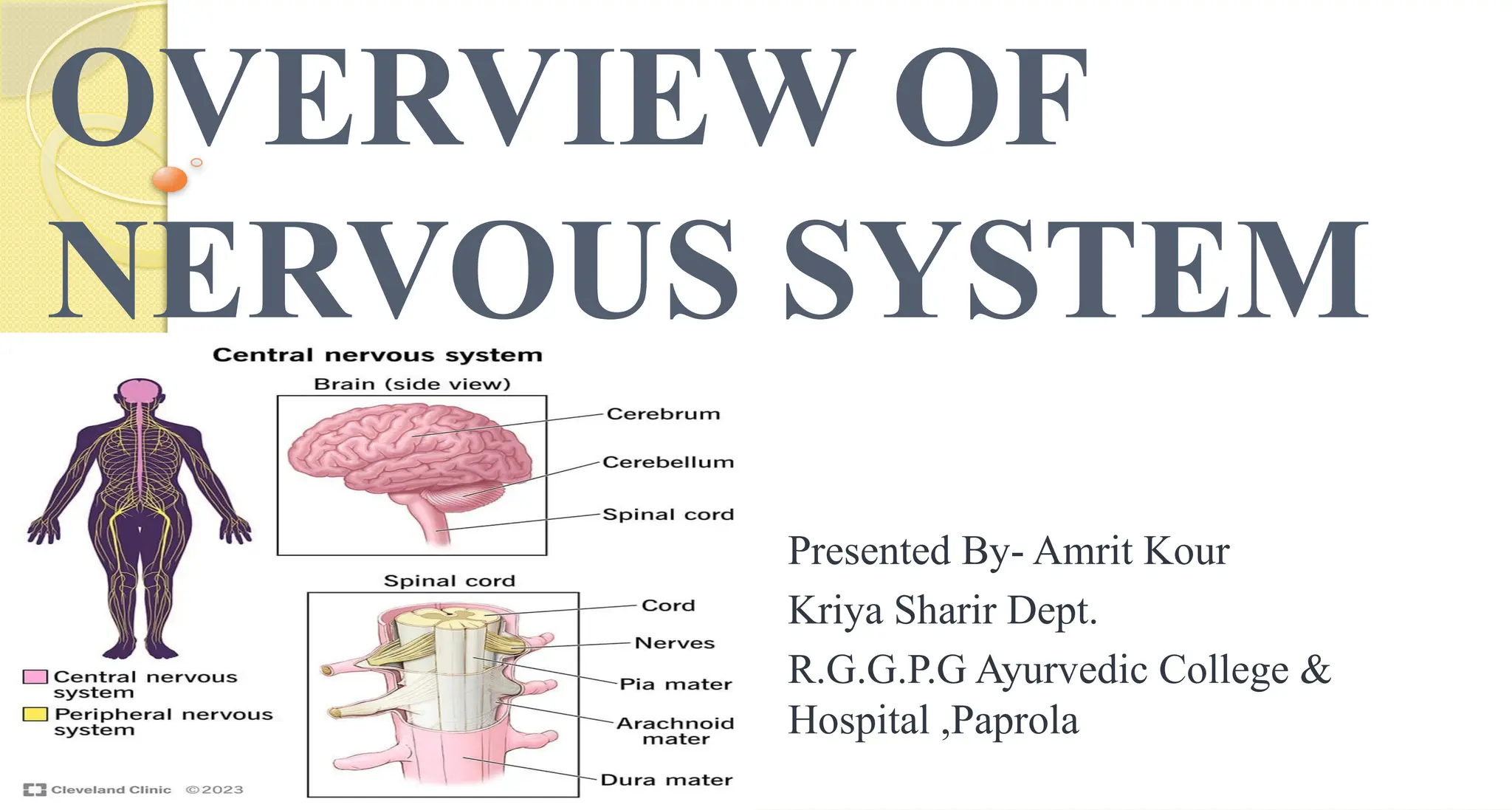
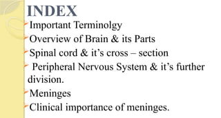
The document provides a comprehensive overview of the nervous system, detailing its structure, functions, and various components including the central and peripheral nervous systems, brain parts, and spinal cord. It discusses the terminology related to gray and white matter, the divisions of the brain, and the meninges, along with their clinical significance. Additionally, it addresses the functionality of sensory and motor pathways, as well as the autonomic nervous system's roles.


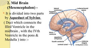
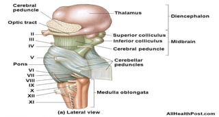
![It is Divided into two parts -
i] Ventral Part, called Cerebral Peduncle , chiefly of
white matter, uniting the Pons with the thalamic region
of the cerebrum.
It comprises the – tegmentum, Substantia nigra & basis
peduncle.
ii] Dorsal part, called tectum which constitutes two
elevations, superior and inferior colliculi.](https://image.slidesharecdn.com/nervoussystempart-1-250128105934-0ac3a6b5/85/NERVOUS-SYSTEM-OVERVIEW-CLASSIFICATION-DIVISION-25-320.jpg)


![Epithelium[1]](https://cdn.slidesharecdn.com/ss_thumbnails/epithelium1-200323141425-thumbnail.jpg?width=640&height=640&fit=bounds)


