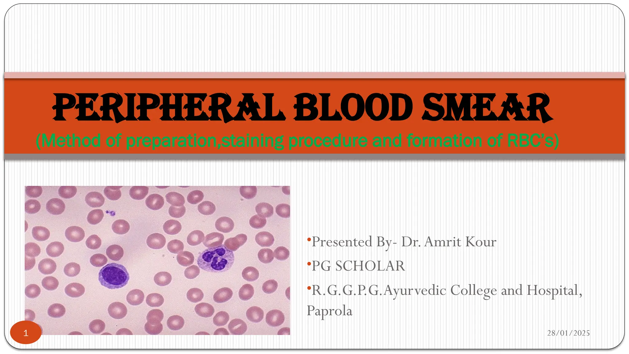
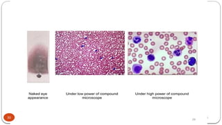
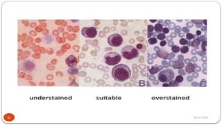
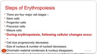
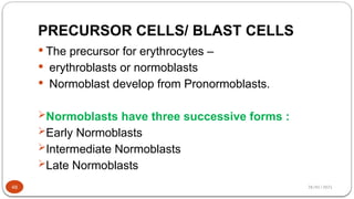
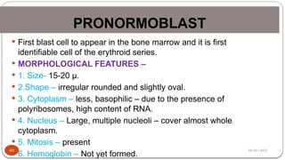
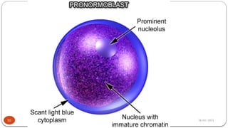
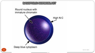
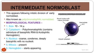
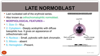
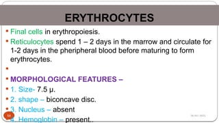
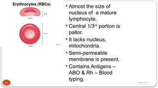
28/01/2025
•Presented By- Dr.Amrit Kour
•PG SCHOLAR
•R.G.G.P.G.Ayurvedic College and Hospital,
Paprola
PERIPHERAL BLOOD SMEAR
(Method of preparation,staining procedure and formation of RBC’s)
1
28/01/2025
Introduction:
 A blood smear or film is a specimen for microscopic
examination
 prepared by spreading a drop of blood across a glass slide
 followed by staining with one of the Romanowsky's stains.
 The peripheral blood slide review is a comprehensive
qualitative examination of the blood film to detect
clinically significant abnormalities in the morphology of
 leukocyte,
 erythrocyte, and
 platelet.
2
28/01/2025
Alternative Names :
Peripheral blood film (PBF)
Blood smear
Peripheral smear
Generalised blood picture(GBP)
3
28/01/2025
USES:
 1. Blood smear is helpful in suggesting the cause of anemia or
thrombocytopenia,
 identifying leukemia and types of leukemia, and
 in diagnosing hemoparasitic infections (malaria, filaria, and
trypanosomiasis).
 It is also helpful in the management of these conditions.
4
28/01/2025
5
 2. To monitor the effect of chemotherapy and
radiotherapy on bone marrow.
 3. To provide direction for further investigations
that will help in arriving at the correct diagnosis
(e.g. in infections, drug toxicity, etc.).
28/01/2025
INDICATIONS OF PBF-
Blood smear examination is therefore indicated in-
 Clinically suspected cases of anemia.
 Thrombocytopenia.
 Hematological malignancies (leukemia, lymphoma,
multiple myeloma).
 Disseminated intravascular coagulation.
 Parasitic infections (like malaria or filaria).
 Infectious mononucleosis.
 Various inflammatory, or malignant diseases.
6
28/01/2025
7
28/01/2025
PREPARATION OF BLOOD SMEAR:
WEDGE TECHNIQUE.
COVERSLIP TECHNIQUE.
AUTOMATED SLIDE MAKING AND
STAINING.
8
28/01/2025
9
28/01/2025
10
28/01/2025
(WEDGE METHOD)
 A small drop of blood (2-3 mm in diameter) is placed in
the center line about 1 cm away from one end of a glass
slide (typical size of slide is 75 × 25 mm; thickness about
1mm) with a wooden stick or glass capillary.
 Blood sample may be venous (anticoagulated with EDTA)
or capillary (finger prick).
 Better blood cell morphology is obtained if smear is made
directly from a skin puncture.
11
28/01/2025
 If EDTA-anticoagulated venous blood is used, smear
should be prepared and stained within 2 hours of blood
collection.
 If venous blood collected in a syringe is used, the last
drop of blood in the needle after withdrawing (or first
drop while dispensing) should be used.
 A 'spreader' slide is placed at an angle of 30° in front of
the drop and then drawn back to touch the drop of blood.
Blood spreads across the line of contact of two slides.
 Smear is made by smooth, forward movement of the
'spreader' along the slide.
12
28/01/2025
13
28/01/2025
 Smear is rapidly dried by waving it in the air or keeping
it under an electric fan.
 Slow drying causes shrinkage artifact of red cells.
 Patient's name or laboratory number and date are
written (with a lead pencil, a permanent marker pen, or
a diamond pencil) on the thicker end of the smear.
14
28/01/2025
15
28/01/2025
16
28/01/2025
17
28/01/2025
18
28/01/2025
19
28/01/2025
20
28/01/2025
21
28/01/2025
22
28/01/2025
23
28/01/2025
24
28/01/2025
25
28/01/2025
26
28/01/2025
27
28/01/2025
28
28/01/2025
29
28/01/2025
30
28/01/2025
31
28/01/2025
32
28/01/2025
33
28/01/2025
A well-stained smear shows following features:
• Red cells: pink-red or deep pink
• Polychromatic cells (Reticulocytes): Gray-blue
• Neutrophils: Pale pink cytoplasm; mauve-
purple granules
• Eosinophils: Pale-pink cytoplasm; orange-red
granules
• Basophils: Blue cytoplasm; dark blue-violet
granules
34
28/01/2025
• Monocytes: Gray-blue cytoplasm; fine reddish (azurophil)
granules
• Small lymphocytes: Dark blue cytoplasm
• Platelets: Purple
• Nuclei of all cells: Purple-violet
35
28/01/2025
ERYTHROPOIESIS
36
28/01/2025
Definition
 Erythropoiesis is the process of the origin, development and
maturation of erythrocytes.
 It is a part of the process of development of blood cells called
Hemopoiesis or hematopoiesis which includes.
 Erythropoiesis
 Leucopoiesis – formation of W.B.Cs
 Thrombopoiesis – formation of platelets
37
28/01/2025
Stages of Erythropoiesis
 There are three stages of erythropoiesis –
 1. Mesoblastic Stage
 2. Hepatic Stage
 2. Myeloid Stage
 1. Mesoblastic Stage –
 During the first two months of intrauterine life, erythropoiesis
takes place in the mesoderm of yolk sac.
 During this stage, erythropoiesis is intravascular.
38
28/01/2025
 2. Hepatic Stage –
 From third month of intrauterine life, the main site for erythropoiesis is
liver.
 Spleen and lymphoid organs are also involved in erythropoiesis.
 3. Myeloid Stage –
 During the last three months of intrauterine life, the RBCs are produced
from red bone marrow and liver.
39
28/01/2025
40
28/01/2025
After birth, bone marrow becomes the sole site of
erythropoiesis.
In young children, active hematopoietic bone marrow is
found in axial skeleton and bones of extremities.
Active hematopoietic bone marrow is red in colour due to
marked cellularity – Red Bone Marrow.
However, there occurs a progressive fatty replacement
throughout the long bones converting red bone marrow to
yellow bone marrow.
41
28/01/2025
 After 20 – 30 years, erythropoiesis is mostly limited
 sternum
 Ribs
 Vertebrae
 Skull
 Pelvis &
 Pelvic Girdle
42
28/01/2025
Steps of Erythropoiesis
 There are four major cell stages –
 Stem cells
 Progenitor cells
 Precursor cells
 Mature cells
During erythropoiesis, following cellular changes occur
–
Cell size progressively decreases
Size of nucleus & number of nucleoli decreases.
Chromatin material condenses & nucleus disappears.
43
28/01/2025
Stem cells
 Pleuripotent stem cells are the mother stem cells for
different cell lines.
It possess two fundamental properties :
 Self Replication – they are capable of giving rise to
more stem cell.
 Differentiation – they have the ability to differentiate
into specalized cells called progenitor cells.
44
28/01/2025
45
28/01/2025
COMMITED STEM CELLS
 These cells develop from pleuripotent stem cells.
 They are of two categories :
 Myeloid stem cell and
 Lymphoid stem cell
 Myeloid stem cell form
 erythroid series,
 monocytic granulocytic series &
 Megakaryoid series.
 Erythroid stem cells - Give rise to progenitor cells for erythroid cell lines.
46
28/01/2025
47
28/01/2025
PRECURSOR CELLS/ BLAST CELLS
 The precursor for erythrocytes –
 erythroblasts or normoblasts
 Normoblast develop from Pronormoblasts.
Normoblasts have three successive forms :
Early Normoblasts
Intermediate Normoblasts
Late Normoblasts
48
28/01/2025
PRONORMOBLAST
49
 First blast cell to appear in the bone marrow and it is first
identifiable cell of the erythroid series.
 MORPHOLOGICAL FEATURES –
 1. Size- 15-20 µ.
 2.Shape – irregular rounded and slightly oval.
 3. Cytoplasm – less, basophilic – due to the presence of
polyribosomes, high content of RNA.
 4. Nucleus – Large, multiple nucleoli – cover almost whole
cytoplasm.
 5. Mitosis – present
 6. Hemoglobin – Not yet formed.
28/01/2025
50
28/01/2025
EARLY/ BASOPHILIC NORMOBLAST
51
 Pronormoblast progresses into early normoblast.
 MORPHOLOGICAL FEATURES –
 1. Size- 12 - 16 µ.
 2.Shape – irregular rounded and slightly oval.
 3. Cytoplasm – scanty, blue and basophilic.
 4. Nucleus –nucleoli disappear & Condensation of
chromatin network occurs.
 5. Mitosis – present
 6. Hemoglobin – Not present.
28/01/2025
52
28/01/2025
INTERMEDIATE NORMOBLAST
53
 This appears following mitotic division of early
normoblast.
 Also known as polychromatophilic normoblast.
 MORPHOLOGICAL FEATURES –
 1. Size- 10 - 14 µ.
 2. Cytoplasm – Polychromatophilic (contains
admixture of basophilic RNA & Acidophilic
Hemoglobin).
 4. Nucleus –coarse, condense, deeply
basophilic with no nucleoli.
 5. Mitosis – present
 6. Hemoglobin – starts appearing.
28/01/2025
LATE NORMOBLAST
54
 Last nucleated cell of the erythroid series.
 Also known as orthochromatophilic normoblast.
 MORPHOLOGICAL FEATURES –
 1. Size- 8 - 10 µ.
 2. Cytoplasm –Deeply acidophilic with diffuse
basophilic hue. It gives an appearance of
orthochromatic cell.
 4. Nucleus – Small, pyknotic with dark chromatin.
 5. Mitosis – absent
 6. Hemoglobin –Present.
28/01/2025
RETICULOCYTE
55
 Immediate precursors of red cells.
 Also known as juvenile red cells.
 MORPHOLOGICAL FEATURES –
 1. Size- 7– 7.5 µ.
 2. Cytoplasm –contain small amount of RNA. With supravital stains
like brilliant cresyl blue, the RNA appears in the form of reticulum
and hence the cell is called reticulocyte.
 4. Nucleus – absent
 5. Mitosis – absent
 6. Hemoglobin – Increases.
28/01/2025
ERYTHROCYTES
56
 Final cells in erythropoiesis.
 Reticulocytes spend 1 – 2 days in the marrow and circulate for
1-2 days in the pheripheral blood before maturing to form
erythrocytes.

 MORPHOLOGICAL FEATURES –
 1. Size- 7.5 µ.
 2. shape – biconcave disc.
 3. Nucleus – absent
 4. Hemoglobin – present..
28/01/2025
57
 Almost the size of
nucleus of a mature
lymphocyte.
 Central 1/3rd
portion is
pallor.
 It lacks nucleus,
mitochondria.
 Semi-permeable
membrane is present.
 Contains Antigens –
ABO & Rh – Blood
typing.
28/01/2025
DURATION
58
 The total period of erythropoiesis is 7 to
9 days.
 It takes 5 – 7 days for progenitor cells to
become reticulocytes and
 another 2 days for reticulocytes to
become red cells.
28/01/2025
APPLIED CONDITIONS
59
 Abnormal count of RBCs –
 1. Increase in erythrocytes – Polycythemia
 2. Decrease in erythrocytes – Anemia
 Abnormal size of RBCs –
 1. Microcytosis – Erythroctes are smaller than 7 µm.
 2. Macrocytosis – Erythroctes are larger than 9 µm.
 3.Anisocytes (cells with different sizes).
 Abnormal shape of RBCs –
 Sickle cell: Crescentic shape as in sickle cell anemia.
 Poikilocytosis: Unusual shapes due to deformed cell membrane.
 The shape will be of flask, hammer or any other unusual shape
28/01/2025
THANK YOU
60

PERIPHERAL BLOOD SMEAR.pptx preparation staining fixation

  • 1.
    28/01/2025 •Presented By- Dr.AmritKour •PG SCHOLAR •R.G.G.P.G.Ayurvedic College and Hospital, Paprola PERIPHERAL BLOOD SMEAR (Method of preparation,staining procedure and formation of RBC’s) 1
  • 2.
    28/01/2025 Introduction:  A bloodsmear or film is a specimen for microscopic examination  prepared by spreading a drop of blood across a glass slide  followed by staining with one of the Romanowsky's stains.  The peripheral blood slide review is a comprehensive qualitative examination of the blood film to detect clinically significant abnormalities in the morphology of  leukocyte,  erythrocyte, and  platelet. 2
  • 3.
    28/01/2025 Alternative Names : Peripheralblood film (PBF) Blood smear Peripheral smear Generalised blood picture(GBP) 3
  • 4.
    28/01/2025 USES:  1. Bloodsmear is helpful in suggesting the cause of anemia or thrombocytopenia,  identifying leukemia and types of leukemia, and  in diagnosing hemoparasitic infections (malaria, filaria, and trypanosomiasis).  It is also helpful in the management of these conditions. 4
  • 5.
    28/01/2025 5  2. Tomonitor the effect of chemotherapy and radiotherapy on bone marrow.  3. To provide direction for further investigations that will help in arriving at the correct diagnosis (e.g. in infections, drug toxicity, etc.).
  • 6.
    28/01/2025 INDICATIONS OF PBF- Bloodsmear examination is therefore indicated in-  Clinically suspected cases of anemia.  Thrombocytopenia.  Hematological malignancies (leukemia, lymphoma, multiple myeloma).  Disseminated intravascular coagulation.  Parasitic infections (like malaria or filaria).  Infectious mononucleosis.  Various inflammatory, or malignant diseases. 6
  • 7.
  • 8.
    28/01/2025 PREPARATION OF BLOODSMEAR: WEDGE TECHNIQUE. COVERSLIP TECHNIQUE. AUTOMATED SLIDE MAKING AND STAINING. 8
  • 9.
  • 10.
  • 11.
    28/01/2025 (WEDGE METHOD)  Asmall drop of blood (2-3 mm in diameter) is placed in the center line about 1 cm away from one end of a glass slide (typical size of slide is 75 × 25 mm; thickness about 1mm) with a wooden stick or glass capillary.  Blood sample may be venous (anticoagulated with EDTA) or capillary (finger prick).  Better blood cell morphology is obtained if smear is made directly from a skin puncture. 11
  • 12.
    28/01/2025  If EDTA-anticoagulatedvenous blood is used, smear should be prepared and stained within 2 hours of blood collection.  If venous blood collected in a syringe is used, the last drop of blood in the needle after withdrawing (or first drop while dispensing) should be used.  A 'spreader' slide is placed at an angle of 30° in front of the drop and then drawn back to touch the drop of blood. Blood spreads across the line of contact of two slides.  Smear is made by smooth, forward movement of the 'spreader' along the slide. 12
  • 13.
  • 14.
    28/01/2025  Smear israpidly dried by waving it in the air or keeping it under an electric fan.  Slow drying causes shrinkage artifact of red cells.  Patient's name or laboratory number and date are written (with a lead pencil, a permanent marker pen, or a diamond pencil) on the thicker end of the smear. 14
  • 15.
  • 16.
  • 17.
  • 18.
  • 19.
  • 20.
  • 21.
  • 22.
  • 23.
  • 24.
  • 25.
  • 26.
  • 27.
  • 28.
  • 29.
  • 30.
  • 31.
  • 32.
  • 33.
  • 34.
    28/01/2025 A well-stained smearshows following features: • Red cells: pink-red or deep pink • Polychromatic cells (Reticulocytes): Gray-blue • Neutrophils: Pale pink cytoplasm; mauve- purple granules • Eosinophils: Pale-pink cytoplasm; orange-red granules • Basophils: Blue cytoplasm; dark blue-violet granules 34
  • 35.
    28/01/2025 • Monocytes: Gray-bluecytoplasm; fine reddish (azurophil) granules • Small lymphocytes: Dark blue cytoplasm • Platelets: Purple • Nuclei of all cells: Purple-violet 35
  • 36.
  • 37.
    28/01/2025 Definition  Erythropoiesis isthe process of the origin, development and maturation of erythrocytes.  It is a part of the process of development of blood cells called Hemopoiesis or hematopoiesis which includes.  Erythropoiesis  Leucopoiesis – formation of W.B.Cs  Thrombopoiesis – formation of platelets 37
  • 38.
    28/01/2025 Stages of Erythropoiesis There are three stages of erythropoiesis –  1. Mesoblastic Stage  2. Hepatic Stage  2. Myeloid Stage  1. Mesoblastic Stage –  During the first two months of intrauterine life, erythropoiesis takes place in the mesoderm of yolk sac.  During this stage, erythropoiesis is intravascular. 38
  • 39.
    28/01/2025  2. HepaticStage –  From third month of intrauterine life, the main site for erythropoiesis is liver.  Spleen and lymphoid organs are also involved in erythropoiesis.  3. Myeloid Stage –  During the last three months of intrauterine life, the RBCs are produced from red bone marrow and liver. 39
  • 40.
  • 41.
    28/01/2025 After birth, bonemarrow becomes the sole site of erythropoiesis. In young children, active hematopoietic bone marrow is found in axial skeleton and bones of extremities. Active hematopoietic bone marrow is red in colour due to marked cellularity – Red Bone Marrow. However, there occurs a progressive fatty replacement throughout the long bones converting red bone marrow to yellow bone marrow. 41
  • 42.
    28/01/2025  After 20– 30 years, erythropoiesis is mostly limited  sternum  Ribs  Vertebrae  Skull  Pelvis &  Pelvic Girdle 42
  • 43.
    28/01/2025 Steps of Erythropoiesis There are four major cell stages –  Stem cells  Progenitor cells  Precursor cells  Mature cells During erythropoiesis, following cellular changes occur – Cell size progressively decreases Size of nucleus & number of nucleoli decreases. Chromatin material condenses & nucleus disappears. 43
  • 44.
    28/01/2025 Stem cells  Pleuripotentstem cells are the mother stem cells for different cell lines. It possess two fundamental properties :  Self Replication – they are capable of giving rise to more stem cell.  Differentiation – they have the ability to differentiate into specalized cells called progenitor cells. 44
  • 45.
  • 46.
    28/01/2025 COMMITED STEM CELLS These cells develop from pleuripotent stem cells.  They are of two categories :  Myeloid stem cell and  Lymphoid stem cell  Myeloid stem cell form  erythroid series,  monocytic granulocytic series &  Megakaryoid series.  Erythroid stem cells - Give rise to progenitor cells for erythroid cell lines. 46
  • 47.
  • 48.
    28/01/2025 PRECURSOR CELLS/ BLASTCELLS  The precursor for erythrocytes –  erythroblasts or normoblasts  Normoblast develop from Pronormoblasts. Normoblasts have three successive forms : Early Normoblasts Intermediate Normoblasts Late Normoblasts 48
  • 49.
    28/01/2025 PRONORMOBLAST 49  First blastcell to appear in the bone marrow and it is first identifiable cell of the erythroid series.  MORPHOLOGICAL FEATURES –  1. Size- 15-20 µ.  2.Shape – irregular rounded and slightly oval.  3. Cytoplasm – less, basophilic – due to the presence of polyribosomes, high content of RNA.  4. Nucleus – Large, multiple nucleoli – cover almost whole cytoplasm.  5. Mitosis – present  6. Hemoglobin – Not yet formed.
  • 50.
  • 51.
    28/01/2025 EARLY/ BASOPHILIC NORMOBLAST 51 Pronormoblast progresses into early normoblast.  MORPHOLOGICAL FEATURES –  1. Size- 12 - 16 µ.  2.Shape – irregular rounded and slightly oval.  3. Cytoplasm – scanty, blue and basophilic.  4. Nucleus –nucleoli disappear & Condensation of chromatin network occurs.  5. Mitosis – present  6. Hemoglobin – Not present.
  • 52.
  • 53.
    28/01/2025 INTERMEDIATE NORMOBLAST 53  Thisappears following mitotic division of early normoblast.  Also known as polychromatophilic normoblast.  MORPHOLOGICAL FEATURES –  1. Size- 10 - 14 µ.  2. Cytoplasm – Polychromatophilic (contains admixture of basophilic RNA & Acidophilic Hemoglobin).  4. Nucleus –coarse, condense, deeply basophilic with no nucleoli.  5. Mitosis – present  6. Hemoglobin – starts appearing.
  • 54.
    28/01/2025 LATE NORMOBLAST 54  Lastnucleated cell of the erythroid series.  Also known as orthochromatophilic normoblast.  MORPHOLOGICAL FEATURES –  1. Size- 8 - 10 µ.  2. Cytoplasm –Deeply acidophilic with diffuse basophilic hue. It gives an appearance of orthochromatic cell.  4. Nucleus – Small, pyknotic with dark chromatin.  5. Mitosis – absent  6. Hemoglobin –Present.
  • 55.
    28/01/2025 RETICULOCYTE 55  Immediate precursorsof red cells.  Also known as juvenile red cells.  MORPHOLOGICAL FEATURES –  1. Size- 7– 7.5 µ.  2. Cytoplasm –contain small amount of RNA. With supravital stains like brilliant cresyl blue, the RNA appears in the form of reticulum and hence the cell is called reticulocyte.  4. Nucleus – absent  5. Mitosis – absent  6. Hemoglobin – Increases.
  • 56.
    28/01/2025 ERYTHROCYTES 56  Final cellsin erythropoiesis.  Reticulocytes spend 1 – 2 days in the marrow and circulate for 1-2 days in the pheripheral blood before maturing to form erythrocytes.   MORPHOLOGICAL FEATURES –  1. Size- 7.5 µ.  2. shape – biconcave disc.  3. Nucleus – absent  4. Hemoglobin – present..
  • 57.
    28/01/2025 57  Almost thesize of nucleus of a mature lymphocyte.  Central 1/3rd portion is pallor.  It lacks nucleus, mitochondria.  Semi-permeable membrane is present.  Contains Antigens – ABO & Rh – Blood typing.
  • 58.
    28/01/2025 DURATION 58  The totalperiod of erythropoiesis is 7 to 9 days.  It takes 5 – 7 days for progenitor cells to become reticulocytes and  another 2 days for reticulocytes to become red cells.
  • 59.
    28/01/2025 APPLIED CONDITIONS 59  Abnormalcount of RBCs –  1. Increase in erythrocytes – Polycythemia  2. Decrease in erythrocytes – Anemia  Abnormal size of RBCs –  1. Microcytosis – Erythroctes are smaller than 7 µm.  2. Macrocytosis – Erythroctes are larger than 9 µm.  3.Anisocytes (cells with different sizes).  Abnormal shape of RBCs –  Sickle cell: Crescentic shape as in sickle cell anemia.  Poikilocytosis: Unusual shapes due to deformed cell membrane.  The shape will be of flask, hammer or any other unusual shape
  • 60.