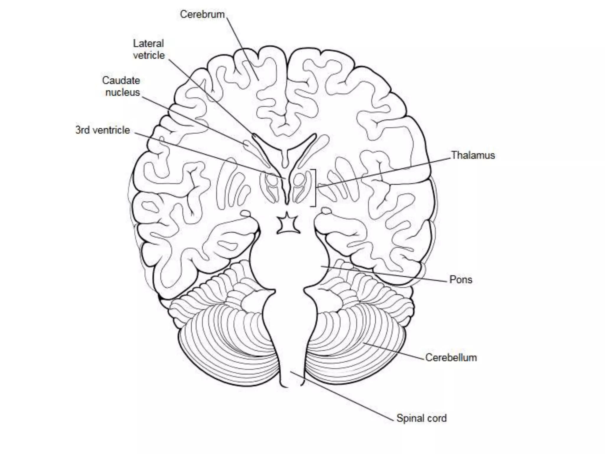
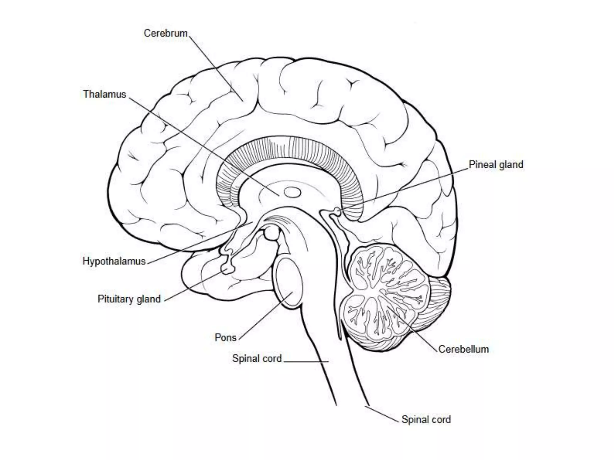
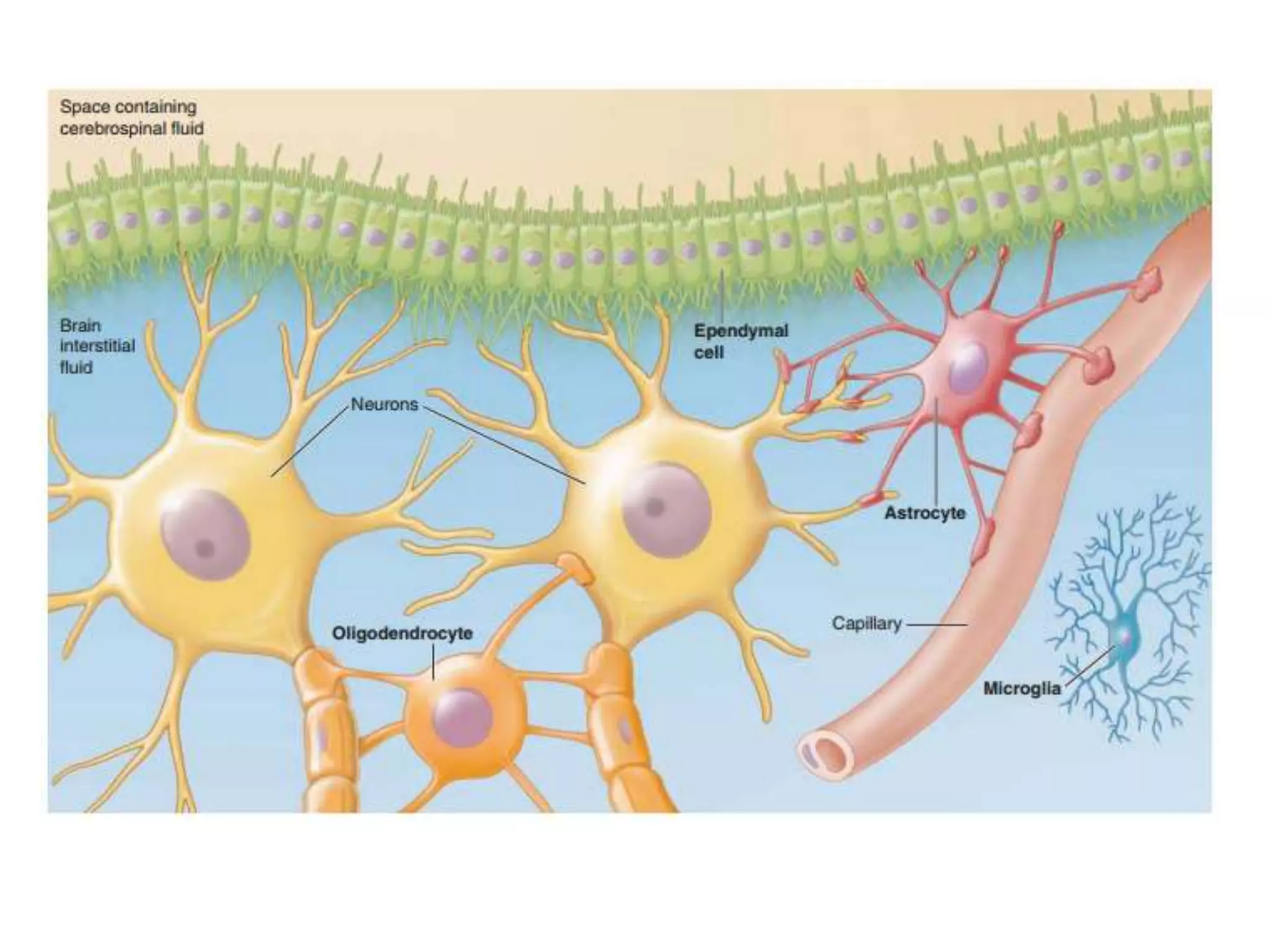
2. Organisation
The central nervous system
Brain and spinal cord
The peripheral nervous system (PNS)- nerve
fibers that carry information between the CNS and
other parts of the body (periphery)
Afferent division- carries information to the CNS,
apprising it of the external environment and providing
status reports on internal activities.
Efferent division- Instructions from the CNS are
transmitted to effector organs- the muscles or glands
Enteric nervous system
3.
4. Central nervous system
The adult human brain weighs between 1,200
to 1,500 g.
Occupies a volume of about 1400 cc
The adult spinal cord is approximately 40 to 50
cm long and occupies about 150 cc
The human nervous system is estimated to
consist of roughly 360 billion non-neural glial
cells and 90 billion nerve cells
5. Morphology:
Grey matter- nerve cell bodies
Superficial in- cerebral cortex & cerebellum
Central in- brain stem and spinal cord
White matter- nerve fibers
Collection of cell bodies in white matter- nuclei
Central nervous system
6. Central nervous system
• The brain and the spinal cord arise in
early development from the neural tube,
which expands to form the three primary
brain divisions:
– The Prosencephalon,
– The Mesencephalon and
– The Rhombencephalon
7.
8. Central nervous system
These three vesicles further differentiate into
five subdivisions:
Prosencephalon-
Telencephalon & Diencephalon
Mesencephalon,
Rhombencephalon-
Metencephalon and the myelencephalon
The mesencephalon, metencephalon, and the
myelencephalon comprise the brain stem.
10. The Brain
Although no part of the brain acts in isolation,
the neurons that work together to accomplish a
given function are organized within a discrete
location. The parts of the brain can be grouped
into-
Forebrain-
Cerebral hemisphere- Cerebral cortex & Basal
nuclei
Diencephalon- Thalamus & Hypothalamus
Brain stem- Mid brain, Pons, Medulla
Cerebellum
11. Cerebral hemispheres
Largest portion of the Brain
Consists of two symmetrical halves separated by
median longitudinal fissure
The surface is highly convoluted with alternate
gyri & sulci
Divided into four lobes- frontal, parietal,
temporal & occipital
Function:
Finer aspects of sensory and motor functions
Seat of higher functions
13. Cerebral hemispheres
Ventricles- hollow cavities within the cerebral
hemispheres
Lateral ventricles- two long cavities
These communicate with the third ventricle in
the midline between two halves of Diencephalon
Limbic system- lies in the deeper parts
Mainly concerned with emotion
14.
15.
16. The Diencephalon
Thalamus
Paired, present on either side of third ventricle
Important relay station. Most ascending sensory
tracts relay in the Thalamus
Laterally related to Basal Ganglia (subcortical
nuclear masses)- have important motor functions
Hypothalamus- lies deep to the Thalamus
Forms floor & part of lateral wall of third ventricle
Important functions- control autonomic function,
endocrine control of pituitary, regulation of thirst,
appetite and body temperature etc.
17.
18. The Brain Stem
Midbrain- contains the cerebral aqueduct in
center which link the third and fourth ventricle
Contains nuclei of Oculomotor and Trochlear
nerves
Pons & Medulla
Contains many important nuclei including nuclei
of many cranial nerves
Contains cardiorespiratory centers
Dorsal aspect forms the floor of the fourth
ventricle
19. Cerebellum
Globular structure lying below the posterior
Part of cerebral hemispheres and on the
posterior aspect of brain stem
Four nuclei are present within the white
matter- fastigial, globose, emboliform &
dentate
Surface is densely fissured
Connected to the brain stem with three pairs of
cerebellar peduncles.
20. Spinal cord
Downward continuation of the medulla
oblongata
Extend down into the vertebral canal up to the
first lumbar vertebra.
Has central canal surrounded by a mass of gray
matter which is divided into anterior, lateral &
posterior horns.
White matter surrounds the gray matter
21.
22. The peripheral nervous system
(PNS)
Consisting of nerve fibers that carry
information between the CNS and other parts
of the body. These include-
Cranial nerves- 12 pairs
Spinal nerves- 31 pairs
The paravertibral sympathetic chain
Preganglionic and postganglionic autonomic
nerves
23. The peripheral nervous system
(PNS)
Afferent division- carries information to the CNS,
Sensory stimuli- apprising it of the external
environment and
Visceral stimuli- providing status reports on internal
activities
Efferent division- transmit instructions from the
CNS to effector organs i.e. the muscles or glands
The somatic nervous system- The fibers of the motor
neurons that supply the skeletal muscles; and
The autonomic nervous system- The fibers that
innervate smooth muscle, cardiac muscle, and
glands.
24. The peripheral nervous system
(PNS)
The enteric nervous system
An extensive nerve network in the wall of the
digestive tract.
Controls digestive activities along with the
autonomic nervous system and hormones.
The enteric nervous system can act
independently of the rest of the nervous
system but is also influenced by autonomic
fibers that terminate on the enteric neurons.
25. Cranial nerves
Arise from nuclei situated in the brain stem
except first two cranial nerves
Olfactory
Optic
Oculomotor- arise from upper half of midbrain
Innervate extrinsic muscles of the eyeball
Supplies parasympathetic fibers arising from Edinger-
Westphal nucleus to ciliary muscles and constrictor
pupillae
Trochlear- arise from lower half of midbrain
Supplies superior oblique muscle
26. Cranial nerves
Abducent (sixth nerve)- arise from its nucleus
from pons and emerges from dorsal aspect of
midbrain.
Supplies lateral rectus
Trigeminal- mixed nerve. Carries sensory
impulses from the face to-
Spinal nucleus (long sensory nucleus), mainly
receives pain sensation
Principal sensory nucleus, especially concerned
with touch
Motor nucleus lies in the pons. Innervate the
muscles of mastication.
27. Facial nerve- mixed nerve
Motor fibers- supply the muscles of face and
scalp
Sensory fibers- carry taste sensation from
anterior two thirds of the tongue
Secretomotor fibers (parasympathetic)-
Submandibular and Sublingual salivary glands
and Lacrimal and palatal glands
Cranial nerves
28. Vestibulocochlear nerve
Vesibular division- receives sensory information
from vestibular apparatus (otolith organs &
semicircular canal)- maintains equilibrium
Cochlear division- hearing
Glossopharyngeal nerve- mixed nerve
Motor fiber from nucleus ambiguus to
stylopharyngeus muscle
Secretomotor fibers to Parotid gland
Cranial nerves
29. Glossopharyngeal nerve
Sensory fibers-
Taste sensation from posterior 1/3rd of tongue,
General sensation- posterior 1/3rd of tongue,
oropharynx, tonsils etc
Innervates the baroreceptors in carotid sinus
& chemoreceptors in carotid body.
Cranial nerves
30. Vagus nerve- longest cranial nerve
Parasympathetic component supplies the heart,
lungs & most of the abdominal viscera
Motor to laryngeal & pharyngeal muscles
Sensory- visceral sensation from thoracic and
abdominal organs via parasympathetic afferents
Supplies the baroreceptors in the aortic arch &
chemoreceptors in the aortic body.
Cranial nerves
31. Accessory nerve- eleventh nerve
Spinal accessory- motor to trapezius &
sternocleidomastoid
Cranial part- supplies laryngeal & pharyngeal
muscles along with vagus
Hypoglossal nerve- motor nerve supplying the
muscles of the tongue
Cranial nerves
32. Spinal nerves
Arise from spinal cord by two roots-
Ventral (anterior)- motor fibers and
Dorsal (posterior)- sensory fibers
The somatic motor fibers in the anterior root
also carry-
Sympathetic efferent- T1- L2
Parasympathetic efferent- S2 – S4
Bell-Magendies law- the sensory fibers enter
through the posterior root & the motor fibers
emerge through anterior root in the spinal
cord.
33.
34. Cellular components of CNS
Glial cells (neuroglia)-
Serve as connective tissue of the CNS and so
support the neurons both physically & metabolically
Maintain homeostasis of fluids that bathe the
neurons
Actively modulate synaptic function & are
considered nearly as important as neurons to
learning and memory.
Neuroglias constitute about 90% of the cells in
CNS but occupy only 50% the volume.
Neurons- transmit impulses in the form of action
potentials
35. Neuroglia
Four major types of glial cells in the CNS:
Astrocytes,
Oligodendrocytes,
Microglia, and
Ependymal cell
Astrocytes- star shaped. Processes of
astrocytes surround the neurons and often
terminate on the wall of blood vessels.
36. Neuroglia
Functions Astrocytes
Physically support neurons in proper spatial
relationships
Serve as a scaffold during fetal brain development
Induce formation of blood–brain barrier
Help transfer nutrients from blood to neurons
Important in the repair of brain injuries and form
neural scar tissue
Take up and degrade released neurotransmitters
(Glutamate & GABA)
Take up excess K+ to help maintain proper brain-ECF
ion concentration and normal neural excitability
37. Neuroglia
Functions Astrocytes:
Enhance synapse formation and strengthen
synaptic transmission via chemical signaling
(thrombospondin) with neurons
Communicate by chemical means with neurons
and among themselves
Oligodendrocytes-
Forms the myelin sheath around axons in the
brain and spinal cord.
Unlike Schwann cells they can myelinate several
nerve cells simultaneously
38. Microglia-
Derived from bone marrow & behave like
macrophages
Release low levels of nerve growth factor, which
help neurons and other glial cells survive and thrive
Ependymal cells
Line the surfaces of the brains ventricles and central
canal of the spinal cord
Posses cilia beating of which contribute to CSF
movement
Contribute to formation of cerebrospinal fluid
Serve as neural stem cells with the potential to form
new neurons and glial cells
Neuroglia
39. Types of Neuroglia
Unlike neurons, glial
cells do not lose the
ability to undergo
cell division, so
most brain tumors
of neural origin
consist of glial cells
(gliomas)
42. Protection of the brain
Four major features help protect the CNS from
injury-
It is enclosed by hard, bony structures. The cranium
(skull)
encases the brain, and the vertebral column
surrounds the spinal cord.
Three protective and nourishing membranes, the
meninges,
lie between the bony covering and the nervous tissue.
The brain “floats” in a special cushioning fluid, the
cerebrospinal fluid (CSF).
A highly selective blood-brain barrier limits access of
blood borne materials into the vulnerable brain tissue
43. The meninges
Duramater- tough, inelastic covering that
consists of two layers, endosteal and
meningeal layers- dural & venous sinuses
lies between these two layers
Venous blood draining from the brain empties into
these sinuses
Cerebrospinal fluid also re-enters the blood at
one of these sinus sites
44. The meninges
Arachnoidmater- richly vascularized layer with
a cobwebby appearance
Arachnoid villi, penetrate through gaps in the
overlying dura and project into the dural sinuses
Subarachnoid space- between arachnoidmater
& piamater- contains CSF
Piamater- most fragile and highly vascular.
Closely adheres to the surfaces of the brain
and spinal cord
45. Ventricles- CSF filled spaces inside the brain
Lateral ventricles- within the cerebral
hemispheres
Connects with the third ventricle between the
thalami through foramina of Monroe
The third ventricle communicates with fourth
ventricle by the aqueduct of Sylvious- runs
through the mid brain
The fourth ventricl communicates with the
subarachnoid space through the foramina of
Magendie (midline) & two foramina of Luschka
(laterally)
Below fourth ventricle is continuous with central
The meninges
46. Surrounds and cushions the brain and spinal cord.
The CSF has about the same density as the
brain itself, so it essentially floats or is suspended
in this fluid environment
Functions:
Serve as a shock-absorbing fluid to prevent the
brain from bumping against the interior of the hard
skull when the head is subjected to sudden, jarring
movements.
Plays an important role in the exchange of materials
between the neural cells and the interstitial fluid
surrounding the brain.
Cerebrospinal fluid
47. Functions:
The composition of the brain interstitial fluid is
influenced more by changes in the composition of
the CSF than by alterations in the blood.
Materials are exchanged fairly freely between the
CSF and brain interstitial fluid, whereas only
limited exchange occurs between the blood and
brain interstitial fluid.
Cerebrospinal fluid
48. Formed primarily by the choroid plexuses found in
particular regions of the ventricles.
Choroid plexuses consist of richly vascularized,
cauliflower-like masses of pia mater tissue that dip
into pockets formed by ependymal cells.
Cerebrospinal fluid forms as a result of selective
transport mechanisms across the membranes of
the choroid plexuses.
The composition of CSF differs from that of blood.
For example, CSF is lower in K+ and slightly higher
in Na+
The biggest difference is the presence of plasma
proteins in the blood but almost no protein is normally
present in the CSF.
Cerebrospinal fluid
49. Cerebrospinal fluid
Circulation of CSF
Lateral
ventricles
Third ventricle
Fourth ventricle
Suarachnoid
space
Foramen of Monro
Aqueduct of Sylvius
Formina of Luschka &
Magendie
Central canal of
spinal canal
50.
51.
52. Absorption of CSF
CSF in the subarachnoid space is absorbed in
the dural venous sinuses by way of arachnoid
villi which project into the superior sagittal sinus
between the layers of duramater
The membrane contains pores which allow
absorption of CSF into venous sinuses when
CSF pressure is high
When venous pressure is higher back flow is
prevented by collapse of the pores.
Much higher osmolality of venous blood draws
CSF into it
53.
54. Composition of CSF
Clear fluid with-
Normal cell conunt- 0-5/cu-mm
Glucose- 50-80 mg/100 ml
Protein- 20-90 mg/100 ml
Chloride- 720-750 mg/100 ml
Na+, K+, Ca+, bicarbonate & urea in trace
amounts
55. Cerebrospinal Fluid- Applied
Lumbar puncture- hollow needle is inserted
into the spinal canal between the third and
fourth lumbar vertebra. Collected CSF is
examined for
Subarachnoid hemmorrhage- blood in CSF
Pyogenic meningitis- turbid with high cell count &
plenty neutrophils. Low glucose & high protein
content
Tubercular meningitis-clear & may show fine clot.
Contains lymphocytes, high protein but low
glucose & Cl+
Viral encephalitis- raised protein, lymphocyte may
also be high
56. Hydrcephalus- excessive accumulation of CSF
with dilatation of ventricles (normal CSF
pressure- 70-180 mm CSF)
Two types-
External or communicating- obstruction outside
the ventricular system. There may be decrease in
reabsorption.
Internal or noncommunicating- there is block
within the ventricles or at the foramina of
Magendie & Luschka.
Cerebrospinal Fluid- Applied
57. The resulting increase in CSF pressure can
lead to brain damage and mental retardation if
untreated.
Treatment consists of surgically shunting the
excess CSF to veins elsewhere in the body.
Cerebrospinal Fluid- Applied
58. Blood Brain Barrier
The brain is carefully shielded from harmful
changes in the blood by a highly selective
blood-brain barrier (BBB)
Unlike the free exchange across capillaries
elsewhere, only selected, carefully regulated
exchanges can be made across the BBB
(between the blood and interstitial fluid)
Features
In brain capillaries, the cells are joined by tight
junctions which completely seal the capillary wall
59. Blood Brain Barrier
Features
The only possible exchanges are through the
capillary cells themselves
Lipid-soluble substances such as O2, CO2,
alcohol, and steroid hormones penetrate these
cells easily by dissolving in their lipid plasma
membrane
Small water molecules also diffuse through
readily by
passing between the phospholipid molecules of
the plasma membrane or through aquaporins
60. Blood Brain Barrier
Features
All other substances exchanged between the blood
and brain interstitial fluid, including such essential
materials as glucose, amino acids, and ions, are
transported by highly selective membrane-bound
carriers
Thus it protects the delicate brain from
Chemical fluctuations in the blood and
Minimizes the possibility that potentially harmful
blood-borne substances might reach the central
neural tissue
Prevents certain circulating hormones that could
also act as neurotransmitters from reaching the brain
61. Blood Brain Barrier
The negative side-
It limits the use of drugs for the treatment of brain
and spinal cord disorders because many drugs are
unable to penetrate this barrier
Astrocytes play two roles in the BBB:
They signal the cells forming the brain capillaries to
“get tight.” Capillary cells do not have an inherent
ability to form tight junctions;
They participate in the cross-cellular transport of
some substances, such as K+ .
Certain areas of the brain, most notably a portion
of the hypothalamus, are not subject to the BBB.
62.
63. Sensory system- receptors
Most activities of the nervous system are initiated
by sensory experiences that excite sensory
receptors
Sensory experience can either cause
immediate reaction from the brain or
memory of the experience can be stored in the brain
for minutes, weeks or years and determine bodily
reactions at some future date
64. Sensory system
This information enters the central nervous
system through peripheral nerves and is
conducted immediately to multiple sensory
areas in-
The spinal cord at all levels;
The reticular substance of the
medulla, pons, and mesencephalon of the brain;
The cerebellum;
The thalamus; and
Areas of the cerebral cortex.
66. Motor system- effectors
Controls
Contraction of appropriate skeletal muscles
throughout the body,
Contraction of smooth muscle in the internal
organs, and
Secretion of active chemical substances by both
exocrine and endocrine glands in many parts of
the body
The muscles and glands are called effectors.
68. Processing of Information-
Integrative Function
One of the most important functions of the
nervous system is to process incoming
information
Necessary for appropriate mental and motor
responses.
More than 99 percent of all sensory information is
discarded by the brain as irrelevant and
unimportant.
Important sensory information's excite the
mind & is channelled into proper integrative
and motor regions of the brain to cause
69. Role of Synapses in Processing
Information
Synapse is the junction point from one neuron
to the next
Synapses determine the directions of spread of
nervous signals through the nervous system
facilitatory and inhibitory signals from other areas
in the nervous system can control synaptic
transmission
some postsynaptic neurons respond with large
numbers of output impulses, and others respond
with only a few.
Thus, the synapses perform a selective action
70. Storage of Information- Memory
The process of storage of information is called
memory- a function of the synapses
After the sensory signals have passed through the
synapses a large number of times, they become
more capable of transmitting the same type of
signal the next time- facilitation
Most storage occurs in the cerebral cortex, but some
can occur in basal regions of the brain and the
spinal cord
They become part of the brain processing
mechanism for future “thinking.”
That is, the thinking processes of the brain compare
new sensory experiences with stored memories
71. MAJOR LEVELS OF
CNS FUNCTION
•The spinal cord level
•The lower brain or subcortical level,
and
•The higher brain or cortical level
72. The spinal cord level
Neuronal circuits in the cord can cause
walking movements,
Reflexes that withdraw portions of the body from painful
objects,
Reflexes that stiffen the legs to support the body against
gravity, and
Reflexes that control local blood vessels, gastrointestinal
movements, or urinary excretion.
The upper levels of the nervous system often
operate not by sending signals directly to the
periphery of the body but by simply “commanding”
the cord centers to perform their functions.
73. The lower brain or subcortical
level
Many subconscious activities of the body are
controlled in the lower areas of the brain- the medulla,
pons, mesencephalon, hypothalamus, thalamus,
cerebellum, and basal ganglia
The medulla & Pons- subconscious control of arterial
pressure and respiration
Control of equilibrium- combined function of the
cerebellum and the reticular substance of the medulla,
pons, and mesencephalon
Feeding reflexes, such as salivation and licking of the lips
in response to the taste of food- controlled by areas in the
medulla, pons, mesencephalon, amygdala, and
hypothalamus
Many emotional patterns, such as anger, excitement,
sexual response, reaction to pain, and reaction to
pleasure, can still occur after destruction of much of the
74. The higher brain or cortical
level
The cortex never functions alone but always in
association with lower centers of the nervous
system
Without the cerebral cortex, the functions of
the lower brain centers are often imprecise
The cerebral cortex is essential for most of our
thought processes, but it cannot function by
itself.
It is the lower brain centers, not the cortex, that
initiate wakefulness in the cerebral cortex