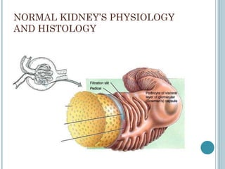
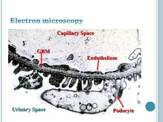
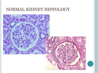
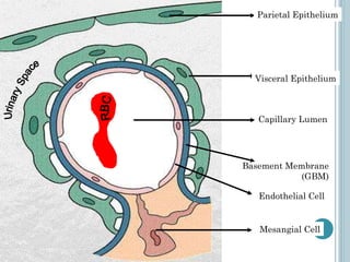
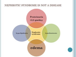
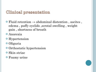
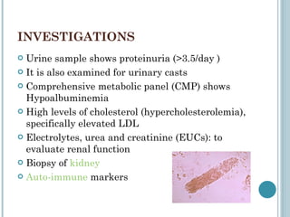
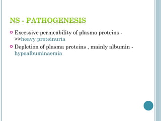
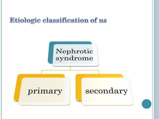
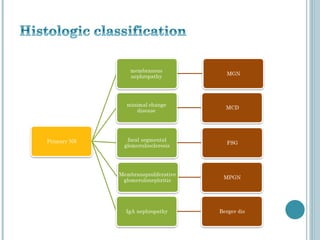
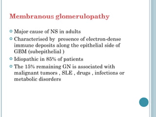
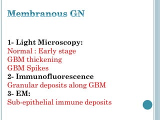
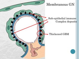
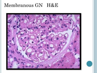
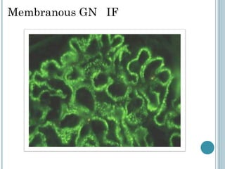
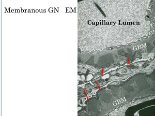
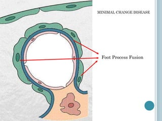
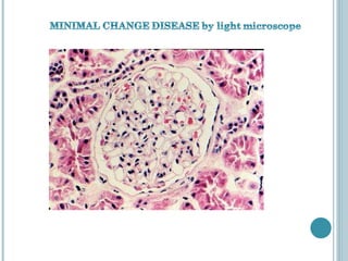
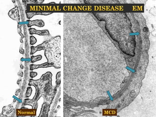
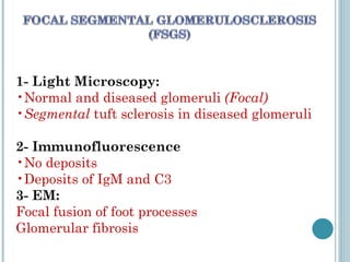
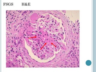
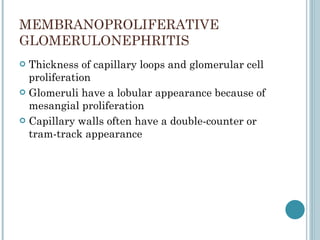
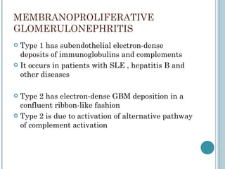
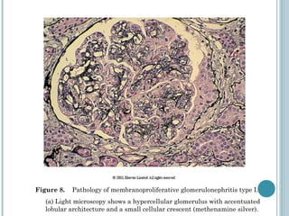
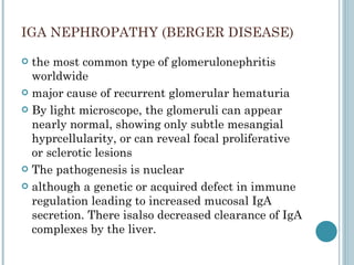
By: Hamza AlGhamdi
NORMAL KIDNEY’S PHYSIOLOGY AND HISTOLOGY
Capillary Space Endothelium Urinary Space GBM Podocyte
NORMAL KIDNEY HISTOLOGY
Parietal Epithelium Visceral Epithelium Capillary Lumen Mesangial Cell Basement Membrane (GBM) Urinary Space RBC Endothelial Cell
NEPHROTIC SYNDROME IS NOT A DISEASE
Fluid retention -> abdominal distention , ascites , edema , puffy eyelids ,scrotal swelling , weight gain , shortness of breath  Anorexia Hypertension  Oliguria  Orthostatic hypertension Skin striae Foamy urine
Periorbital edema Pitting edema of lower limbs
INVESTIGATIONS Urine sample shows proteinuria (>3.5/day ) It is also examined for urinary casts Comprehensive metabolic panel (CMP) shows Hypoalbuminemia High levels of cholesterol (hypercholesterolemia), specifically elevated LDL Electrolytes, urea and creatinine (EUCs): to evaluate renal function Biopsy of  kidney   Auto-immune  markers
Excessive permeability of plasma proteins - >> heavy   proteinuria  Depletion of plasma proteins , mainly albumin -  hypoalbuminaemia
Liver compensates but not successful - >>  reversed A:G ratio Reduced albumin ->  decreased colloid oncotic pressure  of the blood ->  oedema
ADH is stimulated  - >>  oedema  Increased Lipoprotein synthesis and decreased catabolism by liver ->>  Hyperlipidaemia   (mainly VLDL and /or LDL)  HDL is also lost in  urine when heavy  proteinuria  occurs
Loss of body proteins  (immunoglobulins /complement)  -> frequent infection  Loss of anticoagulants antithrombin III , antiplasmin  -  > thrombotic and embolic phenomenon
 
 
Major cause of NS in adults Characterised by  presence of electron-dense  immune deposits along the epithelial side of GBM (subepithelial ) Idiopathic in 85% of patients The 15% remaining GN is associated with malignant tumors , SLE , drugs , infections or metabolic disorders
 
Membranous GN Sub-epithelial immune  Complex deposits Thickened GBM
Membranous GN  H&E
Membranous GN  IF
Capillary Lumen GBM GBM Membranous GN  EM
MINIMAL CHANGE DISEASE the major cause of nephritic syndrome in Children normal  glomeruli on light Microscopy uniform and diffuse effacement of the foot Processes of visceral epithelial  cells on electron microscopy Immunofluorescence  shows no immune deposits The cause and pathogenesis are unknown dramatic response to corticosteroid therapy. long-term prognosis is excellent.
 
MINIMAL CHANGE DISEASE Foot Process Fusion
 
 
sclerosis of some, but not all, glomeruli (thus, it is focal) FSG can be. * Idiopathic. * A secondary evernt, refecting glomerular scarring, consequent to another primary glomerular disease (e.g., IgA nephropathy). * associated to other known disorders (e.g. heroin abuse, HIV infection, obesity). * The result of inherited mutations of proteins present in podocytes (podocin, a –actinin) or in the slit diaphragm between podocytes (nephrin).
1- Light Microscopy:  Normal and diseased glomeruli  (Focal) Segmental  tuft sclerosis in diseased glomeruli  2- Immunofluorescence  No deposits  Deposits of IgM and C3 3- EM:  Focal fusion of foot processes Glomerular fibrosis
FSGS  H&E
MEMBRANOPROLIFERATIVE  GLOMERULONEPHRITIS Thickness of capillary loops and glomerular cell proliferation Glomeruli have a lobular appearance because of mesangial proliferation  Capillary walls often have a double-counter or tram-track appearance
MEMBRANOPROLIFERATIVE  GLOMERULONEPHRITIS Type 1 has subendothelial electron-dense deposits of immunoglobulins and complements It occurs in patients with SLE , hepatitis B and other diseases Type 2 has electron-dense GBM deposition in a confluent ribbon-like fashion Type 2 is due to activation of alternative pathway of complement activation
Figure 8.  Pathology of membranoproliferative glomerulonephritis type I.  (a) Light microscopy shows a hypercellular glomerulus with accentuated lobular architecture and a small cellular crescent (methenamine silver).
IGA NEPHROPATHY (BERGER DISEASE) the most common type of glomerulonephritis worldwide major cause of recurrent glomerular hematuria By light microscope, the glomeruli can appear nearly normal, showing only subtle mesangial hyprcellularity, or can reveal focal proliferative or sclerotic lesions The pathogenesis is nuclear although a genetic or acquired defect in immune regulation leading to increased mucosal IgA secretion. There isalso decreased clearance of IgA complexes by the liver.
Similar IgA deposits are seen in  Henoch-Schönlein purpura  in children hematuria typically lasts for several days chronic renal failure develops in 50% over a period of 20 years
THANK YOU Powerpoint presentation

Nephrotic syndrome

  • 1.
  • 2.
  • 3.
    Capillary Space EndotheliumUrinary Space GBM Podocyte
  • 4.
  • 5.
    Parietal Epithelium VisceralEpithelium Capillary Lumen Mesangial Cell Basement Membrane (GBM) Urinary Space RBC Endothelial Cell
  • 6.
    NEPHROTIC SYNDROME ISNOT A DISEASE
  • 7.
    Fluid retention ->abdominal distention , ascites , edema , puffy eyelids ,scrotal swelling , weight gain , shortness of breath Anorexia Hypertension Oliguria Orthostatic hypertension Skin striae Foamy urine
  • 8.
    Periorbital edema Pittingedema of lower limbs
  • 9.
    INVESTIGATIONS Urine sampleshows proteinuria (>3.5/day ) It is also examined for urinary casts Comprehensive metabolic panel (CMP) shows Hypoalbuminemia High levels of cholesterol (hypercholesterolemia), specifically elevated LDL Electrolytes, urea and creatinine (EUCs): to evaluate renal function Biopsy of kidney Auto-immune markers
  • 10.
    Excessive permeability ofplasma proteins - >> heavy proteinuria Depletion of plasma proteins , mainly albumin - hypoalbuminaemia
  • 11.
    Liver compensates butnot successful - >> reversed A:G ratio Reduced albumin -> decreased colloid oncotic pressure of the blood -> oedema
  • 12.
    ADH is stimulated - >> oedema Increased Lipoprotein synthesis and decreased catabolism by liver ->> Hyperlipidaemia (mainly VLDL and /or LDL) HDL is also lost in urine when heavy proteinuria occurs
  • 13.
    Loss of bodyproteins (immunoglobulins /complement) -> frequent infection Loss of anticoagulants antithrombin III , antiplasmin - > thrombotic and embolic phenomenon
  • 14.
  • 15.
  • 16.
    Major cause ofNS in adults Characterised by presence of electron-dense immune deposits along the epithelial side of GBM (subepithelial ) Idiopathic in 85% of patients The 15% remaining GN is associated with malignant tumors , SLE , drugs , infections or metabolic disorders
  • 17.
  • 18.
    Membranous GN Sub-epithelialimmune Complex deposits Thickened GBM
  • 19.
  • 20.
  • 21.
    Capillary Lumen GBMGBM Membranous GN EM
  • 22.
    MINIMAL CHANGE DISEASEthe major cause of nephritic syndrome in Children normal glomeruli on light Microscopy uniform and diffuse effacement of the foot Processes of visceral epithelial cells on electron microscopy Immunofluorescence shows no immune deposits The cause and pathogenesis are unknown dramatic response to corticosteroid therapy. long-term prognosis is excellent.
  • 23.
  • 24.
    MINIMAL CHANGE DISEASEFoot Process Fusion
  • 25.
  • 26.
  • 27.
    sclerosis of some,but not all, glomeruli (thus, it is focal) FSG can be. * Idiopathic. * A secondary evernt, refecting glomerular scarring, consequent to another primary glomerular disease (e.g., IgA nephropathy). * associated to other known disorders (e.g. heroin abuse, HIV infection, obesity). * The result of inherited mutations of proteins present in podocytes (podocin, a –actinin) or in the slit diaphragm between podocytes (nephrin).
  • 28.
    1- Light Microscopy: Normal and diseased glomeruli (Focal) Segmental tuft sclerosis in diseased glomeruli 2- Immunofluorescence No deposits Deposits of IgM and C3 3- EM: Focal fusion of foot processes Glomerular fibrosis
  • 29.
  • 30.
    MEMBRANOPROLIFERATIVE GLOMERULONEPHRITISThickness of capillary loops and glomerular cell proliferation Glomeruli have a lobular appearance because of mesangial proliferation Capillary walls often have a double-counter or tram-track appearance
  • 31.
    MEMBRANOPROLIFERATIVE GLOMERULONEPHRITISType 1 has subendothelial electron-dense deposits of immunoglobulins and complements It occurs in patients with SLE , hepatitis B and other diseases Type 2 has electron-dense GBM deposition in a confluent ribbon-like fashion Type 2 is due to activation of alternative pathway of complement activation
  • 32.
    Figure 8. Pathology of membranoproliferative glomerulonephritis type I. (a) Light microscopy shows a hypercellular glomerulus with accentuated lobular architecture and a small cellular crescent (methenamine silver).
  • 33.
    IGA NEPHROPATHY (BERGERDISEASE) the most common type of glomerulonephritis worldwide major cause of recurrent glomerular hematuria By light microscope, the glomeruli can appear nearly normal, showing only subtle mesangial hyprcellularity, or can reveal focal proliferative or sclerotic lesions The pathogenesis is nuclear although a genetic or acquired defect in immune regulation leading to increased mucosal IgA secretion. There isalso decreased clearance of IgA complexes by the liver.
  • 34.
    Similar IgA depositsare seen in Henoch-Schönlein purpura in children hematuria typically lasts for several days chronic renal failure develops in 50% over a period of 20 years
  • 35.