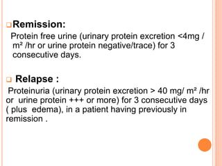
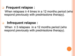
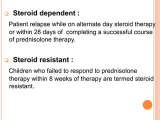
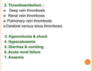
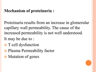
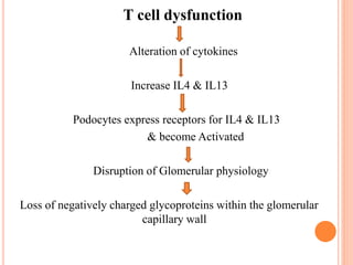
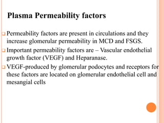
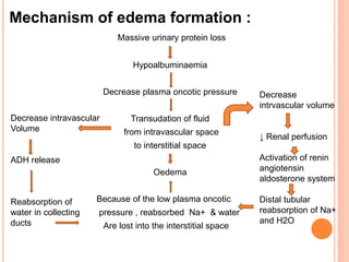
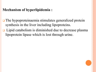
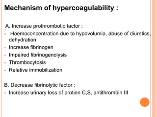
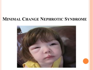
WELCOME
TO
SEMINAR
NEPHROTIC
SYNDROME
PRESENTED BY-
DR. SYED KAMRUL HASAN
IMO, DEPARTMENT OF PAEDIATRICS,
JRRMCH, SYLHET
What is Nephrotic syndrome ?
Nephrotic syndrome is a clinical state characterized by :
Massive proteinuria ( > 40 mg /m²/hour)
Hypoalbuminaemia ( < 2.5 gm/dl)
Generalized edema
Hyperlipidemia ( S. cholesterol >250 mg /dl)
EPIDEMIOLOGY :
2-7/lac, <16 years
In Asia, 7-16/lac, < 16 years
60%-80% present before 6 years
MCNS – M:F 2:1,
Median age 2 and half years
FSGS- M:F 1.3:1
PATHOPHYSIOLOGY
Mechanism of proteinuria :
Proteinuria results from an increase in glomerular
capillary wall permeability. The cause of the
increased permeability is not well understood.
It may be due to :
 T cell dysfunction
 Plasma Permeability factor
 Mutation of genes
T cell dysfunction
Alteration of cytokines
Increase IL4 & IL13
Podocytes express receptors for IL4 & IL13
& become Activated
Disruption of Glomerular physiology
Loss of negatively charged glycoproteins within the glomerular
capillary wall
Plasma Permeability factors
 Permeability factors are present in circulations and they
increase glomerular permeability in MCD and FSGS.
 Important permeability factors are – Vascular endothelial
growth factor (VEGF) and Heparanase.
 VEGF-produced by glomerular podocytes and receptors for
these factors are located on glomerular endothelial cell and
mesangial cells
Mechanism of edema formation :
Massive urinary protein loss
Hypoalbuminaemia
Decrease plasma oncotic pressure
Transudation of fluid
from intravascular space
to interstitial space
Oedema
Because of the low plasma oncotic
pressure , reabsorbed Na+ & water
Are lost into the interstitial space
Decrease intravascular
Volume
ADH release
Reabsorption of
water in collecting
ducts
Decrease
intrvascular volume
↓ Renal perfusion
Activation of renin
angiotensin
aldosterone system
Distal tubular
reabsorption of Na+
and H2O
Mechanism of hyperlipidemia :
 The hypoproteinaemia stimulates generalized protein
synthesis in the liver including lipoproteins.
 Lipid catabolism is diminished due to decrease plasma
lipoprotein lipase which is lost through urine.
Mechanism of hypercoagulability :
A. Increase prothrombotic factor :
 Haemoconcentration due to hypovolumia, abuse of diuretics,
dehydration
 Increase fibrinogen
 Impaired fibrinogenolysis
 Thrombocytosis
 Relative immobilization
B. Decrease fibrinolytic factor :
 Increase urinary loss of protien C,S, antithrombin III
ETIOLOGY
Idiopathic Nephrotic Syndrome: (90%)
a. Minimal change nephrotic syndrome (85%)
b. Focal segmental glomerulo-sclerosis (10%)
c. Mesangial proliferation (5%)
Secondary Nephrotic Syndrome : (10%)
 Infection : Hepatitis B & C, HIV, malaria, syphilis,
toxoplasmosis, cytomegalovirus(CMV), bacterial
endocarditis
 Drugs : Penicillamine, Gold, NSAIDS, Interferone,
Lithium, Captopril, Mercury, Phenidione.
 Allergic Disorder : Bee sting, Food allergens
 Malignant Disease : Chronic lymphocytic
leukaemia, Hodgkin’s lymphoma
 Systemic disease : SLE, HSP, Amyloidosis,
Polyarteritis , Diabetes mellitus
Congenital Nephrotic Syndrome : Rare
Finnish Type
Denys Drash Syndrome
MINIMAL CHANGE NEPHROTIC SYNDROME
• Most commonest type of nephrotic syndrome ,
about 85% of idiopathic nephrotic syndrome.
• Common in male than female (2:1).
• Common in between 2-6 years of age.
• Usually present with mild oedema, initially noted
around the eyes with times oedema become
generalized with development of ascites, pleural
effusion & genital oedema.
• Anorexia, irritability, abdominal pain & diarrhoea are
common.
• Hypertension & gross haematuria are uncommon.
• Bed side heat coagulation test +++/++++
• In light microscopy: No appreciable glomerular
pathology is noted.
• Immunofluroscence microscopy : Typically negative.
• In electron microscopy: Effacement of foot processes
of podocytes in glomerular basement membrane.
• More than 95% response to steroid but high
tendency to relapse.
Minimal Change
Nephrotic Syndrome
FOCAL SEGMENTAL GLOMERULOSCLEROSIS
• About 10% of idiopathic nephrotic syndrome.
• Occurs in older children.
• Male female ratio 1.3:1.
• The clinical profile is similar to MCNS, though
microscopic hematuria & hypertension may be
present.
• Only 20% response to steroid.
• On light microscopy : shows mesangial
proliferation & segmental scarring.
• On immunofluroscence microscopy : shows IgM
& C3 staining in the area of segmental sclerosis.
• On electron microscopy : shows segmental
scarring of glomerulus tuft with the obliteration of
the glomerular capillary lumen.
MESENGIAL PROLIFERATION
o Usually occurs in older children.
o Male & female are equally affected.
o The clinical profile is similar to MCNS with
haematuria & hypertension.
o About 50 % response to steroid.
o On light microscopy : shows diffuse increase in
mesangial cell & matrix.
o Immunofluroscence microscopy : shows slight
deposition of IgM in mesengial cell.
o Electron microscopy : shows increse number of
mesengial cells & matrix as well as effacement of
the epithelial cell foot process.
CONGENITAL NEPHROTIC SYNDROME
 Nephrotic syndrome is present at birth or appears
within three months of life.
 Renal biopsy should be performed in all cases.
 The commonest form is the autosomal recessive
Finnish nephrotic syndrome due to defective
production of Nephrin.
 There is no specific therapy, appropriate supportive
care is instituted.
Causes of congenital nephrotic syndrome:
 Classic' Finnish 'type.
 Diffuse mesangial sclerosis -
*with pseudohermaphroditism (Denys-Drash
syndrome).
*with microcephaly ,development delay and hiatal
hernia(Galloway-Mowat syndrome).
*idiopathic.
 Primary focal segmental glomerulosclerosis
 Congenital infections : cytomegalovirus, syphilis,
rubella, toxoplasma, hepatitis B.
FINNISH TYPE CONGENITAL
NEPHROTIC SYNDROME
 The Finnish type congenital nephrotic syndrome is
an autosomal recessive disease.
 Infants with the Finnish type of congenital nephrotic
syndrome are born prematurely, often with large
placenta.
 Nephrotic syndrome is present at or soon after birth.
Clinical presentation:
 Failure to thrive,
 Repeated infection,
 Delayed development,
 Ascites,
 Spontaneous vascular thrombosis.
Biopsy shows cortical microcysts, representing
dilated proximal convoluted tubules, glomeruli may
show mesangial proliferation and increased
mesangial matrix.
Relation of edema with hypoalbuminaemia :
Serum albumin Oedema
20 - 25 gm/l Periorbital oedema
18 - <20 gm/l Dependent oedema
15 - < 18 gm/l Ascites
< 15 gm/l Genital oedema
Heat Coagulation Test
Interpretation of HCT :
Keep the tube in front of newspaper
Read newspaper Amount of protein
mg/dl
Clearly read Trace 10-20
Can read but with
difficulty
+ 30-100
Can’t read , only
see writing
++ 100-300
Only paper is seen +++ 300-1000
Nothing is seen ++++ >10000
Terminology Related To
Nephrotic Syndrome
Remission:
Protein free urine (urinary protein excretion <4mg /
m² /hr or urine protein negative/trace) for 3
consecutive days.
 Relapse :
Proteinuria (urinary protein excretion > 40 mg/ m² /hr
or urine protein +++ or more) for 3 consecutive days
( plus edema), in a patient having previously in
remission .
 Frequent relapse :
When relapses ≥ 4 times in a 12 months period (who
respond previously with prednisolone therapy).
 Infrequent relapse :
When ≤ 3 relapses in a 12 months period (who
respond previously with prednisolone therapy).
 Steroid dependent :
Patient relapse while on alternate day steroid therapy
or within 28 days of completing a successful course
of prednisolone therapy.
 Steroid resistant :
Children who failed to respond to prednisolone
therapy within 8 weeks of therapy are termed steroid
resistant.
 Late responder :
Patient with initial resistance who responds
later.
 Late resistance :
Initial responder who subsequently fails to
respond to steroid therapy.
INVESTIGATION
For diagnosis :
- Urine R/M/E ( 3+ or 4+ proteinuria , granular &
hyaline casts)
- Spot urine protein creatinine ratio (>2.0)
- 24 hours urinary total protein (>1gm/m2/day )
- Serum albumin (<2.5 gm/dl)
- Serum cholesterol (> 250mg/dl )
For infection screening :
- CBC
- Blood culture
- Urine culture
- Chest X -ray
- HBsAg
- MT
To exclude secondary nephrotic syndrome :
-Serum creatinine
-Serum C3, C4
-Serum electrolyte
-For cause identificatin :
HBsAg, Anti HCV
ANA, Anti ds DNA
HIV screening
VDRL
Renal USG
Renal biopsy
Renal biopsy
Indication :
 Age of onsent < 1 year or >12 years
 Haematuria
 Sustained hypertension
 Renal failure
 Persistent ↓C3 ,C4
 Steroid resistance nephrotic syndrome
 Suspected secondary cause of nephotic
syndrome
 Before cyclosporine or tacrolimus therapy
MANAGEMENT
Criteria for hospital admission :
 First attack ( for counseling)
 With complications
 Need for renal biopsy
 Doubtful compliance
Aim of management :
 Achieve remission
 Prevention of relapse
 Avoidance of complications & side effects
of drugs
Treatment of Nephrotic Syndrome
 Supportive & Symptomatic management
 Specific management
 Prevention and treatment of complications
Supportive & symptomatic treatment:
 Dietary management & daily activities
 Control of oedema
 Prevention and treatment of infections
 Counseling of parents and psychosocial support.
Dietary Management
A balance diet, adequate in protein and calories
 Protein : An adequate protein intake (1.5-2
gram/kg/day)
 Patient with persistent proteinuria should
receive 2-2.5gm /kg/day.
 Fat : <30% of the diet
 Supplements vitamins and minerals
Salt intake restricted up to remission
DAILY ACTIVITIES
 Physical activity as tolerated
 May attend school
Management of Oedema :
Mild to moderate oedema :
Salt restriction up to remission
No fluid restriction no diuretics
Massive oedema :
Salt restriction
 Fluid restriction
 Diuretics :
Oral frusemide (1-3mg/kg) in 1-3 divided
doses or iv 4-10 mg/kg /day.
Spironolactone (1-3mg/kg/day) added in
case of higher or prolonged duration of
treatment.
Hydrochlorthiazide (1-3 mg/kg/day) or
metolazone (0.1-0.5 mg/kg/day) may be
added with frusemide in severe case.
 I/V 20 % human albumin (0.5-1 gm /kg
/dose over 1-2 hours) when fluid restriction
& diuretics are not effective.
Indication of albumin infusion in NS :
Serum albumin < 1.5 gm/dl
Oedema not improve even after maximum
dose of diuretics
Child with signs of hypovolumia
Severe complication of diuretic therapy
 Prevention & treatment of infection by
appropriate antibiotics
Specific Management for
 First Attack
 Relapse
 Steroid dependent nephrotic syndrome
 Steroid resistant nephrotic syndrome
Frequent relapse
Infrequent relapse
First attack
Prednisolone :
60mg/m2/day (max. 80mg) 2-3 divided doses
for 6 weeks. Then
40mg/m2/every alternate day – single morning for
dose for 4 weeks. Then slowly tapered &
discontinued over the next 4-8 weeks.
Relapse
Prednisolone :
60mg/m2/day - till urine become protein free for 3
consecutive days.
Followed by 40mg/m2/every alternate day single
morning for dose for 4 weeks & gradually
tapered over 4-8 weeks
Alternative drugs used in Nephrotic
Syndrome :
o Levamisole
o Cyclophosphamide & chlorambucil
o Cyclosporine
o Mycophenolate mofetil
o Tacrolimus
o Methyl prednisolone
Frequent relapses /
Steroid dependennce
Prednisolone– 60 mg /m²/ day till remission then
Alternate day prednisolone to maintain remission
Steroid threshold
<0.5 mg/kg on
alternate day
Alternate day
prednisolone
for 9-18 months
Threshold >0.5 mg/kg on
alternate day or
Severe complication, or
steroid toxicity
Levamisole ,
Cyclophosphamide,
Tacrolimus,
Cyclosporin A,
Mycophenolate mofetil
Frequent relapses & Steroid dependence :
Following treatment of a relapse, prednisolone is
gradually tapered to maintain the patient in remission
on alternate day dose of 0.5 mg/kg, which is
administered for 9-18 months. If the prednisolone
threshold to maintain remission is higher or if
features of steroid toxicity are seen, additional use of
the following immunomodulators are suggested :
Levamisole :
2-2.5 mg /kg on alternate days for 12-24 months.
Prednisolone 1.5 mg/kg on alternate days for 2-4
weeks.
Gradually reduced by 0.15-0.25 mg/kg every 4 weeks
to a maintenance dose of 0.25-0.5 mg/kg that is
continued for 6 or more months.
Cyclophosphamide :
2-2.5 mg/kg for 12 weeks
Prednisolone 1.5 mg/kg on alternate days for 4 wks.
Followed by 1mg/kg for next 8 weeks.
Steroid therapy tapered & stopped over the next 2-3
months.
Cyclosporine (CsA) :
4-5 mg/kg daily for 12-24 months
Prednisolone 1.5 mg/kg on alternate days for 2-4
weeks .
Gradually reduced by 0.15-0.25 mg/kg every 4 weeks
to a maintenance dose of 0.25-0.5 mg/kg that is
continued for 6 or more months.
Tacrolimus :
0.1-0.2 mg/kg daily for 12 – 24 months.
Mycophenolate mofetil (MMF) :
20-25 mg/kg/day 12 hourly for 12-24 months
tapering doses of prednisolone for 12 -24 months.
Adjunct therapy :
ACE inhibitor
Angiotensin II blocker
These drugs causing efferent arteriolar dilation &
reducing glomerular filtration pressure & thus
reducing proteinuria in steroid resistant nephrotic
syndrome.
Rituximab:
 Is an anti CD-20 monoclonal antibody
 1-2 dose Rituximab significantly reduces relapse
rate in FRNS and SDNS
 Dose : 375 mg/m2 i.v infusion
Steroid Resistance Nephrotic Syndrome :
The choice of therapy in steroid resistant nephrotic
syndrome include:
calcineurin inhibitors with alternate day prednisolone,
IV methylprednisolone followed by cyclophosphamide
or cyclosporine.
Angiotensin converting enzyme inhibitors and
angiotensin receptor blockers reduce the intensity of
proteinuria.
 Hypertension should be controlled, aiming to achieve
reduction of blood pressure to the 50th percentile.
 Statins are used to control hyperlipidaemia
Treatment protocol of steroid
resistant NS of BSMMU
Failure to remission after 8weeks of prednisolone
therapy(60mg/m2)
Do renal biopsy and if it is MCNS with steroid resistant
Oral cyclosporin (100mg/m2/day) or
Tacrolimus (50-70mg/day) plus
alternate day prednisolone(1.5mg/kg/day) for 1-2 years.
 Another protocol-
Pulse methylprednisolone (20-30mg/kg)alternate
day for 5-6 days followed by tapering dose of oral
prednisolone (1.5mg/kg) every alternate day for 1-
2 years.
 For other than MCNS with steroid resistant
Injection cyclophosphamide (500mg/m2/month) for
6 months plus alternate day prednisolone
(1.5mg/kg) for 1-2 years.
CONGENITAL NEPHROTIC SYNDROME
• There is no specific treatment
• The disease is resistant to corticosteroid &
cytotoxic drugs.
Nephrectomy
Dialysis up to weight 10 kg or 1 year of age
Renal transplantation
COMPLICATIONS OF
NEPHROTIC SYNDROME
Complication related to disease :
1.Infections:
 UTI
 Pneumonia
 Spontaneous bacterial peritonitis
 Cellulitis
 Septicemia
 Meningitis
 Bone & joint infection
 Flair up of tuberculosis
 Varicella, Measles
2. Thromboembolism :
 Deep vein thrombosis
 Renal vein thrombosis
 Pulmonary vein thrombosis
 Cerebral venous sinus thrombosis
3. Hypovolumia & shock
4. Hypocalcaemia
5. Diarrhea & vomiting
6. Acute renal failure
7. Anaemia
8. Abdominal pain due to :-
Peritonitis,
Mesenteric ischaemia,
UTI,
Renal vein thrombosis,
Steroid induced gastritis
COMMON ORGANISM CAUSING INFECTION IN
NEPHROTIC SYNDROME
Bacterial :
Streptococcus pneumoniae
Haemophilus influenzae
E.coli
Klebsiella
Pseudomonas
Mycobacterium tuberculosis
Virus :
Measles
Varicella
Prevention of infection
Immunization:
Administration of live vaccines measles,
mumps, rubella, oral polio, varicella is avoided
until steroid therapy discontinued for 4 weeks.
Other vaccines pneumococcal, Hib, Hepatitis
B may be given.
Patient in remission should receive the
varicella vaccine.
If the child with nephrotic syndrome is on
continuous immunosuppression , siblings
should receive the inactivated polio vaccine
instead of OPV.
 Siblings can safely receive the MMR &
varicella vaccines without a risk of
exposing the patient to the attenuated
viruses.
Complication due to drug
STEROID
 Cushingoid face
 Obesity
 Cataract
 Buffalo hump
 Hypertension
 Avascular necrosis
 GF
 More chance to develop infection
 Osteoporosis
 Hyperglycaemia
 Peptic ulcer
Low hair line
Cataract
Facial Plethora
Buffalo Hum
Gynaecomashia
Stria
Hypertension
Abdominal fat
WHAT TO DO?
Stop steroid when toxicity develop
Go for Alternative drug
Counseling the parents
CYCLOPHOSPHAMIDE
Side effect:
 Neutropenia
 Disseminated varicella
 Reversible alopecia
 Hemorrhagic cystitis
 Sterility
 Risk of future malignancy
What to do?
- Blood count –weekly during the first 4 weeks
of treatment than 2 weekly.
-If TC of WBC <5000/ cmm –drug is withheld
till normal
-Maintain high urine output to prevent
hemorrhagic cystitis and use MESNA ( sodium 2-
mercaptoethane sulfanate ) if iv cyclophosphamide
given.
CYCLOSPORINE
Side effect:
Hypertension
Nephrotoxicity
Hirsutism
Tremor
Gum hypertrophy
What to do?
Blood level of urea creatinine should be measured
every 4-6 weeks.
LEVAMISOLE
Side effect:
 GIT upset
 Influenza like symptoms
 Skin rash may associated with leukocytoclastic
vasculitis
 Neutropenia
 Liver toxicity
 Convulsion
What to do?
 Monitor blood leucocyte count for every 2-
4 month
 All the side effect diminishes after drug
withdraw
Tacrolimus :
Side effect :
Septicemia
Cardiac damage
Hypertension
Hepatotoxicity
Nephrotoxicity
Electrolyte imbalance
Hyperglycemia
What to do ?
Blood level of creatinine & glucose should estimated
every 2-3 months
SOME SPECIAL SITUATION
NS WITH TB:
Clinical feature
 low grade fever
 persisting cough
Investigation:
 Chest X-Ray
 MT
Treatment:
 At first start anti tubercular drugs , after 2 weeks
start prednisolone therapy.
NS WITH VARICELA
Treatment:
oral acyclovir 80 mg/kg daily in 4 divided dose for 7-
10 days.
Patient exposed to varicella:
 VZIG 125 unit/10 kg body weight ( minimum
125unit , maximum 625unit) should be given within
96 hours of significant exposure.
 Intravenous immunoglobulin 400mg/kg may be
used.
NS WITH HEPATITIS B
 Lamivudin 2 week before steroid given &
continue 6 weeks to 6 month after the end
of the treatment.
 Interferon should also be given.
NS WITH MEASLES
 Measles causes a serious danger to
patients receiving steroid & cytotoxic drug.
 NS less than <1 year is uncommon , so
most patients are already vaccinated
before they get NS.
 After 2 weeks start prednisolone therapy.
COUNSELING
 Should be trained how to test urine for albumin &
maintain a diary.
 Parents should be explained about the naturel
history of the disease.
 Should be provided with a booklet covering all
information about the condition.
 Normal activity & schooling
 Parents compliance & co operation is essential.
Prognosis :
Depends on whether the patient is steroid
responsive or resistant.
In Steroid sensitive Nephrotic Syndrome:
The final outcome is excellent . Most of the patient
stop getting relapses between the age of 14-20 years
without any residual renal dysfunction. The
subsequent course of steroid responsive NS are-
25-40% have infrequent relapse
40% have frequent relapse
20% steroid dependence
In Steroid resistant Nephrotic Syndrome :
Generally have much poorer prognosis.
Develop ESRD over 10 years requiring dialysis
& or transplantation.
FOLLOW UP
During hospital admission:
Daily- Vital signs,
- Weight
- Fluid intake
- Urine out put
-Abdominal girth
-Edema
-Any sign of infection
.
During discharge:
 Mother should know when to return
 If the child develop edema with any sign of
infection such as fever, cough, diarrhea,
vomiting then mother should test urine for
albumin.
 If the urine for albumin 3+ or more for 3
consecutive days then she must return to
the hospital.
BAD PROGNOSTIC SIGNS OF NS
 Very young (<1 yr) & (>10yr)
 Persistent heavy hematuria
 Hypocomplementemia
 Hypertension
 Renal failure
 No Response within 28 days of adequate
prednisolone regimen
Nephrotic syndrome

Nephrotic syndrome

  • 1.
  • 2.
    NEPHROTIC SYNDROME PRESENTED BY- DR. SYEDKAMRUL HASAN IMO, DEPARTMENT OF PAEDIATRICS, JRRMCH, SYLHET
  • 3.
    What is Nephroticsyndrome ? Nephrotic syndrome is a clinical state characterized by : Massive proteinuria ( > 40 mg /m²/hour) Hypoalbuminaemia ( < 2.5 gm/dl) Generalized edema Hyperlipidemia ( S. cholesterol >250 mg /dl)
  • 4.
    EPIDEMIOLOGY : 2-7/lac, <16years In Asia, 7-16/lac, < 16 years 60%-80% present before 6 years MCNS – M:F 2:1, Median age 2 and half years FSGS- M:F 1.3:1
  • 5.
  • 6.
    Mechanism of proteinuria: Proteinuria results from an increase in glomerular capillary wall permeability. The cause of the increased permeability is not well understood. It may be due to :  T cell dysfunction  Plasma Permeability factor  Mutation of genes
  • 7.
    T cell dysfunction Alterationof cytokines Increase IL4 & IL13 Podocytes express receptors for IL4 & IL13 & become Activated Disruption of Glomerular physiology Loss of negatively charged glycoproteins within the glomerular capillary wall
  • 8.
    Plasma Permeability factors Permeability factors are present in circulations and they increase glomerular permeability in MCD and FSGS.  Important permeability factors are – Vascular endothelial growth factor (VEGF) and Heparanase.  VEGF-produced by glomerular podocytes and receptors for these factors are located on glomerular endothelial cell and mesangial cells
  • 10.
    Mechanism of edemaformation : Massive urinary protein loss Hypoalbuminaemia Decrease plasma oncotic pressure Transudation of fluid from intravascular space to interstitial space Oedema Because of the low plasma oncotic pressure , reabsorbed Na+ & water Are lost into the interstitial space Decrease intravascular Volume ADH release Reabsorption of water in collecting ducts Decrease intrvascular volume ↓ Renal perfusion Activation of renin angiotensin aldosterone system Distal tubular reabsorption of Na+ and H2O
  • 11.
    Mechanism of hyperlipidemia:  The hypoproteinaemia stimulates generalized protein synthesis in the liver including lipoproteins.  Lipid catabolism is diminished due to decrease plasma lipoprotein lipase which is lost through urine.
  • 12.
    Mechanism of hypercoagulability: A. Increase prothrombotic factor :  Haemoconcentration due to hypovolumia, abuse of diuretics, dehydration  Increase fibrinogen  Impaired fibrinogenolysis  Thrombocytosis  Relative immobilization B. Decrease fibrinolytic factor :  Increase urinary loss of protien C,S, antithrombin III
  • 13.
  • 14.
    Idiopathic Nephrotic Syndrome:(90%) a. Minimal change nephrotic syndrome (85%) b. Focal segmental glomerulo-sclerosis (10%) c. Mesangial proliferation (5%)
  • 15.
    Secondary Nephrotic Syndrome: (10%)  Infection : Hepatitis B & C, HIV, malaria, syphilis, toxoplasmosis, cytomegalovirus(CMV), bacterial endocarditis  Drugs : Penicillamine, Gold, NSAIDS, Interferone, Lithium, Captopril, Mercury, Phenidione.  Allergic Disorder : Bee sting, Food allergens  Malignant Disease : Chronic lymphocytic leukaemia, Hodgkin’s lymphoma  Systemic disease : SLE, HSP, Amyloidosis, Polyarteritis , Diabetes mellitus
  • 16.
    Congenital Nephrotic Syndrome: Rare Finnish Type Denys Drash Syndrome
  • 17.
  • 18.
    • Most commonesttype of nephrotic syndrome , about 85% of idiopathic nephrotic syndrome. • Common in male than female (2:1). • Common in between 2-6 years of age. • Usually present with mild oedema, initially noted around the eyes with times oedema become generalized with development of ascites, pleural effusion & genital oedema. • Anorexia, irritability, abdominal pain & diarrhoea are common. • Hypertension & gross haematuria are uncommon. • Bed side heat coagulation test +++/++++
  • 19.
    • In lightmicroscopy: No appreciable glomerular pathology is noted. • Immunofluroscence microscopy : Typically negative. • In electron microscopy: Effacement of foot processes of podocytes in glomerular basement membrane. • More than 95% response to steroid but high tendency to relapse.
  • 20.
  • 22.
  • 23.
    • About 10%of idiopathic nephrotic syndrome. • Occurs in older children. • Male female ratio 1.3:1. • The clinical profile is similar to MCNS, though microscopic hematuria & hypertension may be present. • Only 20% response to steroid.
  • 24.
    • On lightmicroscopy : shows mesangial proliferation & segmental scarring. • On immunofluroscence microscopy : shows IgM & C3 staining in the area of segmental sclerosis. • On electron microscopy : shows segmental scarring of glomerulus tuft with the obliteration of the glomerular capillary lumen.
  • 25.
  • 26.
    o Usually occursin older children. o Male & female are equally affected. o The clinical profile is similar to MCNS with haematuria & hypertension. o About 50 % response to steroid.
  • 27.
    o On lightmicroscopy : shows diffuse increase in mesangial cell & matrix. o Immunofluroscence microscopy : shows slight deposition of IgM in mesengial cell. o Electron microscopy : shows increse number of mesengial cells & matrix as well as effacement of the epithelial cell foot process.
  • 28.
  • 29.
     Nephrotic syndromeis present at birth or appears within three months of life.  Renal biopsy should be performed in all cases.  The commonest form is the autosomal recessive Finnish nephrotic syndrome due to defective production of Nephrin.  There is no specific therapy, appropriate supportive care is instituted.
  • 30.
    Causes of congenitalnephrotic syndrome:  Classic' Finnish 'type.  Diffuse mesangial sclerosis - *with pseudohermaphroditism (Denys-Drash syndrome). *with microcephaly ,development delay and hiatal hernia(Galloway-Mowat syndrome). *idiopathic.  Primary focal segmental glomerulosclerosis  Congenital infections : cytomegalovirus, syphilis, rubella, toxoplasma, hepatitis B.
  • 31.
  • 32.
     The Finnishtype congenital nephrotic syndrome is an autosomal recessive disease.  Infants with the Finnish type of congenital nephrotic syndrome are born prematurely, often with large placenta.  Nephrotic syndrome is present at or soon after birth.
  • 33.
    Clinical presentation:  Failureto thrive,  Repeated infection,  Delayed development,  Ascites,  Spontaneous vascular thrombosis. Biopsy shows cortical microcysts, representing dilated proximal convoluted tubules, glomeruli may show mesangial proliferation and increased mesangial matrix.
  • 34.
    Relation of edemawith hypoalbuminaemia : Serum albumin Oedema 20 - 25 gm/l Periorbital oedema 18 - <20 gm/l Dependent oedema 15 - < 18 gm/l Ascites < 15 gm/l Genital oedema
  • 35.
  • 36.
    Interpretation of HCT: Keep the tube in front of newspaper Read newspaper Amount of protein mg/dl Clearly read Trace 10-20 Can read but with difficulty + 30-100 Can’t read , only see writing ++ 100-300 Only paper is seen +++ 300-1000 Nothing is seen ++++ >10000
  • 38.
  • 39.
    Remission: Protein free urine(urinary protein excretion <4mg / m² /hr or urine protein negative/trace) for 3 consecutive days.  Relapse : Proteinuria (urinary protein excretion > 40 mg/ m² /hr or urine protein +++ or more) for 3 consecutive days ( plus edema), in a patient having previously in remission .
  • 40.
     Frequent relapse: When relapses ≥ 4 times in a 12 months period (who respond previously with prednisolone therapy).  Infrequent relapse : When ≤ 3 relapses in a 12 months period (who respond previously with prednisolone therapy).
  • 41.
     Steroid dependent: Patient relapse while on alternate day steroid therapy or within 28 days of completing a successful course of prednisolone therapy.  Steroid resistant : Children who failed to respond to prednisolone therapy within 8 weeks of therapy are termed steroid resistant.
  • 42.
     Late responder: Patient with initial resistance who responds later.  Late resistance : Initial responder who subsequently fails to respond to steroid therapy.
  • 43.
  • 44.
    For diagnosis : -Urine R/M/E ( 3+ or 4+ proteinuria , granular & hyaline casts) - Spot urine protein creatinine ratio (>2.0) - 24 hours urinary total protein (>1gm/m2/day ) - Serum albumin (<2.5 gm/dl) - Serum cholesterol (> 250mg/dl )
  • 45.
    For infection screening: - CBC - Blood culture - Urine culture - Chest X -ray - HBsAg - MT
  • 46.
    To exclude secondarynephrotic syndrome : -Serum creatinine -Serum C3, C4 -Serum electrolyte -For cause identificatin : HBsAg, Anti HCV ANA, Anti ds DNA HIV screening VDRL Renal USG Renal biopsy
  • 47.
    Renal biopsy Indication : Age of onsent < 1 year or >12 years  Haematuria  Sustained hypertension  Renal failure  Persistent ↓C3 ,C4  Steroid resistance nephrotic syndrome  Suspected secondary cause of nephotic syndrome  Before cyclosporine or tacrolimus therapy
  • 48.
  • 49.
    Criteria for hospitaladmission :  First attack ( for counseling)  With complications  Need for renal biopsy  Doubtful compliance
  • 50.
    Aim of management:  Achieve remission  Prevention of relapse  Avoidance of complications & side effects of drugs
  • 51.
    Treatment of NephroticSyndrome  Supportive & Symptomatic management  Specific management  Prevention and treatment of complications
  • 52.
    Supportive & symptomatictreatment:  Dietary management & daily activities  Control of oedema  Prevention and treatment of infections  Counseling of parents and psychosocial support.
  • 53.
    Dietary Management A balancediet, adequate in protein and calories  Protein : An adequate protein intake (1.5-2 gram/kg/day)  Patient with persistent proteinuria should receive 2-2.5gm /kg/day.  Fat : <30% of the diet  Supplements vitamins and minerals Salt intake restricted up to remission
  • 54.
    DAILY ACTIVITIES  Physicalactivity as tolerated  May attend school
  • 55.
    Management of Oedema: Mild to moderate oedema : Salt restriction up to remission No fluid restriction no diuretics
  • 56.
    Massive oedema : Saltrestriction  Fluid restriction  Diuretics : Oral frusemide (1-3mg/kg) in 1-3 divided doses or iv 4-10 mg/kg /day. Spironolactone (1-3mg/kg/day) added in case of higher or prolonged duration of treatment.
  • 57.
    Hydrochlorthiazide (1-3 mg/kg/day)or metolazone (0.1-0.5 mg/kg/day) may be added with frusemide in severe case.  I/V 20 % human albumin (0.5-1 gm /kg /dose over 1-2 hours) when fluid restriction & diuretics are not effective.
  • 58.
    Indication of albumininfusion in NS : Serum albumin < 1.5 gm/dl Oedema not improve even after maximum dose of diuretics Child with signs of hypovolumia Severe complication of diuretic therapy
  • 59.
     Prevention &treatment of infection by appropriate antibiotics
  • 60.
    Specific Management for First Attack  Relapse  Steroid dependent nephrotic syndrome  Steroid resistant nephrotic syndrome Frequent relapse Infrequent relapse
  • 61.
    First attack Prednisolone : 60mg/m2/day(max. 80mg) 2-3 divided doses for 6 weeks. Then 40mg/m2/every alternate day – single morning for dose for 4 weeks. Then slowly tapered & discontinued over the next 4-8 weeks.
  • 62.
    Relapse Prednisolone : 60mg/m2/day -till urine become protein free for 3 consecutive days. Followed by 40mg/m2/every alternate day single morning for dose for 4 weeks & gradually tapered over 4-8 weeks
  • 63.
    Alternative drugs usedin Nephrotic Syndrome : o Levamisole o Cyclophosphamide & chlorambucil o Cyclosporine o Mycophenolate mofetil o Tacrolimus o Methyl prednisolone
  • 64.
    Frequent relapses / Steroiddependennce Prednisolone– 60 mg /m²/ day till remission then Alternate day prednisolone to maintain remission Steroid threshold <0.5 mg/kg on alternate day Alternate day prednisolone for 9-18 months Threshold >0.5 mg/kg on alternate day or Severe complication, or steroid toxicity Levamisole , Cyclophosphamide, Tacrolimus, Cyclosporin A, Mycophenolate mofetil
  • 65.
    Frequent relapses &Steroid dependence : Following treatment of a relapse, prednisolone is gradually tapered to maintain the patient in remission on alternate day dose of 0.5 mg/kg, which is administered for 9-18 months. If the prednisolone threshold to maintain remission is higher or if features of steroid toxicity are seen, additional use of the following immunomodulators are suggested :
  • 66.
    Levamisole : 2-2.5 mg/kg on alternate days for 12-24 months. Prednisolone 1.5 mg/kg on alternate days for 2-4 weeks. Gradually reduced by 0.15-0.25 mg/kg every 4 weeks to a maintenance dose of 0.25-0.5 mg/kg that is continued for 6 or more months.
  • 67.
    Cyclophosphamide : 2-2.5 mg/kgfor 12 weeks Prednisolone 1.5 mg/kg on alternate days for 4 wks. Followed by 1mg/kg for next 8 weeks. Steroid therapy tapered & stopped over the next 2-3 months.
  • 68.
    Cyclosporine (CsA) : 4-5mg/kg daily for 12-24 months Prednisolone 1.5 mg/kg on alternate days for 2-4 weeks . Gradually reduced by 0.15-0.25 mg/kg every 4 weeks to a maintenance dose of 0.25-0.5 mg/kg that is continued for 6 or more months.
  • 69.
    Tacrolimus : 0.1-0.2 mg/kgdaily for 12 – 24 months. Mycophenolate mofetil (MMF) : 20-25 mg/kg/day 12 hourly for 12-24 months tapering doses of prednisolone for 12 -24 months.
  • 70.
    Adjunct therapy : ACEinhibitor Angiotensin II blocker These drugs causing efferent arteriolar dilation & reducing glomerular filtration pressure & thus reducing proteinuria in steroid resistant nephrotic syndrome.
  • 71.
    Rituximab:  Is ananti CD-20 monoclonal antibody  1-2 dose Rituximab significantly reduces relapse rate in FRNS and SDNS  Dose : 375 mg/m2 i.v infusion
  • 72.
    Steroid Resistance NephroticSyndrome : The choice of therapy in steroid resistant nephrotic syndrome include: calcineurin inhibitors with alternate day prednisolone, IV methylprednisolone followed by cyclophosphamide or cyclosporine. Angiotensin converting enzyme inhibitors and angiotensin receptor blockers reduce the intensity of proteinuria.  Hypertension should be controlled, aiming to achieve reduction of blood pressure to the 50th percentile.  Statins are used to control hyperlipidaemia
  • 73.
    Treatment protocol ofsteroid resistant NS of BSMMU Failure to remission after 8weeks of prednisolone therapy(60mg/m2) Do renal biopsy and if it is MCNS with steroid resistant Oral cyclosporin (100mg/m2/day) or Tacrolimus (50-70mg/day) plus alternate day prednisolone(1.5mg/kg/day) for 1-2 years.
  • 74.
     Another protocol- Pulsemethylprednisolone (20-30mg/kg)alternate day for 5-6 days followed by tapering dose of oral prednisolone (1.5mg/kg) every alternate day for 1- 2 years.  For other than MCNS with steroid resistant Injection cyclophosphamide (500mg/m2/month) for 6 months plus alternate day prednisolone (1.5mg/kg) for 1-2 years.
  • 75.
    CONGENITAL NEPHROTIC SYNDROME •There is no specific treatment • The disease is resistant to corticosteroid & cytotoxic drugs. Nephrectomy Dialysis up to weight 10 kg or 1 year of age Renal transplantation
  • 76.
  • 77.
    Complication related todisease : 1.Infections:  UTI  Pneumonia  Spontaneous bacterial peritonitis  Cellulitis  Septicemia  Meningitis  Bone & joint infection  Flair up of tuberculosis  Varicella, Measles
  • 78.
    2. Thromboembolism : Deep vein thrombosis  Renal vein thrombosis  Pulmonary vein thrombosis  Cerebral venous sinus thrombosis 3. Hypovolumia & shock 4. Hypocalcaemia 5. Diarrhea & vomiting 6. Acute renal failure 7. Anaemia
  • 79.
    8. Abdominal paindue to :- Peritonitis, Mesenteric ischaemia, UTI, Renal vein thrombosis, Steroid induced gastritis
  • 80.
    COMMON ORGANISM CAUSINGINFECTION IN NEPHROTIC SYNDROME Bacterial : Streptococcus pneumoniae Haemophilus influenzae E.coli Klebsiella Pseudomonas Mycobacterium tuberculosis Virus : Measles Varicella
  • 81.
    Prevention of infection Immunization: Administrationof live vaccines measles, mumps, rubella, oral polio, varicella is avoided until steroid therapy discontinued for 4 weeks. Other vaccines pneumococcal, Hib, Hepatitis B may be given. Patient in remission should receive the varicella vaccine.
  • 82.
    If the childwith nephrotic syndrome is on continuous immunosuppression , siblings should receive the inactivated polio vaccine instead of OPV.  Siblings can safely receive the MMR & varicella vaccines without a risk of exposing the patient to the attenuated viruses.
  • 83.
  • 84.
    STEROID  Cushingoid face Obesity  Cataract  Buffalo hump  Hypertension  Avascular necrosis  GF  More chance to develop infection  Osteoporosis  Hyperglycaemia  Peptic ulcer
  • 85.
    Low hair line Cataract FacialPlethora Buffalo Hum Gynaecomashia Stria Hypertension Abdominal fat
  • 86.
    WHAT TO DO? Stopsteroid when toxicity develop Go for Alternative drug Counseling the parents
  • 87.
    CYCLOPHOSPHAMIDE Side effect:  Neutropenia Disseminated varicella  Reversible alopecia  Hemorrhagic cystitis  Sterility  Risk of future malignancy
  • 88.
    What to do? -Blood count –weekly during the first 4 weeks of treatment than 2 weekly. -If TC of WBC <5000/ cmm –drug is withheld till normal -Maintain high urine output to prevent hemorrhagic cystitis and use MESNA ( sodium 2- mercaptoethane sulfanate ) if iv cyclophosphamide given.
  • 89.
    CYCLOSPORINE Side effect: Hypertension Nephrotoxicity Hirsutism Tremor Gum hypertrophy Whatto do? Blood level of urea creatinine should be measured every 4-6 weeks.
  • 90.
    LEVAMISOLE Side effect:  GITupset  Influenza like symptoms  Skin rash may associated with leukocytoclastic vasculitis  Neutropenia  Liver toxicity  Convulsion
  • 91.
    What to do? Monitor blood leucocyte count for every 2- 4 month  All the side effect diminishes after drug withdraw
  • 92.
    Tacrolimus : Side effect: Septicemia Cardiac damage Hypertension Hepatotoxicity Nephrotoxicity Electrolyte imbalance Hyperglycemia What to do ? Blood level of creatinine & glucose should estimated every 2-3 months
  • 93.
    SOME SPECIAL SITUATION NSWITH TB: Clinical feature  low grade fever  persisting cough Investigation:  Chest X-Ray  MT Treatment:  At first start anti tubercular drugs , after 2 weeks start prednisolone therapy.
  • 94.
    NS WITH VARICELA Treatment: oralacyclovir 80 mg/kg daily in 4 divided dose for 7- 10 days. Patient exposed to varicella:  VZIG 125 unit/10 kg body weight ( minimum 125unit , maximum 625unit) should be given within 96 hours of significant exposure.  Intravenous immunoglobulin 400mg/kg may be used.
  • 95.
    NS WITH HEPATITISB  Lamivudin 2 week before steroid given & continue 6 weeks to 6 month after the end of the treatment.  Interferon should also be given.
  • 96.
    NS WITH MEASLES Measles causes a serious danger to patients receiving steroid & cytotoxic drug.  NS less than <1 year is uncommon , so most patients are already vaccinated before they get NS.  After 2 weeks start prednisolone therapy.
  • 97.
    COUNSELING  Should betrained how to test urine for albumin & maintain a diary.  Parents should be explained about the naturel history of the disease.  Should be provided with a booklet covering all information about the condition.  Normal activity & schooling  Parents compliance & co operation is essential.
  • 98.
    Prognosis : Depends onwhether the patient is steroid responsive or resistant. In Steroid sensitive Nephrotic Syndrome: The final outcome is excellent . Most of the patient stop getting relapses between the age of 14-20 years without any residual renal dysfunction. The subsequent course of steroid responsive NS are- 25-40% have infrequent relapse 40% have frequent relapse 20% steroid dependence
  • 99.
    In Steroid resistantNephrotic Syndrome : Generally have much poorer prognosis. Develop ESRD over 10 years requiring dialysis & or transplantation.
  • 100.
    FOLLOW UP During hospitaladmission: Daily- Vital signs, - Weight - Fluid intake - Urine out put -Abdominal girth -Edema -Any sign of infection .
  • 101.
    During discharge:  Mothershould know when to return  If the child develop edema with any sign of infection such as fever, cough, diarrhea, vomiting then mother should test urine for albumin.  If the urine for albumin 3+ or more for 3 consecutive days then she must return to the hospital.
  • 102.
    BAD PROGNOSTIC SIGNSOF NS  Very young (<1 yr) & (>10yr)  Persistent heavy hematuria  Hypocomplementemia  Hypertension  Renal failure  No Response within 28 days of adequate prednisolone regimen