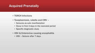
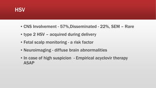
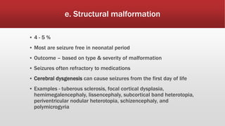
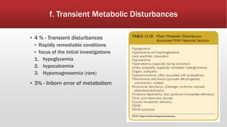
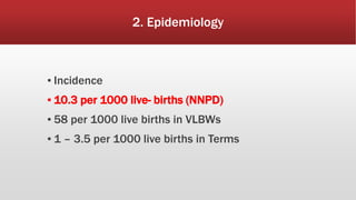
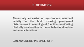
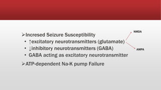
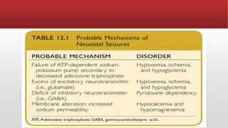
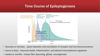
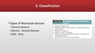
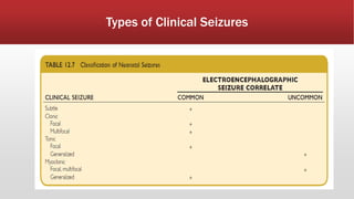
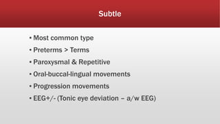
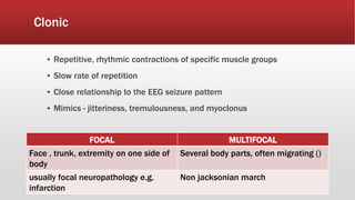
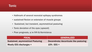
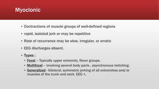
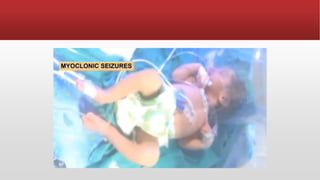
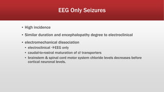
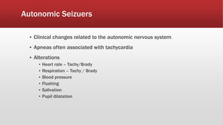
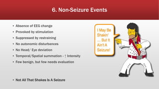
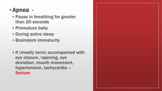
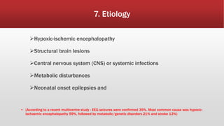
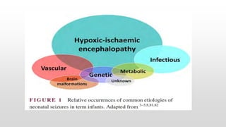
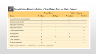
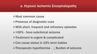
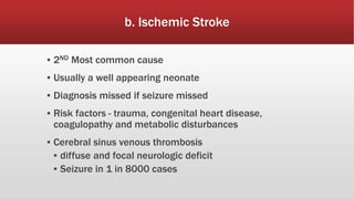
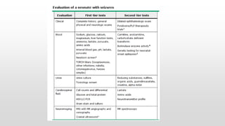
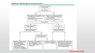
This document provides an overview of neonatal seizures. Key points include:
- Neonatal seizures have an incidence of 10.3 per 1000 live births and are more common in preterm infants.
- They are defined as abnormal excessive neuronal activity causing alterations in motor, behavioral or autonomic functions.
- Common causes include hypoxic-ischemic encephalopathy, structural brain lesions, infections, and metabolic disturbances.
- Seizures are classified based on their clinical presentation and EEG findings as subtle, clonic, tonic, myoclonic or EEG-only.
- Differentiating seizures from non-seizure events like apnea or jitteriness is important.
Neonatal seizures, dr amit vatkar, pediatric neurologistDr Amit Vatkar
In the presentaion i will give you a brief idea to apprach, diagnosis and management of neonatal seizures.
The most prominent feature of neurologic dysfunction in the neonatal period is the occurrence of seizures. Determining the underlying etiology for neonatal seizures is critical. Etiology determines prognosis and outcome and guides therapeutic strategies.
Neonatal seizures, dr amit vatkar, pediatric neurologist
ATAXIA IN CHILDREN -CAUSES, MANAGEMENT, INVESTIGATIONS, TYPES, COMMONEST ATAXIA IN CHILDREN IN DETAIL, HOW WILL YOU FIND OUT THE CAUSE FOR ATAXIA IN CHILDREN FLOWCHART, DEFINITION, TREATMENT
The document provides information on the evaluation of hypotonia in infants. It discusses that hypotonia can be caused by central or peripheral nervous system disorders. The most common central cause is hypoxic ischemic encephalopathy, while the most common peripheral causes are congenital myopathies and spinal muscular atrophy. A thorough history, physical exam, and testing are needed to determine the underlying cause, which guides management and prognosis. The evaluation involves assessing tone, strength, reflexes and other features to localize the problem and rule out various disorders through laboratory and imaging studies.
Neonatal seizures are the most common neurological manifestation in newborns and can be difficult to recognize. They are often caused by hypoxic-ischemic encephalopathy, hypoglycemia, hypocalcemia or infections. When a neonatal seizure is observed, the newborn must be stabilized, underlying causes should be investigated through bloodwork and imaging, and seizures treated aggressively with anticonvulsants like phenobarbital. Identifying and correcting the underlying etiology is important for management and prognosis.
This document discusses epilepsy in children, including types, clinical presentation, investigations, management, and complications. It provides an overview of seizure types and classifications, the etiology and syndromes of epilepsy in children, how epilepsy presents clinically and is evaluated, and guidelines for treatment and counseling to control seizures and prevent complications. Key aspects covered include acute seizure management, anti-epileptic medications, status epilepticus, and the potential neurological and developmental impacts of uncontrolled epilepsy.
The neonatal period involves physiological adjustments outside the womb. Seizures are common and a sign of neurological issues. There are 5 main seizure types seen in neonates. Common causes include hypoxic ischemic encephalopathy, infection, metabolic imbalances. Investigation involves history, exam, labs, imaging and EEG. Treatment focuses on controlling seizures and addressing underlying causes with anticonvulsants like phenobarbital. Prognosis depends on etiology but has improved over time.
This document discusses neonatal hypocalcemia, including its types, causes, and management. There are two types - early onset within 72 hours requiring calcium supplementation, and late onset after 7 days requiring longer treatment. Hypocalcemia is defined as low serum calcium levels. Causes of early onset include prematurity, maternal conditions, and iatrogenic factors. Management of early onset involves calcium supplementation. Late onset in the first week is usually symptomatic and caused by high phosphate intake. Causes also include hypomagnesemia, vitamin D deficiency, and genetic or metabolic syndromes.
Febrile seizures are seizures that occur in young children between 6 months and 5 years old when they have a fever. They are classified as either simple or complex based on features like duration and recurrence. Simple febrile seizures are more common, generalized, and last less than 15 minutes. Risk factors for recurrence include age under 1, duration of fever, and family history. Causes are usually upper respiratory infections or roseola. Evaluation involves history, exam, and occasionally lumbar puncture or EEG. Treatment focuses on bringing the fever down with antipyretics and using benzodiazepines if the seizure lasts more than 5 minutes.
1. A seizure is caused by abnormal excessive neuronal activity in the brain and can be focal, generalized, or febrile.
2. Evaluation of a first seizure involves obtaining a detailed history, physical exam, EEG, and brain imaging to determine the cause and classify the seizure type.
3. Generalized seizures include absence seizures, myoclonic seizures, and generalized tonic-clonic seizures. Focal seizures can become secondary generalized seizures.
This document provides information on evaluating hypotonia in infants. It defines muscle tone and the differences between hypotonia and weakness. Hypotonia can have central or peripheral causes. The differential diagnosis for a floppy infant is extensive and includes central conditions like genetic syndromes or brain insults and peripheral conditions involving the motor unit, nerve, neuromuscular junction or muscle. A thorough evaluation includes the infant's history, development, family history, examination and potentially genetic or metabolic testing to determine the underlying cause.
This document provides information on epilepsy and seizure disorders in children. It defines different types of seizures including generalized, focal, and febrile seizures. Common causes and syndromes of epilepsy in children are discussed. Evaluation, management, and treatment of status epilepticus and febrile seizures are summarized. The goal of treatment is to control seizures with medication while minimizing side effects.
Epilepsy syndromes can be categorized based on age of onset from neonates to childhood. Syndromes include self-limited, developmental and epileptic encephalopathies, and genetic generalized epilepsies. Syndromes are defined by seizure type, EEG findings, development, and treatment response/prognosis. Examples provided include benign familial neonatal epilepsy, Dravet syndrome, childhood absence epilepsy, Lennox-Gastaut syndrome, and Landau-Kleffner syndrome.
The document discusses various epileptic syndromes categorized by age of onset - neonatal, infancy, childhood, adolescence-adult. For each syndrome, it provides information on defining features, age of onset, seizure types, EEG patterns, treatment and prognosis. The syndromes discussed include benign familial neonatal epilepsy, Ohtahara syndrome, West syndrome, Panayiotopoulos syndrome, Lennox-Gastaut syndrome, juvenile myoclonic epilepsy and others.
1. The document discusses different types of seizures including focal onset seizures, generalized onset seizures, and seizures of unknown onset. It provides definitions and examples of motor and non-motor seizures.
2. Etiologies of seizures in the neonatal period and beyond are outlined, including infections, metabolic disorders, brain malformations, drugs/poisons, and epilepsy syndromes.
3. Febrile seizures are defined as seizures associated with fever in children between 6-60 months old. Simple febrile seizures are brief and do not recur within 24 hours, while complex febrile seizures are prolonged or recurrent.
This document discusses various conditions that can mimic epilepsy in children. It notes that epilepsy is sometimes underdiagnosed or overdiagnosed due to unusual symptom presentations or epilepsy mimics. Several common epilepsy mimics are described in detail for different age groups, including breath holding spells in infants, tics and parasomnias in children, and syncope in adolescents. Tables compare features of epilepsy mimics to epileptic seizures during sleep and wakefulness. In conclusion, the document emphasizes taking an age- and state-based approach to differentiating epilepsy from conditions it may imitate.
This document discusses neuronal migration disorders. It begins by introducing neuronal migration as a key feature of nervous system development. It then describes the three stages of neuronal migration and genes involved. Several specific neuronal migration disorders are defined and described in detail, including lissencephaly, schizencephaly, heterotopias, polymicrogyrias, focal cortical dysplasias, and porencephaly. Key features such as imaging findings and clinical presentations are provided for each. The document emphasizes that defects in neuronal migration pathways can lead to developmental problems and neurological issues like seizures or motor impairments.
This document discusses neonatal seizures, including their definition, classification, etiologies, diagnostic approach, treatment, and prognosis. It notes that seizures in newborns can be subtle, clonic, tonic, or myoclonic. Common causes include perinatal conditions like hypoxic-ischemic encephalopathy, intracranial hemorrhages, infections, developmental defects, and various metabolic derangements. The diagnostic approach involves taking a thorough history, conducting examinations, and ordering appropriate investigations. Treatment involves stabilizing the newborn, administering anticonvulsants like phenobarbital, and managing underlying conditions. Outcome depends on factors like etiology, maturity, and neurological status. Complications may
This document discusses neonatal seizures, including:
- Neonatal seizures are a medical emergency that can affect future neurological development. They have several classifications including subtle, clonic, myoclonic, and tonic seizures.
- Common causes include hypoxic-ischemic encephalopathy, intracranial hemorrhage, hypoglycemia, and hypocalcemia. Investigations like blood tests, EEG, and imaging can help determine the underlying cause.
- Treatment involves stabilizing the neonate and treating the underlying condition. If seizures continue, anticonvulsant medications like phenobarbital and phenytoin are used. Prognosis depends on the cause, seizure pattern, and EEG findings
This document discusses the approach to hypoglycemia in childhood. It begins by defining hypoglycemia and describing the importance of glucose for brain development. It then discusses the pathophysiology of hypoglycemia, focusing on how the body maintains blood glucose levels through glycogenolysis, gluconeogenesis, and lipolysis. The clinical features of hypoglycemia are presented, distinguishing between sympathetic overactivity and neuroglycopenic symptoms. Common etiologies like hyperinsulinism, metabolic disorders, and systemic illnesses are outlined. The document concludes with recommendations for investigating hypoglycemia, managing acute episodes, and treating underlying causes to prevent long-term neurological consequences.
This document discusses seizures in children, including febrile seizures. It defines seizures and different types, like generalized seizures and focal seizures. It covers the epidemiology, causes, clinical presentation and diagnosis of seizures. Complications, both acute and chronic, are outlined. Investigations and management approaches are also summarized. The document focuses in particular on febrile seizures, their definition, causes, types, evaluation and treatment in children presenting with fever and seizures.
This document provides a case history, physical examination findings, differential diagnosis, workup, management, and background information on infantile spasms for a 14-month-old male patient presenting with developmental delays, frequent falling, and nonspecific babbling. The patient has axial and appendicular hypotonia on exam. Initial workup including metabolic tests and brain MRI were normal. The patient was started on vigabatrin to treat a presumptive diagnosis of infantile spasms. Further sections provide details on the classification, diagnostic criteria including characteristic EEG patterns, and treatment approaches for infantile spasms.
This document discusses approaches to macrocephaly and microcephaly. Macrocephaly is defined as a head circumference over 2 standard deviations above the mean, while microcephaly is under 3 standard deviations below the mean. Causes of macrocephaly include genetic factors, hydrocephalus, tumors, and metabolic disorders. Hydrocephalus results from abnormal CSF accumulation and can be obstructive or communicating. Microcephaly can be primary/genetic due to syndromes or secondary from infections, drugs, or hypoxia that impact brain development prenatally or in the first two years. Evaluation and treatment depends on the underlying cause.
The document discusses neurodegenerative disorders in children. It describes how these disorders involve the progressive deterioration of neurological function, often with regression of developmental milestones. The disorders can affect gray matter, white matter, or both. Investigations aim to identify the underlying genetic or metabolic cause, while management focuses on treating complications and providing supportive care.
This document provides an overview of hypoxic-ischemic encephalopathy (HIE) in neonates. It defines HIE and discusses its etiology, pathophysiology, diagnosis, clinical staging, assessment tools, and management including hypothermia therapy. HIE results from a hypoxic or ischemic insult to the fetal or neonatal brain. The pathophysiology involves both primary neuronal damage during the insult and secondary damage over subsequent hours and days. Diagnosis is based on evidence of metabolic acidosis during birth and early onset of neurological abnormalities. Hypothermia therapy, involving whole body cooling to 33-35°C for 72 hours, is now standard treatment and reduces mortality and disability by attenuating secondary brain injury.
This document discusses the infant of a diabetic mother. It begins with an introduction stating that diabetes is a common complication of pregnancy and risks to the infant have decreased but still exist. It then covers pathophysiology, epidemiology, complications, management, and prognosis. Key points include: fetal macrosomia is a risk; hypoglycemia is common due to hyperinsulinemia; other risks include hypocalcemia, hypomagnesemia, and congenital heart defects. Management involves monitoring glucose and electrolytes along with imaging tests. Treatment focuses on maintaining normal glucose during labor and delivery along with early breastfeeding to prevent hypoglycemia. Prognosis is generally good but neurodevelopmental risks exist if maternal glucose control was
This document discusses pediatric stroke. It begins with definitions, types, epidemiology, etiology, and pathophysiology of pediatric stroke. The main types are ischemic and hemorrhagic stroke. Risk factors in children include structural heart disease, vasculopathies, hematological disorders, and prothrombotic states. Clinical features can include focal neurological deficits like hemiparesis. Diagnosis involves neuroimaging such as MRI and distinguishing stroke from other conditions. Management aims to prevent recurrence and support rehabilitation.
This document discusses the evaluation of a floppy infant. It begins by defining a floppy infant as one presenting with generalized hypotonia, often arising from an insult during the fetal or neonatal period. It describes the clinical examination of a floppy infant and differential diagnosis, which includes central nervous system causes, spinal cord disorders, peripheral nerve disorders, neuromuscular transmission defects, muscle diseases, and systemic disorders. Key examination findings that help localize the cause of hypotonia are discussed. Common etiologies like cerebral palsy, spinal muscular atrophy, and myasthenia gravis are also summarized.
Neonatal seizures NS in peadiatric populationPooja Khose
This document provides information on neonatal seizures, including:
- Neonatal seizures are defined as sudden, repetitive, evolving episodes of abnormal brain activity lasting at least 10 seconds and are more common in newborns than any other age.
- Common causes of neonatal seizures include hypoxic-ischemic encephalopathy, intracranial hemorrhage, infections, brain malformations, metabolic disorders like hypoglycemia and hypocalcemia, and some rare genetic disorders.
- The immature brain is more excitable and prone to seizures due to an imbalance of excitatory and inhibitory signals, as well as other developmental factors. Therapeutic hypothermia and anti-seizure medications are used to
This document discusses seizures in neonates. It begins by defining seizures, convulsions, and epilepsy. It then notes that seizures are most common in neonates, occurring in 0.5-0.8% of term babies and 6-12% of babies under 1500g. Common causes of neonatal seizures include hypoxic ischemic encephalopathy, intracranial hemorrhage, infections, inborn errors of metabolism like hypoglycemia and hypocalcemia, and epilepsy syndromes. The immature neonatal brain is more excitable and prone to seizures due to reduced connectivity, an imbalance toward excitatory processes, and the excitatory effect of GABA in some regions. The document describes different seizure types and discusses
Overview of neonatal epilepsy syndromes.pptxphilipolielo1
This document provides an overview of several neonatal epilepsy syndromes:
1) Self-limited neonatal epilepsy is characterized by focal tonic seizures in the first weeks that resolve by 6 months, caused by genes KCNQ2 and KCNQ3.
2) Early infantile developmental and epileptic encephalopathy includes Ohtahara syndrome and early myoclonic encephalopathy, presenting with severe, intractable seizures in the first months associated with developmental impairment.
3) Pyridoxine-dependent and pyridoxamine 5'-phosphate oxidase deficiency developmental and epileptic encephalopathy cause seizures beginning prenatally or at birth responsive to pyridoxine/pyridoxamine
Seizures are common in children and can be provoked by factors like fever or infection. They result from abnormal electrical activity in the brain. Partial seizures originate in one area of the brain while generalized seizures involve both hemispheres. Treatment involves identifying and treating the underlying cause, as well as using anticonvulsant medications. It is important to ensure safety during a seizure and provide supportive care afterwards.
This document discusses various epileptic syndromes categorized by age of onset - infantile, childhood, adolescent. Key syndromes described in detail include West syndrome, Dravet syndrome, GEFS+, Panayiotopoulos syndrome, Benign epilepsy with centrotemporal spikes, Electrical status epilepticus in slow sleep, Myoclonic-atonic epilepsy, Lennox-Gastaut syndrome. For each syndrome, the document outlines clinical features, investigations such as common EEG findings and genetic causes, treatment approaches, and typical prognosis.
This document provides information on epileptic encephalopathies that onset in infancy, including definitions and classifications. It describes several specific syndromes - Early Myoclonic Encephalopathy (EME), Ohtahara syndrome, West syndrome, and Dravet syndrome. EME is characterized by erratic myoclonus, focal seizures, and spasms in the first weeks of life, along with a burst suppression pattern on EEG. Ohtahara syndrome involves tonic spasms in the first months with a continuous burst suppression EEG. West syndrome consists of epileptic spasms, developmental delay/regression, and a hypsarrhythmic EEG. Dravet syndrome begins with prolonged febrile
This document discusses neonatal seizures, beginning with an introduction stating they are not uncommon and often the first sign of neurological disorders. It then covers the pathophysiology, incidence, patterns, etiology, diagnosis, management, treatment including anticonvulsants, and prognosis of neonatal seizures over multiple pages with headings and subheadings. Key points include seizures occurring in 1 in 200 healthy newborns, various possible causes like hypoglycemia or infections, treatments involving anticonvulsants like phenobarbital or midazolam, and prognosis varying from normal outcome to neurological sequelae depending on factors like etiology and examination findings.
The neonatal period involves physiologic adjustments outside the womb. Seizures are common during this time, especially in preterm infants. There are 5 main types of neonatal seizures. Common causes include hypoxic-ischemic encephalopathy, infection, metabolic disturbances, and drugs. Treatment involves identifying the underlying cause, administering anticonvulsants like phenobarbital, and supportive care. Prognosis depends on the etiology, with HIE carrying the highest risk of neurodevelopmental issues.
Epilepsy is a chronic neurological condition characterized by recurrent, unprovoked seizures. It can be classified as generalized seizures originating from both hemispheres of the brain or focal seizures arising from one hemisphere. The majority of childhood epilepsy cases are idiopathic, while secondary causes include brain malformations, infections, tumors or trauma. Diagnosis involves clinical history and examination along with an EEG. Treatment primarily consists of antiepileptic drug monotherapy tailored to the seizure type, with the goal of seizure control and prevention of recurrence. Status epilepticus is a medical emergency defined by prolonged or repeated seizures, and requires rapid treatment to prevent neurological injury.
Non-epileptic paroxysmal disorders can mimic seizures. Careful history and examination is often sufficient to differentiate them from epilepsy, though sometimes EEG or other testing is needed. They can be classified by age of presentation and symptoms. Conditions discussed include breath holding spells, prolonged QT syndrome, hyperekplexia, alternating hemiplegia of childhood, motor tics, episodic ataxias, spasmus nutans, opsoclonus-myoclonus syndrome, sleep related disorders like benign sleep myoclonus, sleep transition disorder, and narcolepsy-cataplexy. Differentiation from epilepsy is important to avoid unnecessary treatment while ensuring appropriate management of the underlying
It contains description and salient points to diagnose various epileptic encephalopathies seen during infancy such as early myoclonic encephalopathies, Otahara syndrome, Dravet syndrome, West syndrome.
This document discusses several epileptic encephalopathy syndromes that occur in childhood. It defines epileptic encephalopathies as heterogeneous brain disorders occurring during development where abnormal EEG activity is responsible for cognitive and motor regression beyond what would be expected from the underlying etiology alone. It then describes several recognized epileptic encephalopathy syndromes in children including Ohtahara syndrome, early myoclonic encephalopathy, West syndrome, Dravet syndrome, Lennox-Gastaut syndrome, epileptic encephalopathy with continuous spike and wave during sleep, and Landau-Kleffner syndrome. For each syndrome, it provides details on age of onset, common seizure types and EEG patterns, potential etiologies,
This document discusses neonatal seizures. It begins by defining seizures and describing the different types seen in neonates. The most common type is subtle seizures. Hypoxic ischemic encephalopathy is usually the most common cause, especially within the first 24 hours. Other common causes include intracranial hemorrhage and metabolic disorders. Phenobarbital is the first-line treatment, with phenytoin and benzodiazepines as second-line options. Seizures from subarachnoid hemorrhage or late-onset hypocalcemia typically carry a good prognosis, while those associated with hypoxic ischemic encephalopathy, cerebral malformations or meningitis usually have a poorer neurological outcome.
Neonatal-Seizures diagnosis and managementFelixBoamah3
This document discusses neonatal seizures. It begins by defining seizures and describing the different types seen in neonates. The most common cause is hypoxic ischemic encephalopathy. Other common causes include intraventricular hemorrhage and acute metabolic disorders. Phenobarbital is the first-line treatment, with phenytoin and benzodiazepines as subsequent options. Prognosis depends on the underlying etiology, with focal clonic seizures and those from subarachnoid hemorrhage or late hypocalcemia having better outcomes. Anti-seizure medications should be tapered slowly after seizure control is achieved.
Autoimmune encephalitis a term used in children presenting with neurological syndrome associated with serum and/ or cerebrospinal fluid antibodies directed against ion channels, receptors and associated proteins.
It comprises group of clinical syndrome that can occur at all ages but preferentially affect younger adult and children.
Auto antibodies against:
Neuronal cell surface protein
Synaptic receptors involved in transmission ,plasticity ,excitability.
Triggers: Tumors, Post viral infections, Post vaccination.
Autoimmune encephalitis includes ADEM
Anti NMDAR encephalitis
Encephalitis a/w GABA R antibodies
NMOSD
Opsoclonus-myoclonus & cerebellar - brainstem encephalitis
Bickerstaff encephalitis
Hashimoto encephalitis
Rasmussen encephalitis
Basal ganglia encephalitis
CLIPPERS
ROHHAD.
ADEM is MC cause of autoimmune encephalitis in children and adolescents.
Acute onset of polyfocal neurological deficit accompanied by encephalopathy and changes compatible with demyelination on MRI brain.
This document discusses neonatal seizures. It defines seizures and neonatal seizures, describing the pathophysiology of seizures in newborns. The main causes of neonatal seizures are then outlined, with hypoxic-ischemic encephalopathy, brain malformations, and infections listed as common etiologies. The document proceeds to describe the different types of neonatal seizures and considerations for differentiating seizures from non-convulsive movements in newborns. Diagnosis and management of neonatal seizures is then covered, including acute treatment and duration of treatment.
1) Antimicrobial stewardship programs aim to optimize antibiotic use and prevent resistance by coordinating actions to improve prescribing. They promote appropriate use through guidelines, education, and monitoring of antibiotic use and outcomes.
2) In NICUs, antimicrobial stewardship faces unique challenges due to non-specific signs of infection in neonates and difficulties obtaining cultures. Programs seek to minimize broad-spectrum antibiotic exposure and duration to reduce resistance and side effects.
3) Effective strategies for neonatal antimicrobial stewardship include developing unit-specific treatment guidelines, prospectively auditing antibiotic use, educating providers, streamlining therapy based on culture results, and implementing bundles to prevent infections.
Total parenteral nutrition in the nicu Total parenteral nutrition in the nicuDr Praman Kushwah
This document provides information on total parenteral nutrition (TPN) in the neonatal intensive care unit (NICU). It discusses indications for TPN, nutritional composition including dextrose, amino acids, lipids, additives, and calculations. Potential complications are outlined including parenteral nutrition associated liver disease. Guidelines are provided for calcium, phosphorus, and minimizing metabolic bone disease and cholestasis.
Chromosome abnormalities are common in live births, stillbirths, and early fetal losses. Cytogenetic testing is indicated for recurrent miscarriages, familial chromosome abnormalities, clinical findings suggestive of abnormalities, and chromosome breakage syndromes. Analysis methods include karyotyping, FISH, aCGH to detect deletions/duplications. Trisomy 21 is the most common genetic cause of intellectual disability. It is usually due to an extra maternal chromosome 21 and causes characteristic physical and developmental features. Prenatal testing options include invasive and noninvasive methods to diagnose trisomy 21.
Diabetes in pregnancy poses risks to both mother and baby. Good glycemic control through nutrition, lifestyle changes, and possibly medication can help reduce risks. Babies of diabetic mothers may be large with potential birth injuries, and face risks of low blood sugar, breathing issues, and heart and metabolic problems. Close monitoring and management throughout pregnancy aims to deliver healthy babies.
This document discusses the use of steroids in neonatology, including both antenatal and postnatal applications. It begins by introducing the current recommendations for antenatal steroid therapy to reduce neonatal morbidity and mortality. It then covers the classification, mechanisms of action, benefits and considerations for administration of antenatal corticosteroids. It also discusses the use of postnatal steroids in the NICU to treat conditions like chronic lung disease. While noting both the benefits and risks, it concludes that postnatal steroids should be reserved for high-risk preterm infants who remain ventilator-dependent beyond 2-3 weeks of age.
This document discusses intrauterine growth restriction (IUGR), including definitions, health burden, classification, etiology, pathophysiology, screening, prevention, diagnosis, interventions, management, and long-term outcomes. IUGR is defined as a fetus that does not achieve expected in utero growth potential due to genetic or environmental factors. It affects about 10% of live births and is a leading cause of perinatal morbidity and mortality. Causes include fetal, maternal, placental, and environmental factors. Screening involves ultrasound and Doppler assessments. Management involves timing of delivery based on gestational age and severity. IUGR is associated with short and long-term complications.
This document discusses various methods used to monitor fetal well-being during labor, including fetal heart rate monitoring, fetal blood sampling, fetal pulse oximetry, fetal electrocardiography, and ultrasound. It provides details on interpreting fetal heart rate patterns, the results of fetal blood sampling, and evidence regarding electronic fetal monitoring versus intermittent auscultation. While electronic fetal monitoring has increased cesarean rates compared to intermittent auscultation, studies have not found it reduces rates of hypoxic ischemic encephalopathy or cerebral palsy.
This document discusses antenatal screening for fetal aneuploidy. It introduces cytogenetics and explains that chromosomal abnormalities occur in approximately 1-2% of live births. The most common types of aneuploidy are trisomy 21, trisomy 18, trisomy 13 and sex chromosome aneuploidies. Screening methods discussed include biochemical screening, ultrasound screening for soft markers and structural abnormalities, and non-invasive prenatal testing using cell-free DNA. Biochemical markers analyzed include PAPP-A, beta-hCG, AFP, uE3, and inhibin A. Soft markers seen on ultrasound include increased nuchal translucency, absent nasal bone, ech
This document discusses thyroid disorders in newborns. It covers thyroid physiology in the fetus and newborn, causes of congenital hypothyroidism including thyroid dysgenesis and dyshormonogenesis, clinical features of hypothyroidism, and methods for screening and diagnosis of congenital hypothyroidism in newborns. It also addresses transient hypothyroidism, sick euthyroid syndrome, and the importance of newborn screening to detect congenital hypothyroidism.
This document provides an overview of pulmonary graphics and ventilator waveforms. It discusses the basics of how ventilator monitoring systems work and the importance of interpreting graphic displays. The key ventilator waveforms of pressure-time, volume-time, flow-time and pressure-volume loops are described. Specific features of each waveform are defined, and they can be analyzed to evaluate lung mechanics, respiratory system pressures, gas trapping, leaks, asynchrony, and other factors. Understanding ventilator graphics is critical for optimizing ventilator settings and managing mechanically ventilated patients.
This document summarizes fluid homeostasis and water balance in the fetus and neonate. It discusses the following key points:
1. Water exists in intracellular and extracellular fluid compartments separated by semipermeable membranes. Total body water decreases with gestational age from 95% early in gestation to 75% at term.
2. Amniotic fluid volume is tightly regulated between 350-800mL and is derived mainly from fetal urine production by the third trimester.
3. At birth, term infants lose approximately 10% of their body weight in the first week due to redistribution of body water from intracellular to extracellular spaces and increased renal water and sodium excretion.
Retinopathy of prematurity (ROP) is a developmental vascular disorder of the retina that occurs in preterm infants. The retina is incompletely vascularized at birth for infants born before 30 weeks gestation. Premature birth interrupts normal retinal vascularization, exposing the retina to abnormal oxygen levels. This can cause vasoconstriction and arrest of blood vessel growth. Later, abnormal neovascularization may develop, potentially leading to retinal detachment and blindness if not treated. Screening guidelines recommend examinations starting at 4 weeks of age for infants born before 30 weeks, with treatment indicated for "threshold" or "pre-threshold" ROP. Laser photocoagulation is the primary treatment, which ablates the av
1. MRI is highly sensitive for detecting white matter injuries in preterm neonates such as periventricular leukomalacia (PVL). PVL appears as small, symmetrically distributed hyperintense lesions on T1-weighted MRI near the ventricles.
2. Diffusion-weighted MRI can detect white matter injuries even earlier by showing hyperintensity and reduced diffusion in injured areas.
3. MRI is also very good for detecting germinal matrix and intraventricular hemorrhages. Acute hemorrhages appear as areas of normal or increased signal on T1-weighted MRI and distinct hypointensity on T2-weighted MRI.
This document discusses the history and development of growth charts used to monitor infant growth. Some key points:
- The first growth curves were published in the 18th century but modern growth charts began in the late 19th/early 20th century with work by Bowditch and Lubchenco.
- Fetal growth is most rapid between 22-40 weeks gestation and influenced by fetal, placental and maternal factors. Postnatal growth depends on genetics and internal/external factors.
- Growth charts graphically represent growth of a reference population over time and are important for monitoring development. The two main types are growth references and growth standards.
- Major studies that developed widely used growth charts include Fenton
This document discusses the importance of follow-up care for high-risk newborns after discharge from the NICU. It outlines that follow-up should include regular medical examinations to monitor growth and development, screening tests, and neurobehavioral assessments. Follow-up care is most intensive for high-risk infants born prematurely or with very low birth weight, and can involve multiple specialists. The long-term goal is to identify any disabilities or delays early and provide intervention to prevent or reduce impairments.
This document describes several pain scales used in neonatal intensive care units (NICUs), including N-PASS, NIPS, and PIPP-R. N-PASS assesses both pain and sedation in infants by scoring behavioral and physiological criteria. Higher scores indicate more pain or deeper sedation. NIPS and PIPP-R also score facial expressions, extremities, vital signs, and other indicators to assess pain, with scores above 3 indicating pain. PIPP-R modified the original PIPP scale to better account for how gestational age and behavioral state influence pain responses. These scales provide standardized ways to evaluate pain in nonverbal infants to determine needed treatment and intervention.
This document discusses the pathophysiology of preterm birth, which accounts for 6-10% of births and is a major cause of neonatal death and impairment. Risk factors include race, age, socioeconomic status, BMI, smoking, and stress. Preterm birth is caused by inflammation/infection, hormonal changes, cervical insufficiency, and genetic factors. It can be predicted using tests for fetal fibronectin and cervical length. Prevention methods include cervical cerclage, progesterone supplementation, and antibiotics in some cases. The goal of treatment is to inhibit preterm labor when possible and ensure delivery occurs in a facility equipped for neonatal care.
This document discusses developmentally supportive care for preterm infants in the NICU. It aims to improve outcomes by supporting brain development, motor skills, sensory systems and family involvement through careful handling, positioning, feeding practices and control of the neonatal environment. Key aspects of care include protecting sleep, minimizing pain and stress, developmental activities like feeding and skin care, family centered care, and providing appropriate light, sound, and a caring staff. This holistic approach can help preterm infants heal and develop normally.
The document discusses hydrops fetalis, which refers to fluid accumulation in fetal tissues. It describes the two main types - immune and non-immune hydrops fetalis. Immune hydrops is caused by maternal-fetal blood group incompatibilities producing antibodies, while non-immune has many potential causes including genetic conditions, infections, cardiac abnormalities, and tumors. The document provides details on mechanisms, risk factors, pathophysiology, diagnostic features, and management approaches for different causes of hydrops fetalis.
Rh isoimmunization occurs when a Rh-negative mother develops antibodies against Rh-positive fetal red blood cells. This can cause hemolytic disease in future Rh-positive pregnancies. Prophylaxis with Rh immunoglobulin is highly effective at preventing sensitization. For sensitized pregnancies, maternal anti-D titers and fetal middle cerebral artery Doppler are used to monitor for anemia. Severe anemia may require cordocentesis or intrauterine transfusions. After delivery, affected newborns may need exchange transfusions or other supportive care to manage hyperbilirubinemia and anemia. Prompt and specialized treatment can help prevent complications in isoimmunized pregnancies.
Basavarajeeyam is an important text for ayurvedic physician belonging to andhra pradehs. It is a popular compendium in various parts of our country as well as in andhra pradesh. The content of the text was presented in sanskrit and telugu language (Bilingual). One of the most famous book in ayurvedic pharmaceutics and therapeutics. This book contains 25 chapters called as prakaranas. Many rasaoushadis were explained, pioneer of dhatu druti, nadi pareeksha, mutra pareeksha etc. Belongs to the period of 15-16 century. New diseases like upadamsha, phiranga rogas are explained.
Promoting Wellbeing - Applied Social Psychology - Psychology SuperNotesPsychoTech Services
A proprietary approach developed by bringing together the best of learning theories from Psychology, design principles from the world of visualization, and pedagogical methods from over a decade of training experience, that enables you to: Learn better, faster!
Does Over-Masturbation Contribute to Chronic Prostatitis.pptxwalterHu5
In some case, your chronic prostatitis may be related to over-masturbation. Generally, natural medicine Diuretic and Anti-inflammatory Pill can help mee get a cure.
TEST BANK For Community Health Nursing A Canadian Perspective, 5th Edition by...Donc Test
TEST BANK For Community Health Nursing A Canadian Perspective, 5th Edition by Stamler, Verified Chapters 1 - 33, Complete Newest Version Community Health Nursing A Canadian Perspective, 5th Edition by Stamler, Verified Chapters 1 - 33, Complete Newest Version Community Health Nursing A Canadian Perspective, 5th Edition by Stamler Community Health Nursing A Canadian Perspective, 5th Edition TEST BANK by Stamler Test Bank For Community Health Nursing A Canadian Perspective, 5th Edition Pdf Chapters Download Test Bank For Community Health Nursing A Canadian Perspective, 5th Edition Pdf Download Stuvia Test Bank For Community Health Nursing A Canadian Perspective, 5th Edition Study Guide Test Bank For Community Health Nursing A Canadian Perspective, 5th Edition Ebook Download Stuvia Test Bank For Community Health Nursing A Canadian Perspective, 5th Edition Questions and Answers Quizlet Test Bank For Community Health Nursing A Canadian Perspective, 5th Edition Studocu Test Bank For Community Health Nursing A Canadian Perspective, 5th Edition Quizlet Test Bank For Community Health Nursing A Canadian Perspective, 5th Edition Stuvia Community Health Nursing A Canadian Perspective, 5th Edition Pdf Chapters Download Community Health Nursing A Canadian Perspective, 5th Edition Pdf Download Course Hero Community Health Nursing A Canadian Perspective, 5th Edition Answers Quizlet Community Health Nursing A Canadian Perspective, 5th Edition Ebook Download Course hero Community Health Nursing A Canadian Perspective, 5th Edition Questions and Answers Community Health Nursing A Canadian Perspective, 5th Edition Studocu Community Health Nursing A Canadian Perspective, 5th Edition Quizlet Community Health Nursing A Canadian Perspective, 5th Edition Stuvia Community Health Nursing A Canadian Perspective, 5th Edition Test Bank Pdf Chapters Download Community Health Nursing A Canadian Perspective, 5th Edition Test Bank Pdf Download Stuvia Community Health Nursing A Canadian Perspective, 5th Edition Test Bank Study Guide Questions and Answers Community Health Nursing A Canadian Perspective, 5th Edition Test Bank Ebook Download Stuvia Community Health Nursing A Canadian Perspective, 5th Edition Test Bank Questions Quizlet Community Health Nursing A Canadian Perspective, 5th Edition Test Bank Studocu Community Health Nursing A Canadian Perspective, 5th Edition Test Bank Quizlet Community Health Nursing A Canadian Perspective, 5th Edition Test Bank Stuvia
Cell Therapy Expansion and Challenges in Autoimmune DiseaseHealth Advances
There is increasing confidence that cell therapies will soon play a role in the treatment of autoimmune disorders, but the extent of this impact remains to be seen. Early readouts on autologous CAR-Ts in lupus are encouraging, but manufacturing and cost limitations are likely to restrict access to highly refractory patients. Allogeneic CAR-Ts have the potential to broaden access to earlier lines of treatment due to their inherent cost benefits, however they will need to demonstrate comparable or improved efficacy to established modalities.
In addition to infrastructure and capacity constraints, CAR-Ts face a very different risk-benefit dynamic in autoimmune compared to oncology, highlighting the need for tolerable therapies with low adverse event risk. CAR-NK and Treg-based therapies are also being developed in certain autoimmune disorders and may demonstrate favorable safety profiles. Several novel non-cell therapies such as bispecific antibodies, nanobodies, and RNAi drugs, may also offer future alternative competitive solutions with variable value propositions.
Widespread adoption of cell therapies will not only require strong efficacy and safety data, but also adapted pricing and access strategies. At oncology-based price points, CAR-Ts are unlikely to achieve broad market access in autoimmune disorders, with eligible patient populations that are potentially orders of magnitude greater than the number of currently addressable cancer patients. Developers have made strides towards reducing cell therapy COGS while improving manufacturing efficiency, but payors will inevitably restrict access until more sustainable pricing is achieved.
Despite these headwinds, industry leaders and investors remain confident that cell therapies are poised to address significant unmet need in patients suffering from autoimmune disorders. However, the extent of this impact on the treatment landscape remains to be seen, as the industry rapidly approaches an inflection point.
Integrating Ayurveda into Parkinson’s Management: A Holistic ApproachAyurveda ForAll
Explore the benefits of combining Ayurveda with conventional Parkinson's treatments. Learn how a holistic approach can manage symptoms, enhance well-being, and balance body energies. Discover the steps to safely integrate Ayurvedic practices into your Parkinson’s care plan, including expert guidance on diet, herbal remedies, and lifestyle modifications.
Basavarajeeyam is a Sreshta Sangraha grantha (Compiled book ), written by Neelkanta kotturu Basavaraja Virachita. It contains 25 Prakaranas, First 24 Chapters related to Rogas& 25th to Rasadravyas.
ABDOMINAL TRAUMA in pediatrics part one.drhasanrajab
Abdominal trauma in pediatrics refers to injuries or damage to the abdominal organs in children. It can occur due to various causes such as falls, motor vehicle accidents, sports-related injuries, and physical abuse. Children are more vulnerable to abdominal trauma due to their unique anatomical and physiological characteristics. Signs and symptoms include abdominal pain, tenderness, distension, vomiting, and signs of shock. Diagnosis involves physical examination, imaging studies, and laboratory tests. Management depends on the severity and may involve conservative treatment or surgical intervention. Prevention is crucial in reducing the incidence of abdominal trauma in children.
8 Surprising Reasons To Meditate 40 Minutes A Day That Can Change Your Life.pptxHolistified Wellness
We’re talking about Vedic Meditation, a form of meditation that has been around for at least 5,000 years. Back then, the people who lived in the Indus Valley, now known as India and Pakistan, practised meditation as a fundamental part of daily life. This knowledge that has given us yoga and Ayurveda, was known as Veda, hence the name Vedic. And though there are some written records, the practice has been passed down verbally from generation to generation.
3. 1. INTRODUCTION
▪ RED ALERT
▪ SECONDARY BRAIN INJURY
▪ FOCAL OR MULTIFOCAL : SINGLE HEMISPHERE
▪ MAJORITY SUBCLINICAL OR SUBTLE
▪ LESS SEIZURE THRESHOLD BUT BETTER PROGNOSIS.
4. 2. Epidemiology
▪ Incidence
▪ 10.3 per 1000 live- births (NNPD)
▪ 58 per 1000 live births in VLBWs
▪ 1 – 3.5 per 1000 live births in Terms
5. 3. DEFINITION
Abnormally excessive or synchronous neuronal
activity in the brain causing paroxysmal
disturbances in neurological function manifesting
clinically as alteration in motor, behavioral and/or
autonomic functions
CAN ANYONE DEFINE EPILEPSY ?
9. ▪ Onset is focal /multifocal, spread within one hemisphere only
▪ Immature mylenation of cortical efferent systems & commissure.
▪ Advanced Cortical development in Limbic system - Subtle
10. Energy dependent ion
pumping
Decreased ATP &
Phosphocreatine
Stimulation of
glycolysis and ↑ Lactate
Vasodilation & ↑ blood
supply
SEIZURE EPISODE
Incresed
blood
pressure
Increased
Cerebral
blood flow
This fall in brain glucose
concentration and rise in
brain lactate are directly
reminiscent of a hypoxic-
ischemic brain insult
11.
12. Time Course of Epileptogenesis
▪ Seconds to minutes – gene induction and translation of receptor and ion-channel proteins
▪ hours to days - neuronal death, inflammation, and altered transcriptional regulation
▪ weeks to months - mossy fiber sprouting, gliosis, neurogenesis
13. 5. Classification
▪ Types of Neonatal seizure
▪ Clinical Seizure
▪ Electro - clinical Seizure
▪ EEG - Only
15. Subtle
▪ Most common type
▪ Preterms > Terms
▪ Paroxysmal & Repetitive
▪ Oral-buccal-lingual movements
▪ Progression movements
▪ EEG+/- (Tonic eye deviation – a/w EEG)
16.
17. Clonic
▪ Repetitive, rhythmic contractions of specific muscle groups
▪ Slow rate of repetition
▪ Close relationship to the EEG seizure pattern
▪ Mimics - jitteriness, tremulousness, and myoclonus
FOCAL MULTIFOCAL
Face , trunk, extremity on one side of
body
Several body parts, often migrating ()
usually focal neuropathology e.g.
infarction
Non jacksonian march
18.
19. Tonic
▪ Hallmark of several neonatal epilepsy syndromes
▪ sustained flexion or extension of muscle groups
▪ Sustained, but transient, asymmetrical posturing
▪ Tonic deviation of the eyes (upward)
▪ Poor prognosis, a/w IVH & Kernicterus
FOCAL GENERALIZED
Sustained, asymmetrical Posturing Decerebrate/decorticate like posturing
Mostly EEG discharges + 15% EEG +
20. Myoclonic
▪ Contractions of muscle groups of well-defined regions
▪ rapid, isolated jerk or may be repetitive
▪ Rate of recurrence may be slow, irregular, or erratic
▪ EEG discharges absent.
▪ Types :
▪ Focal – Typically upper extremity, flexor groups.
▪ Multifocal – involving several body parts , asynchronous twitching.
▪ Generalized - bilateral, symmetric jerking of all extremities and/or
muscles of the trunk and neck. EEG +,
21.
22. EEG Only Seizures
▪ High incidence
▪ Similar duration and encephalopathy degree to electroclinical
▪ electromechanical dissociation
▪ electroclinical EEG only
▪ caudal-to-rostral maturation of cl- transporters
▪ brainstem & spinal cord motor system chloride levels decreases before
cortical neuronal levels.
23. Autonomic Seizuers
▪ Clinical changes related to the autonomic nervous system
▪ Apneas often associated with tachycardia
▪ Alterations
▪ Heart rate – Tachy/Brady
▪ Respiration – Tachy / Brady
▪ Blood pressure
▪ Flushing
▪ Salivation
▪ Pupil dilatation
24. 6. Non-Seizure Events
▪ Absence of EEG change
▪ Provoked by stimulation
▪ Suppressed by restraining
▪ No autonomic disturbances
▪ No Head/ Eye deviation
▪ Temporal/Spatial summation - ↑ Intensity
▪ Few benign, but few needs evaluation
▪ Not All That Shakes Is A Seizure
25. ▪Apnea -
▪ Pause in breathing for greater
than 20 seconds
▪ Premature baby
▪ During active sleep
▪ Brainstem immaturity
▪ If (mostly term) accompanied with
eye closure /opening, eye
deviation, mouth movement,
hypertension, tachycardia –
Seizure
26. ▪ Jitteriness
▪ Very Common
▪ Excessive response to
stimulation
▪ Back-and-forth oscillation ,
Same amplitude &
frequency
▪ Hypocalcemia,
hypoglycemia, and hypoxic-
ischemic encephalopathy
▪ Head or eye deviation during
an episode → seizure
27. ▪ Benign neonatal sleep
myoclonus
▪ during sleep – NREM
▪ jerks are typically bilateral,
symmetric
▪ can be stopped by rousing the
infant
▪ does not involve the face
▪ Normal neurologically
▪ BZDs provokes / exacerbates
▪ Spontaneous resolution ~2 mo
28. ▪ Hyperekplexia (startle disease)
▪ Congenital stiff-man syndrome
▪ Startle & sustained tonic spasms response to stimuli
▪ Nocturnal myoclonus.
▪ “minor” excessive startle
▪ “major” stiffness while awake, nocturnal myoclonus, risk of SIDS
▪ Glycine receptor gene mutations
▪ Clonazepam - effective treatment
▪ Resolution by 2 yr age
29. Normal Motor Activities
▪ Roving, sometimes dysconjugate eye movements, with occasional
nonsustained nystagmoid jerks at the extremes of horizontal
movement
▪ Sucking, puckering – NO ocular fixation or deviation
▪ While - Asleep
▪ Fragmentary myoclonic jerks (may be multiple)
▪ Isolated, generalized myoclonic jerk as infant wakes from sleep
30. 7. Etiology
Hypoxic-ischemic encephalopathy
Structural brain lesions
Central nervous system (CNS) or systemic infections
Metabolic disturbances
Neonatal onset epilepsies and
▪ (According to a recent multicentre study - EEG seizures were confirmed 35%. Most common cause was hypoxic-
ischaemic encephalopathy 59%, followed by metabolic/genetic disorders 21% and stroke 13%)
31.
32.
33. a. Hypoxic Ischemic Encephalopathy
Most common cause
Presence of diagnostic cues
With short, frequent and refractory episodes
>50% - have subclinical seizures
Treatment is urgent & complicated
Can cause status in 15% term babies
Therapeutic hypothermia - ↓ Burden of seizures
34. b. Ischemic Stroke
▪ 2ND Most common cause
▪ Usually a well appearing neonate
▪ Diagnosis missed if seizure missed
▪ Risk factors - trauma, congenital heart disease,
coagulopathy and metabolic disturbances
▪ Cerebral sinus venous thrombosis
▪ diffuse and focal neurologic deficit
▪ Seizure in 1 in 8000 cases
35.
36. c. Intracranial Hemorrhage
▪ 10 – 15%
▪ Instrumentation, Breech, Large for gestation
▪ Well appearing Neonates
▪ Subduaral – Falx tears
37.
38.
39. ▪ Preterms Neonates–
▪ IVH/GM
▪ Persistent tonic
▪ b/w 3-7 days of life
▪ Poor outcome
▪ First 3 days in GM and
Severe IVH
• Term Neonates
1. Subarachnoid hemorrhage
• infant delivered vaginally.
• second day focal seizure
• resolve rapidly
• good prognosis.
2. Subdural hematomas
• a/w trauma and cerebral
contusion.
• Focal seizures
• First 2 days
40. d. INFECTIONS
▪ 4 % of total
▪ Term = Preterms
▪ Acquired Prenatally –
▪ Intra Uterine Infections – TORCH
▪ Microcephaly; poor intrauterine growth; prematurity; and skin,
ophthalmic, and systemic findings
▪ Acquire postnatally –
▪ Meningitis – Bacterial / Viral / Fungal
▪ Associated clinical features
41. Acquired Prenatally
▪ TORCH Infections
▪ Toxoplasmosis, rubella and CMV –
▪ Seizures as sole manifestation
▪ Occur in first 3 days in the neonatal period
▪ Specific diagnostic clues
▪ HSV & Enterovirus causing encephalitis
▪ HSV – Seizure after 7 days
42. HSV
▪ CNS Involvement - 57%,Disseminated - 22%, SEM – Rare
▪ type 2 HSV – acquired during delivery
▪ Fetal scalp monitoring - a risk factor
▪ Neuroimaging - diffuse brain abnormalities
▪ In case of high suspicion - Empirical acyclovir therapy
ASAP
43. Acquired Postnatally
▪ Bacterial meningitis - seizures later in the first post natal week
▪ Group B streptococci, listeria, Escherichia coli and other GM-
44. e. Structural malformation
▪ 4 - 5 %
▪ Most are seizure free in neonatal period
▪ Outcome – based on type & severity of malformation
▪ Seizures often refractory to medications
▪ Cerebral dysgenesis can cause seizures from the first day of life
▪ Examples - tuberous sclerosis, focal cortical dysplasia,
hemimegalencephaly, lissencephaly, subcortical band heterotopia,
periventricular nodular heterotopia, schizencephaly, and
polymicrogyria
45. f. Transient Metabolic Disturbances
▪ 4 % - Transient disturbances
▪ Rapidly remediable conditions
▪ focus of the initial investigations
1. hypoglycemia
2. hypocalcemia
3. Hypomagnesemia (rare)
▪ 3% - Inborn error of metabolism
46. Hypoglycemia
▪ Most frequent in SGAs and IDMs
▪ Can be a part of other etiologies
▪ Symptoms of neuroglycopenia should prompt treatment
▪ Determinant for seizure probability – time elapsed
▪ Neurological symptoms commonly - jitteriness, stupor, hypotonia,
apnea, and seizures.
▪ Treatment at <40 mg/dL <24 hours and <50 after 24 hours
47. Hypocalcemia
▪ Has two major peaks
▪ 1st Peak -DOL 2-3 LBWs, IDMs, IUGRs, Perinatal Asphyxia
▪ 2nd Peak- Late neonatal age in large, full-term
▪ Consumption of cow milk or wrong formula
▪ Hypomagnesemia is a frequent accompaniment
▪ a/w endocrinopathy (hypoparathyroidism)
▪ Jitteriness, muscle jerks, seizures, and rarely laryngospasm.
▪ Therapeutic response to IV calcium correlating with etiology
48. g. Inborn Errors of Metabolism
▪ 1-3% seizures, seizures may occur in virtually all IEM
▪ enzyme defect in the metabolic pathways and accumulation of
toxic products
▪ initially appear well - placental clearance
▪ seizures > 2 to 3 days
▪ ↓Glucose, ↑pH,↑ ammonia are few of biochemical markers
▪ Diagnosis f/b counseling for later pregnancies
▪ Clues - family history of consanguinity, early sibling death along
with organomegaly on examination
49. Cont.
▪ Presentation –
▪ Vomiting and anorexia or failure to feed
▪ Lethargy that can progress to coma
▪ Seizures
▪ Rapid, deep breathing that can progress to apnea
▪ Hypothermia
▪ Rhabdomyolysis
▪ Unexpected infant death
50. Amino acid or organic acid metabolism Defect
▪ Most common
1. Non-ketotic hyperglycinemia (Burst suppression)
2. Sulfite oxidase deficiency,
3. Multiple carboxylase deficiency,
4. Multiple acyl-coenzyme A dehydrogenase deficiency
5. Urea cycle defect
▪ Hyperammonemia or/and acidosis present
▪ glycine cleavage enzyme defect
▪ CSF glycine levels diagnostic.
▪ resolves spontaneously after ~6 weeks of age
51. Pyridoxine dependency
▪ Rare but treatable cause
▪ defect in the ALDH7A1/antiquitin gene
▪ Deficiency alpha amino-adipic semialdehyde (α-AASA)
dehydrogenase
▪ Impact the metabolism of the neurotransmitters glutamate and
GABA.
▪ Seizures present early, sometimes “in utero”
▪ seizure cessation and resolution of EEG abnormalities after IV
Pyridoxine 100 mg is diagnostic.
52. Pyridoxamine phosphate oxidase deficiency (PNPO)
▪ Fetal seizures
▪ Encephalopathy as well as seizures in premature
▪ Treatment with pyridoxal-5 phosphate
Folinic acid-responsive seizures
▪ Onset as early as the first hours of life
▪ Responsiveness to oral Folinic Acid 2-20 mg twice daily
53. h. Neonatal Epilepsy Syndromes
▪ Rare, accounting for about 1% of cases of seizures
▪ ILAE defines as “a complex of clinical features, signs, and
symptoms that together define a distinctive, recognizable clinical
disorder.”
▪ Major Epilepsy Syndromes With Onset in the Neonatal Period
1. Benign Familial Neonatal Epilepsy
2. Benign nonfamilial neonatal convulsions (fifth-day fits)
3. Early Myoclonic Epilepsy
4. Early Infantile Epileptic Encephalopathy (Ohtahara syndrome)
5. Malignant migrating partial seizures
54. Benign Familial Neonatal Epilepsy
▪ Otherwise well infants on DOL 2 or 3.
▪ Focal clonic or tonic (usually asymmetrical).
▪ Family history of seizures present
▪ Resolves usually within 6 months
▪ Abnormality of voltage-gated K channels, KCNQ2 & KCNQ3.
▪ Developmental outcome is normal
▪ 5% to 15% may have later non-febrile convulsions
55. Benign infantile neonatal seizures
▪ a/k/a “fifth day fits”
▪ suddenly on DOL 4 to 6.
▪ frequent seizures status epilepticus.
▪ Seizures are focal clonic often with apnea
▪ Seizures resolves within 2 weeks. The etiology is unknown.
56. Early myoclonic epilepsy (EME)
▪ first few days of life
▪ focal motor seizures and myoclonus
▪ seizures are very refractory to medications.
▪ EEG - burst-suppression pattern - often exacerbated by sleep.
▪ Syndrome associated with underlying metabolic disorders
▪ Severely affected development
57. Early infantile epileptic encephalopathy
▪ a/k/a Ohtahara syndrome
▪ Early onset & refractory Seizures
▪ tonic spasms along with focal motor seizures.
▪ Burst-suppression pattern on EEG
▪ Brain structural lesions usually associated .
▪ Developmental prognosis poor
▪ Chaotic epileptiform pattern known as hypsarrhythmia on EEG
58. Malignant migrating partial seizures in infancy
▪ a/k/a Coppola syndrome
▪ Present from 1st to 10th month of age.
▪ Aggressively escalating Focal motor seizures
▪ Highly refractory to anticonvulsant medications.
▪ Developmental status is acutely affected
▪ Poor prognosis for normal outcome
▪ Etiology is unknown
59. Time Of Onset and Likely Etiology
Age of Onset Likely Etiology
<24 hrs HIE, Severe Birth Trauma, Congenital CNS
Anomalies, Pyridoxine Dependency, Hypoglycaemia
24 – 48 hrs All the above + milder birth trauma, hypocalcaemia,
hypomagnesaemia, infarcts, some IEMs
<48 – 72 hrs All the above + dys-electrolytaemias, sepsis,
other Encephalopathies
>72 hsr – 1 week All the above + benign neonatal seizures
<1 – 4 weeks Late hypocalcaemia, sepsis, progressive hydrocephalus,
cerebral dysgenesis, epileptic syndromes, herpes encephalitis, some I
EMs
60. 8. Approach to management
▪ expedited evaluation for the etiology is warranted
▪ Most are symptomatic manifestations of acute brain injury
▪ many require urgent, specific treatment
Diagnosis
History Examination Investigations
62. History
Attempted to identify risk factors for seizures and
clues to the underlying etiology
Gestational and birth history –
Anoxic injury nuchal cord or cord thrombosis, fetal
heart rate decelerations, meconium, low Apgar scores,
and placental abnormalities.
Nature of the delivery operative vaginal delivery
ICH
birth injury macrosomia, maternal obesity, abnormal
fetal lie
63. Maternal history
miscarriages congenital anomalies
gestational diabetes neonatal hypoglycemia
STD or other infections neonatal Sepsis
Illness during pregnancy e.g. maternal rash and
fever could suggest in utero viral infection
clotting or bleeding tendencies (neonatal stroke or
hemorrhage).
64. Family history
Early sibling death from unknown causes
Consanguinity inborn errors of metabolism)
Family history of epilepsy particularly neonatal(BFNC)
65. Perinatal history
▪ PERINATAL ASPHYXIA as the commonest cause
▪ History of
▪ fetal distress,
▪ decreased fetal movements,
▪ instrumental delivery,
▪ need for resuscitation in the labor room,
▪ Apgar scores, and
▪ abnormal cord pH (<7) & base deficit (>10 mEq/L)
66. Seizure History
▪ Description Of The Seizure
▪ History
▪ Associated Eye Movements,
▪ Change In Color Of Skin (Mottling Or Cyanosis)
▪ Conscious Or Sleeping
▪ Day Of Life
▪ Day 0-3 Perinatal Asphyxia, ICH, Metabolic Causes,
▪ Day 4-7 Sepsis, Meningitis, Metabolic Causes, Developmental
Defects
67. Feeding history
▪ Inborn Errors Of Metabolism
▪ Lethargy, poor activity, drowsiness, and vomiting after
initiation of breast-feeding
▪ Top Feeding With Cow’s Milk
▪ Late onset hypocalcemia considered
68. Examination
Vital signs:
Heart rate, respiration, blood pressure, capillary refill
time and temperature
General examination:
Gestation, birth weight, and weight for age seizures
in a term ‘well baby’ or in large for date baby may be
different
presence of any obvious malformations or dysmorphic
features.
69. Systemic examination
CNS :
▪ Bulging fontanel meningitis or intracranial haemorrhage
▪ Neurological examination
▪ assessment of consciousness (alert/drowsy/comatose)
▪ tone (hypotonia or hypertonia), and
▪ Fundus examination for chorioretinitis.
Other systems
▪ Organomegaly or an abnormal urine odor may be suggestive of IEM
▪ Skin neuro-cutaneous markers (e.g. hypopigmented macules or ash-
leaf spot in Tuberous sclerosis.)
73. Additional Investigations
▪ Considered in neonates who do not responding to first line
antiepileptics or neonates with specific features.
▪ Neuroimaging (ct, mri),
▪ Screen for congenital infections (TORCH)
▪ Screen for inborn errors of metabolism
▪ An ARTERIAL BLOOD GAS should be performed if IEM is strongly
suspected
74. Neuroimaging
1. NEUROSONOGRAPHY
▪ Excellent bedside tool for detection of intraventricular and
parenchymal hemorrhage. (But not SAH & SDH)
▪ done in all infants with seizures.
2. CT scan
▪ Should generally be avoided
▪ 2nd tier investigation
▪ subarachnoid hemorrhage and developmental malformations
75. ▪ Magnetic resonance imaging (MRI)
▪ all neonates with seizures to evaluate
▪ Useful in hypoxic-ischemic injury, ICH , Stroke, brain malformations.
▪ MR angiography ischemic stroke or vascular malformation
▪ MR venography venous sinus thrombosis
▪ MR spectroscopy evaluate metabolites like
▪ glycine (nonketotic hyperglycinemia),
▪ lactate (mitochondrial disorders), or
▪ loss of creatine (disorder of brain creatine metabolism)
76. EEG
▪ To determine the risk and presence of seizures
▪ Seizure is defined as abnormal EEG pattern which evolves, is of >2
microvolt amplitude, and has a duration of ≥10 seconds
▪ Video EEG monitoring — The gold standard for neonatal seizure
diagnosis is multi-channel video EEG monitoring
▪ EEG should be performed for at least one hour.
▪ Background abnormality in both term and preterm high risk for
neurological sequelae.
▪ emphasis on continuous EEG monitoring to aid in management of
seizures in newborns.
77. ▪ excess of sharp waves nonspecific indicator of encephalopathy.
▪ Bursts of repetitive or short, stereotyped evolving rhythmic bursts
of sharp waves increased seizure risk
▪ Evolving rhythmic discharges longer than 10 seconds seizures
78. Amplitude-integrated EEG (aEEG)
▪ widespread use at the bedside
▪ reduced number of electrodes single channel (2 electrodes) or
dual-channel (4 electrodes) EEG tracing
▪ Final display showing several hours of aEEG data on a single
screen generated.
▪ Electrographic seizures are characterized
▪ by upward arches
79. Management
▪ Who to treat
▪ Identify first gold standard, conventional video EEG, or, limited
channel aEEG
▪ potential adverse effects of seizures on ventilatory function, circulation,
cerebral metabolism, and subsequent brain development, considered
▪ repeated seizures should be stopped
▪ World Health Organization recommended treatment of all clinical and
electrographic seizures
▪ goal of therapy is the elimination of electrical seizure activity
80. Stabilization
1. Thermoneutral environment
2. ensure airway, breathing, and circulation (TABC).
3. Oxygen should be started, IV access should be secured, and
blood should be collected for glucose and other investigations.
4. A brief relevant history should be obtained and quick clinical
examination should be performed.
5. All this should not require more than 2-5 minutes
81. Easily correctable causes
▪ Correction of hypoglycemia hypocalcemia and hypomagnesemia :
▪ Hypoglycemia
▪ 2 mL/kg of 10% dextrose bolus injection followed by a continuous
infusion
▪ hypoglycemia - treated / excluded
▪ Give 2 mL/kg of 10% calcium gluconate IV over 10 minutes under
strict cardiac monitoring.
▪ If hypocalcemia
▪ Give additional calcium gluconate at 8 mL/kg/d for 3 days.
▪ If seizures continue despite hypocalcemia, 0.25 mL/kg of 50%
magnesium sulfate should be given intramuscularly.
83. Anti-epileptic drug therapy (AED)
▪ Even in a single clinical seizure and facilities for continuous EEG
monitoring not available.
▪ Eliminating all electrical seizure activity should be the goal of AED
therapy
▪ seizures persist even after metabolic correction
84. PHENOBARBITONE
▪ Drug of choice in neonatal seizures
▪ Loading -20 mg/kg/IV slowly over 20 minutes
▪ seizures persist additional doses of phenobarbitone 10 mg/kg every
20-30 minutes until a total dose of 40 mg/kg
▪ maintenance dose - 3-5 mg/kg/day in 1-2 divided doses, started 12 hours
after the loading dose.
85. PHENYTOIN
▪ maximal dose of phenobarbitone failed
▪ or appearance of adverse effects like respiratory
depression, hypotension, Bradycardia
▪ Loading Dose is 20 mg/kg IV
▪ Only NS dilution
▪ refractory seizures repeat dose 10 mg/kg
▪ maintenance dose 3-5 mg/kg/d in 2-4 divided doses.
86. BENZODIAZEPINES
▪ may be required in up to 15-20% of neonatal seizures.
▪ lorazepam and midazolam.
▪ Diazepam - prolonged sedative effect, narrow therapeutic
index, sodium benzoate as a preservative.
▪ Lorazepam - longer duration of action and less adverse effects
▪ Midazolam is faster acting than lorazepam and may be
administered as an infusion.
▪ It causes less respiratory depression and sedation than
lorazepam.
87. ▪ The doses of these drugs are given below:
▪ Lorazepam: 0.05 mg/kg IV bolus over 2-5 minutes; may be repeated
▪ Midazolam: 0.15 mg/kg IV bolus followed by infusion of 0.1 to 0.4mg/kg/hour.
88. LEVETIRACETAM
▪ Benign side effect profile, and limited interactions
▪ Loading doses vary from 10 to 20 mg/kg to as high as 40 to 50 mg/kg.
▪ Maintenance doses 10 to 80 mg/kg/day with most providers starting at 20
mg/kg/day, whereas others suggest 40 mg/kg/day.
▪ Although twice daily dosing is usual, three-times daily dosing has been
suggested.
89. Antiepileptic drugs for refractory seizures
LIDOCAINE:
▪ It is usually administered as a bolus dose of 4 mg/kg IV followed by an infusion
rate of 2 mg/kg/hr.
▪ Adverse effects include arrhythmias, hypotension, and seizures.
▪ It should not be administered with phenytoin.
PARALDEHYDE:
▪ A dose of 0.1-0.2 mL/kg/dose may be given IM
▪ 0.3 mL/kg/dose mixed with coconut oil in 3:1 may be used by per rectal route
▪ pulmonary hemorrhage, pulmonary edema, hypotension, and liver injury.
90. ▪ SODIUM VALPROATE:
▪ Per rectal or IV route may be used in acute condition.
▪ The dose is 20-25 mg/kg/d followed by 5-10 mg/kg every 12 hours.
▪ Used with caution in newborns given the uncertain risk of hepatotoxicity
following its use.
▪ VIGABATRIN:
▪ It has been used in neonates with infantile spasms.
▪ The dose is 50mg/kg/day.
91. ▪ TOPIRAMATE:
▪ potential neuroprotective effect against injury caused by seizures.
▪ For refractory infantile spasms in infants.
▪ initial and maintenance doses of approximately 3 mg/kg
92. Other therapies
▪ PYRIDOXINE:
▪ A therapeutic trial of pyridoxine is reserved as a last resort in refractory seizures.
▪ Intravenous route is the preferred method
▪ intramuscular (IM) route be used
▪ hypotension and apnea can occur.
93.
94. ▪ EXCHANGE TRANSFUSION:
▪ Indicated in
▪ life-threatening metabolic disorders,
▪ accidental injection of local anesthetic,
▪ trans-placental transfer of maternal drugs (e.g. chlorpropamide)
▪ bilirubin encephalopathy.
95. Maintenance anti-epileptic therapy
▪ Monotherapy is the most appropriate strategy to control seizures.
▪ Attempts should be made to stop all anti-epileptic drugs and wean
the baby to only phenobarbitone at 3-5 mg/kg/day.
▪ If seizures are uncontrolled or if clinical toxicity appears, a second
AED may be added.
96. When to discontinue AED
▪ no specific guidelines
▪ Discontinue phenobarbitone as early as possible
▪ try to discontinue all medications at discharge if clinical examination is normal, irrespective
of etiology and EEG
▪ If neurological examination is persistently abnormal at discharge, AED is continued and the
baby is reassessed at one month.
▪ If the baby is normal on examination and seizure free at 1 month, phenobarbitone is
discontinued over 2 weeks.
▪ If neurological assessment is not normal, an EEG is obtained. If EEG is not overtly
paroxysmal, phenobarbitone is tapered and stopped.
▪ If EEG is overtly abnormal, the infant is reassessed in the same manner at 3 months and
then 3 monthly till 1 year of age
97. Take home message
▪ Neonatal seizures are common in both the preterm and term born infant.
▪ • Electroencephalography is essential to diagnose and treat neonatal seizures
because of the frequency with which clinical signs are misinterpreted as
seizures OR seizures or status epilepticus can be clinically silent.
▪ • Seizures are a sign of neurologic dysfunction from a variety of etiologies,
including hypoxic ischemia, such as stroke, and metabolic and infectious
etiologies. Investigation to determine the etiology of neonatal seizures is
necessary and often requires blood and cerebrospinal fluid sampling,
electroencephalography, and magnetic resonance imaging.
▪ • Therapy for neonatal seizures is important to limit both the short-term
physiologic impact of the seizures and the potential contribution to long-term
outcomes. Phenobarbitol remains first-line anticonvulsant therapy.
▪ • Length of time with anticonvulsant therapy for neonatal seizures remains
unknown, although more recent trends are to minimize exposure to
anticonvulsants unless a protracted risk for seizures persists
98. REFERENCES
1. Cloherty and starks manual of neonatal care 8th edition.
2. Aiims protocols in neonatology
3. Avery’s diseases of the newborn. -- 9th ed. / [Edited by] christine
A. Gleason, sherin U. Devask
4. Fanaroff and martin’s neonatal-perinatal medicine : diseases of
the fetus and infant / [edited by] richard J. Martin, avroy A.
Fanaroff, michele C. Walsh.—10th edition.