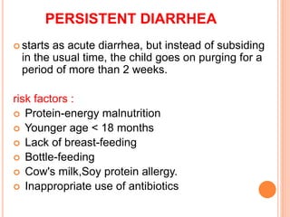
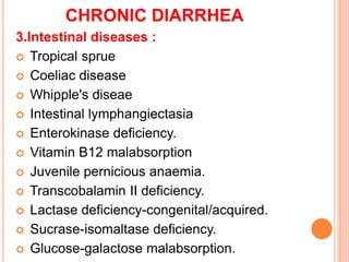
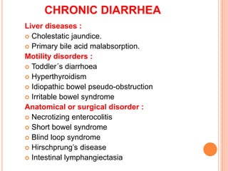
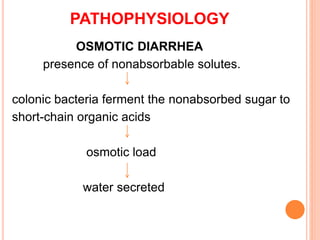
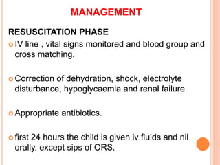
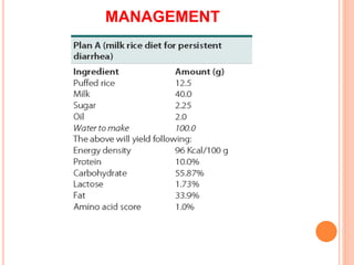
CHRONIC DIARRHEA
Dr M.Sanjeevappa
M.D.(Paeds)
Asst.Professor,
Dept. of Paediatrics
Govt.Medical College
Ananthapuramu.
 Most diarrheal disorders resolve within the first
week of the illness.
 1 to 3% of acute diarrhoeas become chronic, With
a high mortality and morbidity.
 Chronic diarrhea has been defined as an episode
that begins acutely but lasts for 14 days or longer.
 Two entities :
1) Persistent diarrhea.
2) Chronic diarrhea.
PERSISTENT DIARRHEA
 starts as acute diarrhea, but instead of subsiding
in the usual time, the child goes on purging for a
period of more than 2 weeks.
risk factors :
 Protein-energy malnutrition
 Younger age < 18 months
 Lack of breast-feeding
 Bottle-feeding
 Cow's milk,Soy protein allergy.
 Inappropriate use of antibiotics
PERSISTENT DIARRHEA
Risk factors :
 Improper therapy of ADD.
 Use of antimotility drugs like loperamide.
 Starvation during ADD.
 Vitamin A deficiency.
 Zinc deficiency.
 Poor hygiene leading to reinfection.
 Certain extra intestinal infections, e.g :UTI
CHRONIC DIARRHEA
Definition :
 Chronic diarrhea is defined as diarrhea greater than
2 weeks duration, with an insidious onset and
usually due to noninfectious cause. Almost all
patients need a complete workup for underlying
malabsorptive state.
1. Inflammatory causes :
 Tuberculosis.
 Eosinophilic gastroenteritis.
 Crohn's disease.
 Necrotising enterocolitis.
 Allergic colitis.
 Henoch-Schonlein vasculitis.
CHRONIC DIARRHEA
2. Malabsorption states :
 Pancreatic diseases.
 Cystic fibrosis.
 Chronic pancreatitis.
 Hereditary pancreatitis.
 Congenital lipase deficiency.
 Congenital trypsin deficiency.
CHRONIC DIARRHEA
3.Intestinal diseases :
 Tropical sprue
 Coeliac disease
 Whipple's diseae
 Intestinal lymphangiectasia
 Enterokinase deficiency.
 Vitamin B12 malabsorption
 Juvenile pernicious anaemia.
 Transcobalamin II deficiency.
 Lactase deficiency-congenital/acquired.
 Sucrase-isomaltase deficiency.
 Glucose-galactose malabsorption.
CHRONIC DIARRHEA
4.Metabolic disorders :
 Darrow's syndrome (congenital chloride diarrhea).
 Abetalipoproteinaemia.
 Acrodermatitis enteropathica.
5.Endocrine causes :
 Hypoparathyroidism, Hyperthyroidism.
 Diabetes mellitus.
 Adrenal insufficiency.
6.Congenital alterations in electrolyte transport :
 Congenital chloride diarrhea
CHRONIC DIARRHEA
7.Immune defects :
 Agammaglobulinaemia.
 Isolated IgA deficiency.
 Defective CMI.
 Combined immunodeficiency.
8.Neoplasms :
 lmmunoproliferative small intestinal disease
(IPSID orMediterranean lymphoma).
 Western lymphoma.
 Ganglioneuroma.
 Vernor-Morrison syndrome (pancreatic cholera)
 Zollinger-Ellison syndrome.
CHRONIC DIARRHEA
Liver diseases :
 Cholestatic jaundice.
 Primary bile acid malabsorption.
Motility disorders :
 Toddler´s diarrhoea
 Hyperthyroidism
 Idiopathic bowel pseudo-obstruction
 Irritable bowel syndrome
Anatomical or surgical disorder :
 Necrotizing enterocolitis
 Short bowel syndrome
 Blind loop syndrome
 Hirschprung’s disease
 Intestinal lymphangiectasia
PATHOPHYSIOLOGY
OSMOTIC DIARRHEA
presence of nonabsorbable solutes.
colonic bacteria ferment the nonabsorbed sugar to
short-chain organic acids
osmotic load
water secreted
CAUSES FOR OSMOTIC DIARRHEA
 malabsorption of water-soluble nutrients
-glucose-galactose malabsorption
-congenital, acquired disaccharidase deficiencies.
 excessive intake of carbonated fluid.
 excessive intake of nonabsorbable solutes.
-sorbitol, lactulose, magnesium hydroxide.
 stops with fasting.
 has a low pH.
 positive for reducing substances
PATHOPHYSIOLOGY
SECRETORY DIARRHEA
activation cAMP, cGMP, and intracellular calcium.
active chloride secretion (crypt cells)+ inhibit
the neutral coupled sodium chloride absorption.
alter the paracellular ion flux( toxin-mediated
injury to the tight junctions)
secretory diarrhea
CAUSES OF SECRETORY DIARRHEA
ACTIVATION OF CYCLIC AMP
 Bacterial toxins: enterotoxins of cholera, Escherichia coli
(heat-labile), Shigella, Salmonella, Campylobacter jejuni,
Pseudomonas aeruginosa
 Hormones: vasoactive intestinal peptide, gastrin, secretin
 Anion surfactants: bile acids.
ACTIVATION OF CYCLIC GMP
 Bacterial toxins: E. coli (heat-stable) enterotoxin, Yersinia
enterocolitica toxin
CALCIUM-DEPENDENT
 Bacterial toxins: Clostridium difficile enterotoxin
 Neurotransmiters: acetylcholine, serotonin
 Paracrine agents: bradykinin
EVALUATION OF A CHILD WITH
CHRONIC DIARRHEA
STOOL HISTORY :
 SBD : profuse, watery, usually offensive stools,
without blood.
 LBD: small quantity, frequently with blood and
mucus.
 Odourless blood tinged stools - shigellosis
 Frequent mucoid stools in a healthy child without blood -
IBS
 Nocturnal diarrhoea favours organic disease over IBS.
 History of delayed passage of meconiurn and if
constipation preceded diarrhoea,- Hirschsprung's
disease
DIETETIC HISTORY
 It may provide vital clues to the aetiology, e.g., cow's milk
protein intolerance, lactose intolerance, gluten enteropathy,
soy protien intolerance, egg protien enteropathy.
 Overfeeding, concentrated formula feeds> osmotic
diarrhoea.
 Chewing gums and chocolates.
 plenty of undiluted fruit juices (e.g., pineapple juice has
an osmolality of 900 mOsm/L and apple juice 650 mOsm/L
TREATMENT HISTORY
 If achild on antibiotics develops diarrhea, -
pseudomembranous colitis.
 Drugs - neomycin, colchicine, cholestyrarnine, digitalis,
and propranolol.
 Laxatives abuse(Factitious diarrhoea by proxy)
 A family history- IBS.
DIAGNOSIS
 A complete clinical history is mandatory.
 Age of onset
 Nutritional assessment
 Associated symptoms: fever, vomiting, abdominal pain,
anorexia.
 Stool characteristics: blood, mucous, non digested
substances, steatorrhea.
 Physical examination: FTT, abdominal distension,
 visceromegaly, tenderness, presence of abdominal
masses.
 Other organs affected, e.g. skin, respiratory system.
DIAGNOSIS
 Hyperpigmentation- Addison's disease, coeliac
disease ,Whipple's disease.
 Generalized lymphadenopathy- lymphoma, AIDS or
Whipple's disease.
 In a child born of consanguinous parents with
malabsorption and chronic lung disease, cystic
fibrosis should be ruled out.
DIAGNOSIS
 Recurrent fever- tuberculosis, lynphoma, and IBD
 Marked loss of weight- malabsorption, lymphomas,
IBD, TB.
 periorifcial lesions : acrodermatitis emeropathica.
 perianal fistula - Crohn's disease.
 Clubbing - malabsorption syndromes, IBD.
 Chronic liver disease- IBD
 Hepatomegaly -lymphomas, metastatic carcinoid,
IBD and Whipple's disease.
 Ascites - TB and lymphoma.
INVESTIGATION
STOOL EXAMINATION
Microscopy :
 Polymorphs and RBCs - bacterial colitis, whipworm
colitis, amoebic colitis and in IBD.
 Eosinophils are seen in milk or soya protein intolerance.
pH and Reducing Substance :
 A stool pH < 5.5 (on cow's milk) or < 5 (on breast milk) is
suggestive of carbohydrate malabsorption and proximal
small bowel damage.
 Stool pH gives a clue to the amount of organic acids in
stool while the increased amounts of reducing substances
indicate the presence of unabsorbed sugars.
DEMONSTRATION OF REDUCING SUGARS IN STOOL
 Benedict's test : 1 ml of distilled water is added to 0.5
ml liquid stool and shaken well. 8 drops of this are
added to 5 ml of preboiled Benedict's solution and
boiled for 1 minute.The solution is cooled and the
precipitate is examined for colour change.
 Stool Culture :
Stool culture is positive only in 20% of patients with
acute diarrhoea and it is even lower in PD.
 Occult Blood :
In acute diarrhoea- bacterial or parasitic colitis.
Chronic diarrhoea- IBD like ulcerative colitis and
Crohn's colitis and IPSID(Immunoproliferative small
intestinal desease).
INVESTIGATION
Peripheral Blood Picture
 iron deficiency anaemia or dimorphic anaemia.
 abetalipoproteinaemia (acanthocytes)
Biochemical Investigations
 Serum electrolytes,
 blood urea,
 RBS and plasma proteins.
Blood and Urine Culture
 Systemic infections are important causes of CD in
infancy. Cultures from various sites, before starting
antibiotics, are extremely useful in detecting these
infections.
INVESTIGATION
 Barium meal follow through:
This will detect ulcers and strictures of small bowel.
 Small bowel biopsy:
tropicalsprue, coeliac disease, tuberculosis,
lymphoma,abetalipoproteinaetnia, Whipple's
disease, amyloidosis, lymphangicetasia.
INVESTIGATION
 Proctosigmoidoscopy:
 To differentiate SBD from LBD(colitis).
 To visualize pseudomernbrane/polyps/ulcers/tumours.
 Direct swabs for microscopy and culture.
 Rectal biopsy.
 Rectal Biopsy :
 Ulcerative colitis.
 Crohn's disease.
 Schistosomiasis.
 Trichuriasis.
 Amyloidosis.
 Whipple's disease
INVESTIGATION
Tests for Tuberculosis :
 Mantoux test.
 X-ray chest.
 Barium meal follow-through for ulcers, strictures,
malabsorption pattern etc.
 Barium enema-if colonic lesion is suspected.
 Duodenal, jejunal or colonic biopsy-for tissue
diagnosis.
MANAGEMENT
 About 30% of patients with PD require
hospitalization, if they have 1 or more of the
following:
 Age: Less than 4 months and not breast feed.
 Severe PEM.
 Dehydration
 Presence of systemic infections.
MANAGEMENT
The management of PD consists of 3 phases:
 Resuscitation phase (24-48 hours).
 Control of diarrhoea (up to 7 days).
 Rehabilitation phase (up to 8 weeks).
MANAGEMENT
RESUSCITATION PHASE
 IV line , vital signs monitored and blood group and
cross matching.
 Correction of dehydration, shock, electrolyte
disturbance, hypoglycaemia and renal failure.
 Appropriate antibiotics.
 first 24 hours the child is given iv fluids and nil
orally, except sips of ORS.
MANAGEMENT
CONTROL OF DIARRHEA
The major factors responsible for PD
 Bacterial contamination of the gut.
 Systemic infections.
 Food allergen (cow milk, soy protein, egg protein).
 Lactose intolerance.
 Toxins.
 Bile acids.
MANAGEMENT
 Many infants with PD are very sick and have features of
systemic infections like septicaemia and
bronchopneumonia.
 Combination of I.V. Amikacin and I.V. cefotaxime is
extremely effective in sick infants.
 In infants less than 3 months I.V. Amikacin and I.V.
Ampicillin are more effective.
 In a recent study cotrimoxazole was found to be very
useful in children with PD.
 Albendazole.
 Shigellosis – ciprofloxcacin.
 Amebiasis – metronidazole.
MANAGEMENT
REHABILITATION PHASE AND AIMS
 To improve the general health and nutritional
status.
 To correct nutritional deficiencies.
 For catch-up growth.
 To educate the parents, especially to prevent future
relapse.
MANAGEMENT
DIETARY MANAGEMENT AND GOALS :
 Small frequent feeds.
 Start with a high carbohydrate, low protein, and no fat
regime, as the patient improves, coconut oil may be
added.
 Always avoid those food substances, which may be
responsible for PD e.g., milk and milk products in
cow's milk allergy.
 Provide adequate micronutrients and vitamins.
 The diet should not be hyperosmolar.
MANAGEMENT
MANAGEMENT
MANAGEMENT
MANAGEMENT
INDICATORS OF TREATMENT FAILURE :
 Passage of >7 stools per day at the end of one
week.
 Weight loss or poor weight gain, in spite of an oral
intake of at least 100 ml/kg/day, over the previous
3 days.
 If the child develops dehydration at anytime.
 Significant increase in diarrhoea with in 48 hr
MANAGEMENT
Problems and Remedies :
 Anorexia - try nasogastric feeding.
 Intolerance - change diet, postpone alimentation.
 Poor weight gain - add fat and pancreatic enzymes.
 Trace element deficiency - oral zinc, Mg.
 Hypothermia - wrap the baby well.
THANK YOU
Chronic diarrhea in children

Chronic diarrhea in children

  • 1.
    CHRONIC DIARRHEA Dr M.Sanjeevappa M.D.(Paeds) Asst.Professor, Dept.of Paediatrics Govt.Medical College Ananthapuramu.
  • 2.
     Most diarrhealdisorders resolve within the first week of the illness.  1 to 3% of acute diarrhoeas become chronic, With a high mortality and morbidity.  Chronic diarrhea has been defined as an episode that begins acutely but lasts for 14 days or longer.  Two entities : 1) Persistent diarrhea. 2) Chronic diarrhea.
  • 3.
    PERSISTENT DIARRHEA  startsas acute diarrhea, but instead of subsiding in the usual time, the child goes on purging for a period of more than 2 weeks. risk factors :  Protein-energy malnutrition  Younger age < 18 months  Lack of breast-feeding  Bottle-feeding  Cow's milk,Soy protein allergy.  Inappropriate use of antibiotics
  • 4.
    PERSISTENT DIARRHEA Risk factors:  Improper therapy of ADD.  Use of antimotility drugs like loperamide.  Starvation during ADD.  Vitamin A deficiency.  Zinc deficiency.  Poor hygiene leading to reinfection.  Certain extra intestinal infections, e.g :UTI
  • 5.
    CHRONIC DIARRHEA Definition : Chronic diarrhea is defined as diarrhea greater than 2 weeks duration, with an insidious onset and usually due to noninfectious cause. Almost all patients need a complete workup for underlying malabsorptive state. 1. Inflammatory causes :  Tuberculosis.  Eosinophilic gastroenteritis.  Crohn's disease.  Necrotising enterocolitis.  Allergic colitis.  Henoch-Schonlein vasculitis.
  • 6.
    CHRONIC DIARRHEA 2. Malabsorptionstates :  Pancreatic diseases.  Cystic fibrosis.  Chronic pancreatitis.  Hereditary pancreatitis.  Congenital lipase deficiency.  Congenital trypsin deficiency.
  • 7.
    CHRONIC DIARRHEA 3.Intestinal diseases:  Tropical sprue  Coeliac disease  Whipple's diseae  Intestinal lymphangiectasia  Enterokinase deficiency.  Vitamin B12 malabsorption  Juvenile pernicious anaemia.  Transcobalamin II deficiency.  Lactase deficiency-congenital/acquired.  Sucrase-isomaltase deficiency.  Glucose-galactose malabsorption.
  • 8.
    CHRONIC DIARRHEA 4.Metabolic disorders:  Darrow's syndrome (congenital chloride diarrhea).  Abetalipoproteinaemia.  Acrodermatitis enteropathica. 5.Endocrine causes :  Hypoparathyroidism, Hyperthyroidism.  Diabetes mellitus.  Adrenal insufficiency. 6.Congenital alterations in electrolyte transport :  Congenital chloride diarrhea
  • 9.
    CHRONIC DIARRHEA 7.Immune defects:  Agammaglobulinaemia.  Isolated IgA deficiency.  Defective CMI.  Combined immunodeficiency. 8.Neoplasms :  lmmunoproliferative small intestinal disease (IPSID orMediterranean lymphoma).  Western lymphoma.  Ganglioneuroma.  Vernor-Morrison syndrome (pancreatic cholera)  Zollinger-Ellison syndrome.
  • 10.
    CHRONIC DIARRHEA Liver diseases:  Cholestatic jaundice.  Primary bile acid malabsorption. Motility disorders :  Toddler´s diarrhoea  Hyperthyroidism  Idiopathic bowel pseudo-obstruction  Irritable bowel syndrome Anatomical or surgical disorder :  Necrotizing enterocolitis  Short bowel syndrome  Blind loop syndrome  Hirschprung’s disease  Intestinal lymphangiectasia
  • 11.
    PATHOPHYSIOLOGY OSMOTIC DIARRHEA presence ofnonabsorbable solutes. colonic bacteria ferment the nonabsorbed sugar to short-chain organic acids osmotic load water secreted
  • 12.
    CAUSES FOR OSMOTICDIARRHEA  malabsorption of water-soluble nutrients -glucose-galactose malabsorption -congenital, acquired disaccharidase deficiencies.  excessive intake of carbonated fluid.  excessive intake of nonabsorbable solutes. -sorbitol, lactulose, magnesium hydroxide.  stops with fasting.  has a low pH.  positive for reducing substances
  • 13.
    PATHOPHYSIOLOGY SECRETORY DIARRHEA activation cAMP,cGMP, and intracellular calcium. active chloride secretion (crypt cells)+ inhibit the neutral coupled sodium chloride absorption. alter the paracellular ion flux( toxin-mediated injury to the tight junctions) secretory diarrhea
  • 14.
    CAUSES OF SECRETORYDIARRHEA ACTIVATION OF CYCLIC AMP  Bacterial toxins: enterotoxins of cholera, Escherichia coli (heat-labile), Shigella, Salmonella, Campylobacter jejuni, Pseudomonas aeruginosa  Hormones: vasoactive intestinal peptide, gastrin, secretin  Anion surfactants: bile acids. ACTIVATION OF CYCLIC GMP  Bacterial toxins: E. coli (heat-stable) enterotoxin, Yersinia enterocolitica toxin CALCIUM-DEPENDENT  Bacterial toxins: Clostridium difficile enterotoxin  Neurotransmiters: acetylcholine, serotonin  Paracrine agents: bradykinin
  • 16.
    EVALUATION OF ACHILD WITH CHRONIC DIARRHEA STOOL HISTORY :  SBD : profuse, watery, usually offensive stools, without blood.  LBD: small quantity, frequently with blood and mucus.  Odourless blood tinged stools - shigellosis  Frequent mucoid stools in a healthy child without blood - IBS  Nocturnal diarrhoea favours organic disease over IBS.  History of delayed passage of meconiurn and if constipation preceded diarrhoea,- Hirschsprung's disease
  • 17.
    DIETETIC HISTORY  Itmay provide vital clues to the aetiology, e.g., cow's milk protein intolerance, lactose intolerance, gluten enteropathy, soy protien intolerance, egg protien enteropathy.  Overfeeding, concentrated formula feeds> osmotic diarrhoea.  Chewing gums and chocolates.  plenty of undiluted fruit juices (e.g., pineapple juice has an osmolality of 900 mOsm/L and apple juice 650 mOsm/L
  • 18.
    TREATMENT HISTORY  Ifachild on antibiotics develops diarrhea, - pseudomembranous colitis.  Drugs - neomycin, colchicine, cholestyrarnine, digitalis, and propranolol.  Laxatives abuse(Factitious diarrhoea by proxy)  A family history- IBS.
  • 19.
    DIAGNOSIS  A completeclinical history is mandatory.  Age of onset  Nutritional assessment  Associated symptoms: fever, vomiting, abdominal pain, anorexia.  Stool characteristics: blood, mucous, non digested substances, steatorrhea.  Physical examination: FTT, abdominal distension,  visceromegaly, tenderness, presence of abdominal masses.  Other organs affected, e.g. skin, respiratory system.
  • 20.
    DIAGNOSIS  Hyperpigmentation- Addison'sdisease, coeliac disease ,Whipple's disease.  Generalized lymphadenopathy- lymphoma, AIDS or Whipple's disease.  In a child born of consanguinous parents with malabsorption and chronic lung disease, cystic fibrosis should be ruled out.
  • 21.
    DIAGNOSIS  Recurrent fever-tuberculosis, lynphoma, and IBD  Marked loss of weight- malabsorption, lymphomas, IBD, TB.  periorifcial lesions : acrodermatitis emeropathica.  perianal fistula - Crohn's disease.  Clubbing - malabsorption syndromes, IBD.  Chronic liver disease- IBD  Hepatomegaly -lymphomas, metastatic carcinoid, IBD and Whipple's disease.  Ascites - TB and lymphoma.
  • 22.
    INVESTIGATION STOOL EXAMINATION Microscopy : Polymorphs and RBCs - bacterial colitis, whipworm colitis, amoebic colitis and in IBD.  Eosinophils are seen in milk or soya protein intolerance. pH and Reducing Substance :  A stool pH < 5.5 (on cow's milk) or < 5 (on breast milk) is suggestive of carbohydrate malabsorption and proximal small bowel damage.  Stool pH gives a clue to the amount of organic acids in stool while the increased amounts of reducing substances indicate the presence of unabsorbed sugars.
  • 23.
    DEMONSTRATION OF REDUCINGSUGARS IN STOOL  Benedict's test : 1 ml of distilled water is added to 0.5 ml liquid stool and shaken well. 8 drops of this are added to 5 ml of preboiled Benedict's solution and boiled for 1 minute.The solution is cooled and the precipitate is examined for colour change.  Stool Culture : Stool culture is positive only in 20% of patients with acute diarrhoea and it is even lower in PD.  Occult Blood : In acute diarrhoea- bacterial or parasitic colitis. Chronic diarrhoea- IBD like ulcerative colitis and Crohn's colitis and IPSID(Immunoproliferative small intestinal desease).
  • 24.
    INVESTIGATION Peripheral Blood Picture iron deficiency anaemia or dimorphic anaemia.  abetalipoproteinaemia (acanthocytes) Biochemical Investigations  Serum electrolytes,  blood urea,  RBS and plasma proteins. Blood and Urine Culture  Systemic infections are important causes of CD in infancy. Cultures from various sites, before starting antibiotics, are extremely useful in detecting these infections.
  • 25.
    INVESTIGATION  Barium mealfollow through: This will detect ulcers and strictures of small bowel.  Small bowel biopsy: tropicalsprue, coeliac disease, tuberculosis, lymphoma,abetalipoproteinaetnia, Whipple's disease, amyloidosis, lymphangicetasia.
  • 26.
    INVESTIGATION  Proctosigmoidoscopy:  Todifferentiate SBD from LBD(colitis).  To visualize pseudomernbrane/polyps/ulcers/tumours.  Direct swabs for microscopy and culture.  Rectal biopsy.  Rectal Biopsy :  Ulcerative colitis.  Crohn's disease.  Schistosomiasis.  Trichuriasis.  Amyloidosis.  Whipple's disease
  • 27.
    INVESTIGATION Tests for Tuberculosis:  Mantoux test.  X-ray chest.  Barium meal follow-through for ulcers, strictures, malabsorption pattern etc.  Barium enema-if colonic lesion is suspected.  Duodenal, jejunal or colonic biopsy-for tissue diagnosis.
  • 28.
    MANAGEMENT  About 30%of patients with PD require hospitalization, if they have 1 or more of the following:  Age: Less than 4 months and not breast feed.  Severe PEM.  Dehydration  Presence of systemic infections.
  • 29.
    MANAGEMENT The management ofPD consists of 3 phases:  Resuscitation phase (24-48 hours).  Control of diarrhoea (up to 7 days).  Rehabilitation phase (up to 8 weeks).
  • 30.
    MANAGEMENT RESUSCITATION PHASE  IVline , vital signs monitored and blood group and cross matching.  Correction of dehydration, shock, electrolyte disturbance, hypoglycaemia and renal failure.  Appropriate antibiotics.  first 24 hours the child is given iv fluids and nil orally, except sips of ORS.
  • 31.
    MANAGEMENT CONTROL OF DIARRHEA Themajor factors responsible for PD  Bacterial contamination of the gut.  Systemic infections.  Food allergen (cow milk, soy protein, egg protein).  Lactose intolerance.  Toxins.  Bile acids.
  • 32.
    MANAGEMENT  Many infantswith PD are very sick and have features of systemic infections like septicaemia and bronchopneumonia.  Combination of I.V. Amikacin and I.V. cefotaxime is extremely effective in sick infants.  In infants less than 3 months I.V. Amikacin and I.V. Ampicillin are more effective.  In a recent study cotrimoxazole was found to be very useful in children with PD.  Albendazole.  Shigellosis – ciprofloxcacin.  Amebiasis – metronidazole.
  • 33.
    MANAGEMENT REHABILITATION PHASE ANDAIMS  To improve the general health and nutritional status.  To correct nutritional deficiencies.  For catch-up growth.  To educate the parents, especially to prevent future relapse.
  • 34.
    MANAGEMENT DIETARY MANAGEMENT ANDGOALS :  Small frequent feeds.  Start with a high carbohydrate, low protein, and no fat regime, as the patient improves, coconut oil may be added.  Always avoid those food substances, which may be responsible for PD e.g., milk and milk products in cow's milk allergy.  Provide adequate micronutrients and vitamins.  The diet should not be hyperosmolar.
  • 35.
  • 36.
  • 37.
  • 38.
    MANAGEMENT INDICATORS OF TREATMENTFAILURE :  Passage of >7 stools per day at the end of one week.  Weight loss or poor weight gain, in spite of an oral intake of at least 100 ml/kg/day, over the previous 3 days.  If the child develops dehydration at anytime.  Significant increase in diarrhoea with in 48 hr
  • 39.
    MANAGEMENT Problems and Remedies:  Anorexia - try nasogastric feeding.  Intolerance - change diet, postpone alimentation.  Poor weight gain - add fat and pancreatic enzymes.  Trace element deficiency - oral zinc, Mg.  Hypothermia - wrap the baby well.
  • 40.