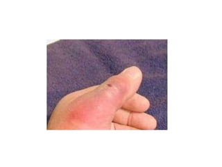
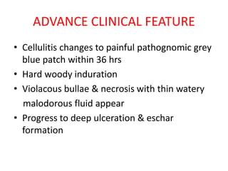
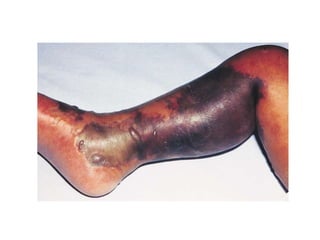
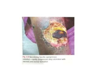
NECROTISING
FASCITIS
ABHISHEK BHARTI
Roll no 17005
INTRODUCTION
• Rapidly spreading necrotising infection of
subcutaneous tissue & fascia
• Alsoknown as: “flesh-eating” bacteria.
• Infection rapidly destroy the skinand soft tissue
beneath it
• Other names: β-hemolytic streptococcal
gangrene, Meleneyulcer, acute dermal gangrene,
hospital gangrene, and necrotizing cellulitis.
Types of NF.
• Type I : apolymicrobial flora
• Type II Group A β-Streptococcus bacteria
Transmission of bacteria
• From outside due to penetrating trauma,
surgery
• From inside due to transient bacteremia
RISK FACTORS
• Old age
• Diabetes
• Alcoholism
• Immuno-suppression-steroids/HCV
• Severe illnesses:heart, lung, or liverdisease
• Malnourished
• Obesity
PATHOPHYSIOLOGY
• Bacteria eat away at tissue between skin and
muscle
• Inflammatory response by immune system
• Bacterial toxins released
• Cytokines impede function of phagocytic cells
• Anaerobes thrive speeding up necrotic process
• Endothelium damaged leading to poor blood
supply
• Poor blood supply inhibits inflammatoy
resopnse & cause hypoxia
EARLY CLINICAL FEATURE
• Flu like symptoms that include fever, chills,
nausea, weakness, dizziness, aches
• Skinbecomes tender, warm, red in color, and
start to swell
• Patients mayexperience pain greater than
expected from the appearance of the wound
• Subcutaneous tissue mayalso havea hard feel on
palpation
ADVANCE CLINICAL FEATURE
• Cellulitis changes to painful pathognomic grey
blue patch within 36 hrs
• Hard woody induration
• Violacous bullae & necrosis with thin watery
malodorous fluid appear
• Progress to deep ulceration & eschar
formation
• High fever with chills ,malaise,tachytardia
• Toxic shock syndrome may occur
• Lesion may become anaesthetic due to
cutaneous nerve damage
• MOST Common site are extremities
INVESTIGATION & DIAGNOSIS
• Blood investigation:
TLC,DLC,CPK, elevatedor lowered
creatinine, glucose, CPK, bicarbonate, albumin,
andcalcium levels
• MRI toknowdepth
• BacterialCulture & Drugsensitivity
TREATMENT
• Early diagnosis & treatment is vital
• Emergency debridement
• IV antibiotics
• Hyperbaric oxygen therapy for anaerobes
• Morphine drip & patient controlled analgesia
pump
Necrotising fascitis

Necrotising fascitis

  • 1.
  • 2.
    INTRODUCTION • Rapidly spreadingnecrotising infection of subcutaneous tissue & fascia • Alsoknown as: “flesh-eating” bacteria. • Infection rapidly destroy the skinand soft tissue beneath it • Other names: β-hemolytic streptococcal gangrene, Meleneyulcer, acute dermal gangrene, hospital gangrene, and necrotizing cellulitis.
  • 3.
    Types of NF. •Type I : apolymicrobial flora • Type II Group A β-Streptococcus bacteria
  • 4.
    Transmission of bacteria •From outside due to penetrating trauma, surgery • From inside due to transient bacteremia
  • 5.
    RISK FACTORS • Oldage • Diabetes • Alcoholism • Immuno-suppression-steroids/HCV • Severe illnesses:heart, lung, or liverdisease • Malnourished • Obesity
  • 6.
    PATHOPHYSIOLOGY • Bacteria eataway at tissue between skin and muscle • Inflammatory response by immune system • Bacterial toxins released • Cytokines impede function of phagocytic cells • Anaerobes thrive speeding up necrotic process • Endothelium damaged leading to poor blood supply • Poor blood supply inhibits inflammatoy resopnse & cause hypoxia
  • 7.
    EARLY CLINICAL FEATURE •Flu like symptoms that include fever, chills, nausea, weakness, dizziness, aches • Skinbecomes tender, warm, red in color, and start to swell • Patients mayexperience pain greater than expected from the appearance of the wound • Subcutaneous tissue mayalso havea hard feel on palpation
  • 9.
    ADVANCE CLINICAL FEATURE •Cellulitis changes to painful pathognomic grey blue patch within 36 hrs • Hard woody induration • Violacous bullae & necrosis with thin watery malodorous fluid appear • Progress to deep ulceration & eschar formation
  • 10.
    • High feverwith chills ,malaise,tachytardia • Toxic shock syndrome may occur • Lesion may become anaesthetic due to cutaneous nerve damage • MOST Common site are extremities
  • 13.
    INVESTIGATION & DIAGNOSIS •Blood investigation: TLC,DLC,CPK, elevatedor lowered creatinine, glucose, CPK, bicarbonate, albumin, andcalcium levels • MRI toknowdepth • BacterialCulture & Drugsensitivity
  • 14.
    TREATMENT • Early diagnosis& treatment is vital • Emergency debridement • IV antibiotics • Hyperbaric oxygen therapy for anaerobes • Morphine drip & patient controlled analgesia pump