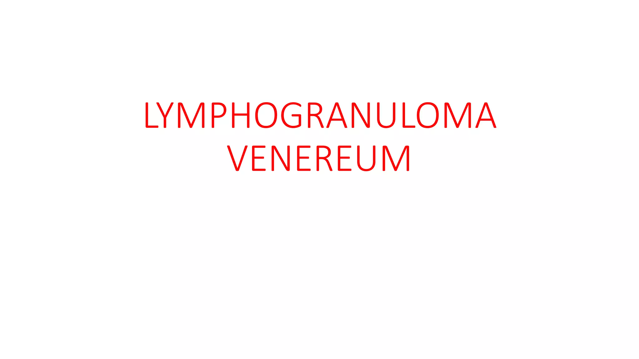
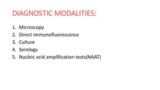
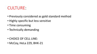
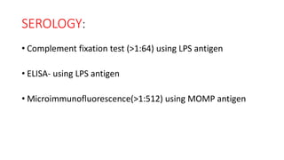
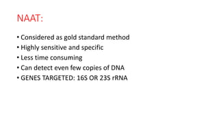
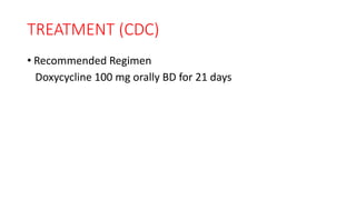
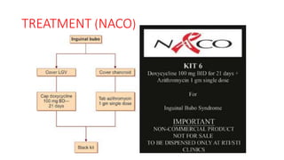
This document discusses laboratory diagnosis of Lymphogranuloma venereum (LVG). It outlines various diagnostic modalities including microscopy, direct immunofluorescence, culture, serology, and nucleic acid amplification tests. Microscopy has low sensitivity for detecting Chlamydia. Culture was previously the gold standard but NAAT is now considered the most sensitive and specific method as it can detect DNA copies and is less time consuming. Serology tests for antibodies and NAAT targets rRNA genes. Treatment recommended by CDC and NACO involves a 21 day course of doxycycline.


![CTEV [ clubfoot] DR ARUN LAL ,DR MOHAMED ASHRAF travancore medical college k...](https://cdn.slidesharecdn.com/ss_thumbnails/ctevclubfootdrarunlaldrmohamedashraftravancoremedicalcollegekollamkeralaindia-260208063247-18fc466c-thumbnail.jpg?width=640&height=640&fit=bounds)

![ONFH[AVN HIP] -TRIPLE REGIME -A NOVAL SURGICAL CONCEPT .pptx](https://cdn.slidesharecdn.com/ss_thumbnails/onfhavnhip2026koaconcalicutdrgokuldevdrmashraf-260210064517-213ec005-thumbnail.jpg?width=640&height=640&fit=bounds)