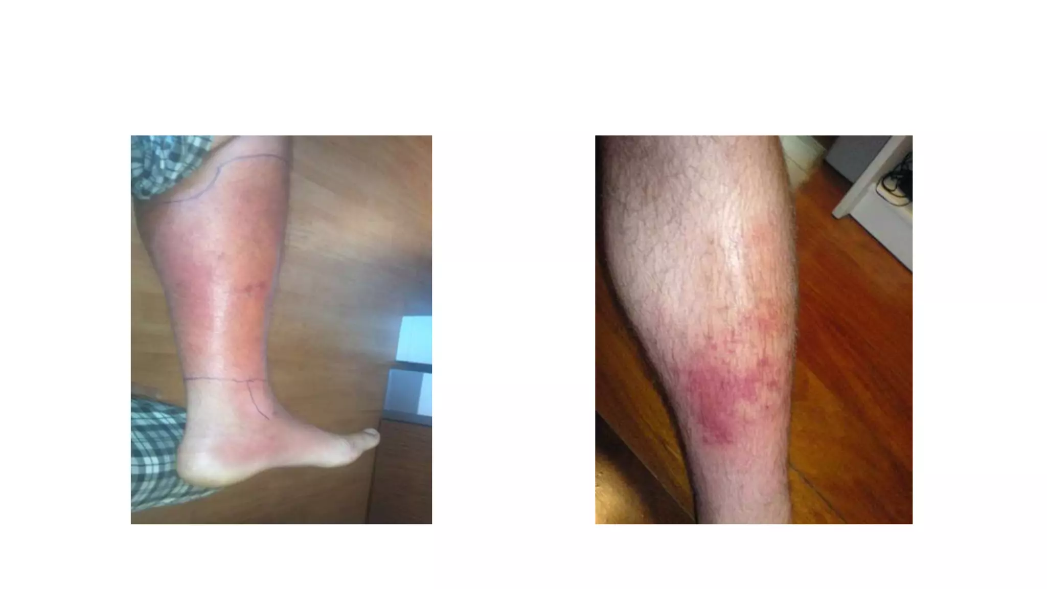
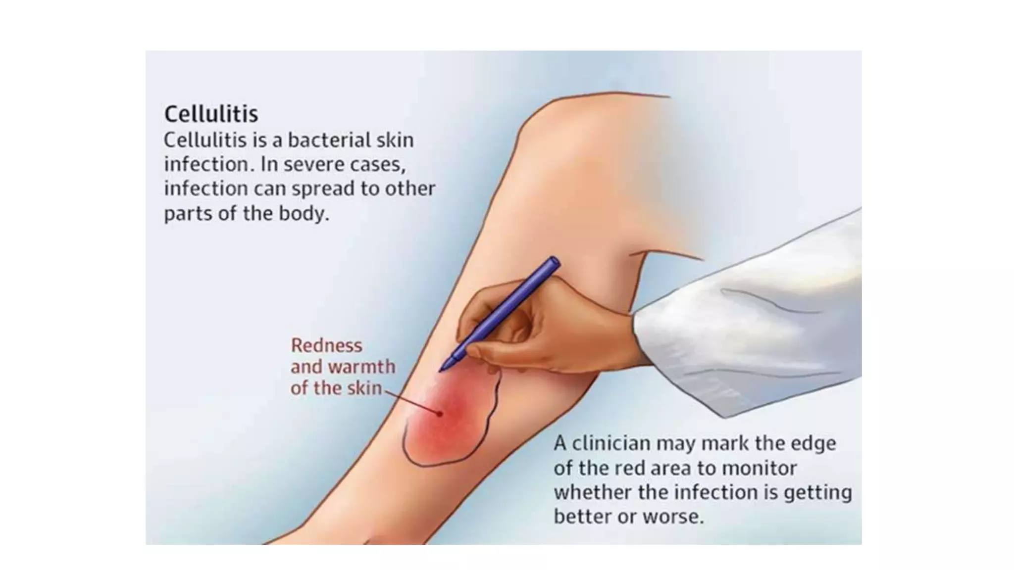
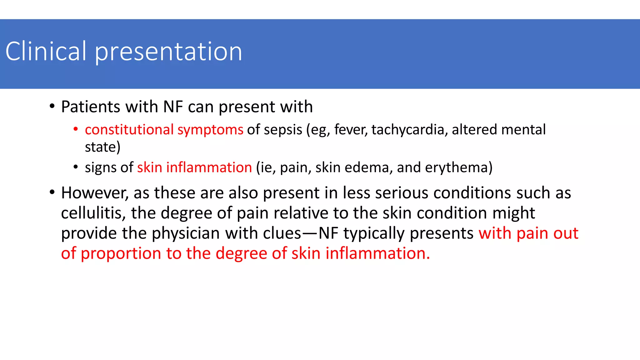
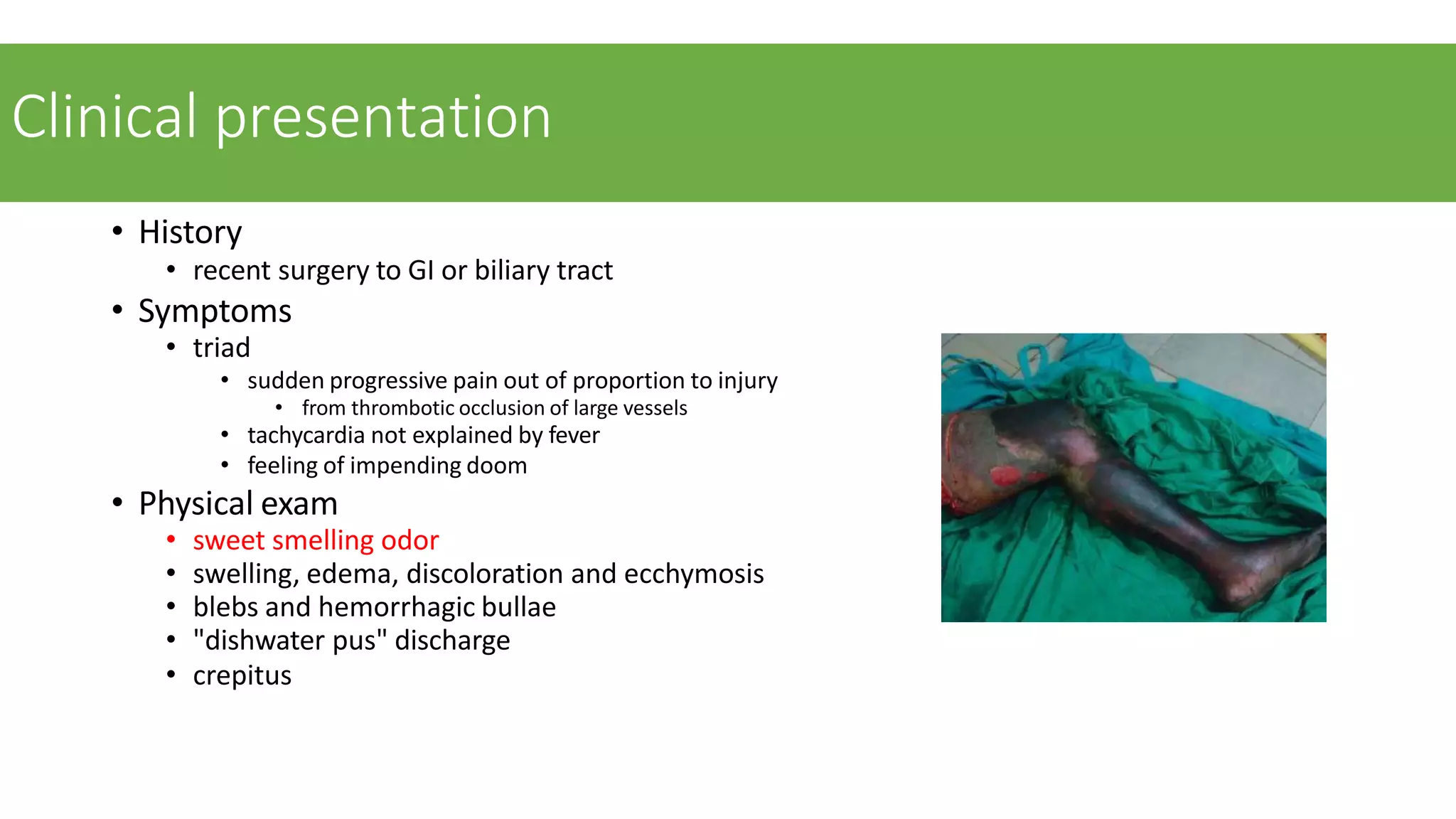
Cellulitis
And
Necrotising Fascitis
• Definition
• Risk factors
• Etiology
• Pathogenesis
• Microbiology
• Clinical presentation
• Workup
• Management
• Prognosis
Cellulitis
Definitation- It is spreading infl ammation of subcutaneous tissue and fascial
planes.
Infection may follow a small scratch or wound or incision or
insect/snake/scorpion bite.
Causative agents
• Commonly due to Streptococcus pyogenes and other Gram +ve organisms.
Release of streptokinase and hyaluronidase cause spread of infection.
•Often Gram –ve organisms like Klebsiella, Pseudo monas, E. coli are also involved
(usually Gram –ve organisms cause secondary infection).
• It can be superfi cial or deep. More common superfi cial type is easier to
diagnose.
• It is common in diabetics, immunosuppressed people and old age.
• It is common in face, lower limb, upper limb and scrotum.
•Cellulitis occurring in children is never primary but secondary to an underlying
bone infection.
Sequelae of disease.
•Infection can get localised to form pyogenic abscess.
•Infection can spread to cause bacteraemia, septicaemia, pyaemia.
•Often infection can lead to local gangrene.
•Extensive necrosis of skin and subcutaneous tissue— necrotizing fasciitis.
Clinical Features
• Fever, toxicity (tachycardia, hypotension).
•Swelling is diffuse and spreading in nature.
•Pain and tenderness, red, shiny area with stretched warm skin.
•Cellulitis will progress rapidly in diabetic and immunosup- pressed individuals.
•Tender regional lymph nodes may be palpable which signify severity of the
infection.
•No edge; no pus; no fluctuation; no limit.
Investigations
•CBC
•RBS
•Serum Creatinine
•X ray – to rule out underline bone infection
•Ultrasonography- to see for underlying abscess formation (pus collection)
Management
•Elevation of limb or part to reduce oedema so as to increase the circulation
•Antibiotics—penicillins, cephalosporins.
•Dressing (often glycerine dressing is used as it reduces the oedema because of
its hygroscopic action glycerine magnesium sulphate dressing).
•Bandaging.
Necrotising Fasciitis
• Definition -
Necrotizing fasciitis is a necrotizing soft tissue infection spreading
along fascial planes with or without overlying cellulitis.
•Diabetes
•Chronic disease
•Immunosuppressive drugs (eg, prednisolone)
•Malnutrition
•Age > 60 years
•Intravenous drug misuse
•Peripheral vascular disease
•Renal failure
•Underlying malignancy
•Obesity
Risk factors
bacterial introduction
IV drug use
hypodermic therapeutic injections
insect bites
skin abrasions
abdominal and perineal surgery
TYPES
Type 1 : Polymicrobial
Causes:-combinations of aerobic and anaerobic organisms
Most common anaerobic bacteria – Bacteroids ,clostridium ,
peptostreptococcus
Enterobacteria-E coli, klebsiela, proteus
Facultative anaerobic streptococci
Most Common site – Perineum ( Fournier’s gangrene), Cervical (head and
neck ) due to oral or dental infection
Type 2: Monomicrobial
Causes :- Group A streptococci, other beta haemolytic streptococci,
Staphylococcus aureus.
Type 3: Salt water infection
Causes: - Vibrio Vulnificus
Rare
Type 4 : Fungal infection
Etiology
Clinical presentation
• Patients with NF can present with
• constitutional symptoms of sepsis (eg, fever, tachycardia, altered mental
state)
• signs of skin inflammation (ie, pain, skin edema, and erythema)
• However, as these are also present in less serious conditions such as
cellulitis, the degree of pain relative to the skin condition might
provide the physician with clues—NF typically presents with pain out
of proportion to the degree of skin inflammation.
• Necrotizing fasciitis typically presents with patchy discolouration of
the skin with pain and swelling, but without a defined margin
• Progression of NF is marked with the development of tense edema, a
grayish-brown discharge, vesicles, bullae, necrosis, and crepitus
Workup
• Laboratory
• Cultures
• Imaging
Laboratory risk indicator for NF (LRINEC) score
score > 6 has PPV of 92% of
having necrotizing fasciitis
• CRP (mg/L)
• ≥150: 4 points
• WBC count (×103/mm3)
• <15: 0 points
• 15–25: 1 point
• >25: 2 points
• Hemoglobin (g/dL)
• >13.5: 0 points
• 11–13.5: 1 point
• <11: 2 points
• Sodium (mmol/L)
<135: 2 points
• Creatinine (umol/L)
>141: 2 points
• Glucose (mmol/L)
>10: 1 point
Cultures
• Blood cultures
• Intraoperative tissue cultures
Imaging
• The common plain radiographic findings non-specific with increased soft-
tissue thickness and opacity. Radiographs can be normal until the advanced
stages of infection and necrosis. The characteristic finding of gas in the soft
tissues is seen in only a minority of cases
• imaging plays a very limited role in diagnosis and management of necrotising
fasciitis.
Treatment
• Antibiotics
• Operative
Antibiotics
• initial antibiotics
• start empirically with penicillin, clindamycin, metronidazole, and an
aminoglycoside
• definitive antibiotics
• penicillin G
• for strep or clostridium
• imipenem or doripenem or meropenem
• for polymicrobial
• add vancomycin or daptomycin
• if MRSA suspected
Operative
• emergency radical debridement with broad-spectrum IV antibiotics
operative findings
• liquefied subcutaneous fat
• dishwater pus
• muscle necrosis
• venous thrombosis
• noncontracting muscle,
• and a positive “probe test” result, which is characterized by lack of resistance to finger
dissection in normally adherent tissues
Gas gangrene
Definition
• necrotizing soft tissue infection of skeletal muscle caused by toxin-
and gas-producing Clostridium species.
• The synonym clostridial myonecrosis better describes both the
causative agent and the target tissue.
Risk factors
• risk factors
• posttraumatic (associated with C perfringens)
• car accidents (most common)
• crush injuries
• gunshot wounds with foreign bodies
• burns and frostbite
• IV drug abuse
• postoperative
• bowel resection or perforation
• biliary surgery
• premature wound closure
• spontaneous
• colon cancer (associated with C. septicum)
Etiology
Clostridial species
• Clostridium perfringens (most common),
• Clostridium novyi
• Clostridium septicum
found in soil and gut flora
• gram-positive obligate anaerobic spore-forming rods that produce exotoxins (e.g.
C. perfringens alpha toxin)
• gas produced by fermentation of glucose
• other bacteria include E. coli, Pseudomonas aeruginosa, Proteus
species, Klebsiella pneumoniae
Clinical presentation
• History
• recent surgery to GI or biliary tract
• Symptoms
• triad
• sudden progressive pain out of proportion to injury
• from thrombotic occlusion of large vessels
• tachycardia not explained by fever
• feeling of impending doom
• Physical exam
• sweet smelling odor
• swelling, edema, discoloration and ecchymosis
• blebs and hemorrhagic bullae
• "dishwater pus" discharge
• crepitus
workup
• Laboratory
• Cultures
• Imaging
Radiographs
• findings
• linear streaks of gas in soft tissues
Labs
• Elevated WCC
Histology :
• Gram stain reveals Gram-positive bacilli
Culture
• blood culture rarely grows Clostridial species
• Intraoperative tissue, muscle cultures
treatment
Antibiotics
• high dose IV antibiotics
• 1st line is penicillin G and clindamycin
• alternative treatment is erythromycin, tetracycline or ceftriaxone
• clindamycin and tetracycline inhibit toxin synthesis
Operative
• radical surgical debridement with fasciotomies
Intraoperative
• Non viable muscle, myonecrosis
references
• https://www.orthobullets.com/trauma/1007/necrotizing-fasciitis
• https://www.ncbi.nlm.nih.gov/pmc/articles/PMC2762295/
• https://radiopaedia.org/cases/gas-gangrene-of-lower-limb
• https://www.orthobullets.com/trauma/1067/gas-gangrene
THANK YOU

CELLULITIS AND NECROTISING FASCITIS PPT.pptx

  • 1.
  • 2.
    • Definition • Riskfactors • Etiology • Pathogenesis • Microbiology • Clinical presentation • Workup • Management • Prognosis
  • 3.
    Cellulitis Definitation- It isspreading infl ammation of subcutaneous tissue and fascial planes. Infection may follow a small scratch or wound or incision or insect/snake/scorpion bite.
  • 4.
    Causative agents • Commonlydue to Streptococcus pyogenes and other Gram +ve organisms. Release of streptokinase and hyaluronidase cause spread of infection. •Often Gram –ve organisms like Klebsiella, Pseudo monas, E. coli are also involved (usually Gram –ve organisms cause secondary infection). • It can be superfi cial or deep. More common superfi cial type is easier to diagnose. • It is common in diabetics, immunosuppressed people and old age. • It is common in face, lower limb, upper limb and scrotum. •Cellulitis occurring in children is never primary but secondary to an underlying bone infection.
  • 5.
    Sequelae of disease. •Infectioncan get localised to form pyogenic abscess. •Infection can spread to cause bacteraemia, septicaemia, pyaemia. •Often infection can lead to local gangrene. •Extensive necrosis of skin and subcutaneous tissue— necrotizing fasciitis.
  • 6.
    Clinical Features • Fever,toxicity (tachycardia, hypotension). •Swelling is diffuse and spreading in nature. •Pain and tenderness, red, shiny area with stretched warm skin. •Cellulitis will progress rapidly in diabetic and immunosup- pressed individuals. •Tender regional lymph nodes may be palpable which signify severity of the infection. •No edge; no pus; no fluctuation; no limit.
  • 8.
    Investigations •CBC •RBS •Serum Creatinine •X ray– to rule out underline bone infection •Ultrasonography- to see for underlying abscess formation (pus collection)
  • 9.
    Management •Elevation of limbor part to reduce oedema so as to increase the circulation •Antibiotics—penicillins, cephalosporins. •Dressing (often glycerine dressing is used as it reduces the oedema because of its hygroscopic action glycerine magnesium sulphate dressing). •Bandaging.
  • 11.
    Necrotising Fasciitis • Definition- Necrotizing fasciitis is a necrotizing soft tissue infection spreading along fascial planes with or without overlying cellulitis.
  • 12.
    •Diabetes •Chronic disease •Immunosuppressive drugs(eg, prednisolone) •Malnutrition •Age > 60 years •Intravenous drug misuse •Peripheral vascular disease •Renal failure •Underlying malignancy •Obesity Risk factors bacterial introduction IV drug use hypodermic therapeutic injections insect bites skin abrasions abdominal and perineal surgery
  • 13.
    TYPES Type 1 :Polymicrobial Causes:-combinations of aerobic and anaerobic organisms Most common anaerobic bacteria – Bacteroids ,clostridium , peptostreptococcus Enterobacteria-E coli, klebsiela, proteus Facultative anaerobic streptococci Most Common site – Perineum ( Fournier’s gangrene), Cervical (head and neck ) due to oral or dental infection Type 2: Monomicrobial Causes :- Group A streptococci, other beta haemolytic streptococci, Staphylococcus aureus. Type 3: Salt water infection Causes: - Vibrio Vulnificus Rare Type 4 : Fungal infection
  • 14.
  • 15.
    Clinical presentation • Patientswith NF can present with • constitutional symptoms of sepsis (eg, fever, tachycardia, altered mental state) • signs of skin inflammation (ie, pain, skin edema, and erythema) • However, as these are also present in less serious conditions such as cellulitis, the degree of pain relative to the skin condition might provide the physician with clues—NF typically presents with pain out of proportion to the degree of skin inflammation.
  • 16.
    • Necrotizing fasciitistypically presents with patchy discolouration of the skin with pain and swelling, but without a defined margin • Progression of NF is marked with the development of tense edema, a grayish-brown discharge, vesicles, bullae, necrosis, and crepitus
  • 17.
  • 18.
    Laboratory risk indicatorfor NF (LRINEC) score score > 6 has PPV of 92% of having necrotizing fasciitis • CRP (mg/L) • ≥150: 4 points • WBC count (×103/mm3) • <15: 0 points • 15–25: 1 point • >25: 2 points • Hemoglobin (g/dL) • >13.5: 0 points • 11–13.5: 1 point • <11: 2 points • Sodium (mmol/L) <135: 2 points • Creatinine (umol/L) >141: 2 points • Glucose (mmol/L) >10: 1 point
  • 19.
    Cultures • Blood cultures •Intraoperative tissue cultures
  • 20.
    Imaging • The commonplain radiographic findings non-specific with increased soft- tissue thickness and opacity. Radiographs can be normal until the advanced stages of infection and necrosis. The characteristic finding of gas in the soft tissues is seen in only a minority of cases • imaging plays a very limited role in diagnosis and management of necrotising fasciitis.
  • 21.
  • 22.
    Antibiotics • initial antibiotics •start empirically with penicillin, clindamycin, metronidazole, and an aminoglycoside • definitive antibiotics • penicillin G • for strep or clostridium • imipenem or doripenem or meropenem • for polymicrobial • add vancomycin or daptomycin • if MRSA suspected
  • 24.
    Operative • emergency radicaldebridement with broad-spectrum IV antibiotics operative findings • liquefied subcutaneous fat • dishwater pus • muscle necrosis • venous thrombosis • noncontracting muscle, • and a positive “probe test” result, which is characterized by lack of resistance to finger dissection in normally adherent tissues
  • 25.
  • 26.
    Definition • necrotizing softtissue infection of skeletal muscle caused by toxin- and gas-producing Clostridium species. • The synonym clostridial myonecrosis better describes both the causative agent and the target tissue.
  • 27.
    Risk factors • riskfactors • posttraumatic (associated with C perfringens) • car accidents (most common) • crush injuries • gunshot wounds with foreign bodies • burns and frostbite • IV drug abuse • postoperative • bowel resection or perforation • biliary surgery • premature wound closure • spontaneous • colon cancer (associated with C. septicum)
  • 28.
    Etiology Clostridial species • Clostridiumperfringens (most common), • Clostridium novyi • Clostridium septicum found in soil and gut flora • gram-positive obligate anaerobic spore-forming rods that produce exotoxins (e.g. C. perfringens alpha toxin) • gas produced by fermentation of glucose • other bacteria include E. coli, Pseudomonas aeruginosa, Proteus species, Klebsiella pneumoniae
  • 29.
    Clinical presentation • History •recent surgery to GI or biliary tract • Symptoms • triad • sudden progressive pain out of proportion to injury • from thrombotic occlusion of large vessels • tachycardia not explained by fever • feeling of impending doom • Physical exam • sweet smelling odor • swelling, edema, discoloration and ecchymosis • blebs and hemorrhagic bullae • "dishwater pus" discharge • crepitus
  • 30.
  • 31.
    Radiographs • findings • linearstreaks of gas in soft tissues
  • 32.
    Labs • Elevated WCC Histology: • Gram stain reveals Gram-positive bacilli Culture • blood culture rarely grows Clostridial species • Intraoperative tissue, muscle cultures
  • 33.
    treatment Antibiotics • high doseIV antibiotics • 1st line is penicillin G and clindamycin • alternative treatment is erythromycin, tetracycline or ceftriaxone • clindamycin and tetracycline inhibit toxin synthesis Operative • radical surgical debridement with fasciotomies Intraoperative • Non viable muscle, myonecrosis
  • 35.
    references • https://www.orthobullets.com/trauma/1007/necrotizing-fasciitis • https://www.ncbi.nlm.nih.gov/pmc/articles/PMC2762295/ •https://radiopaedia.org/cases/gas-gangrene-of-lower-limb • https://www.orthobullets.com/trauma/1067/gas-gangrene
  • 36.