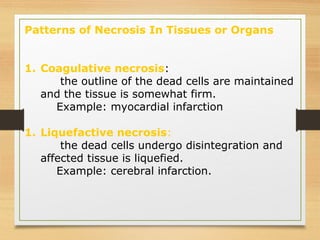
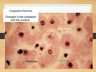
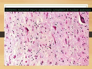
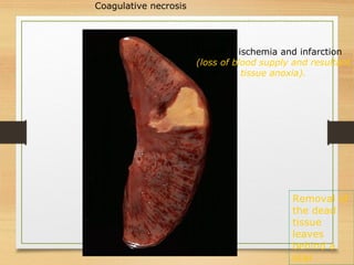
Necrosis
By- Sheril S. Philip
15BCB0120
• Define necrosis and apoptosis
• List the different types of necrosis, examples
of each and its features
• List the different conditions associated with
apoptosis, its morphology and its mechanism
• Know the difference between apoptosis and
necrosis
CONTENTS
Define necrosis and apoptosis
 List the different types of necrosis, examples
of each and its features
 List the different conditions associated with
apoptosis, its morphology and its mechanism
 Know the difference between apoptosis and
necrosis
Cell Death
Death of cells occurs in two ways:
1. Necrosis--(irreversible injury) changes produced by
enzymatic digestion of dead cellular elements
2. Apoptosis--vital process that helps eliminate unwanted
cells--an internally programmed series of events
effected by dedicated gene products
Morphology of necrosis :
1. Cellular swelling or rupture
2. Denaturation and coagulation of
cytoplasmic proteins
3. Breakdown of cell organelles
4. Breakdown of nuclear DNA
Patterns of Necrosis In Tissues or Organs
As a result of cell death the tissues or organs display
one of these six macroscopic changes:
1. Coagulative necrosis
2. Liquifactive necrosis
3. Caseous necrosis
4. Fat necrosis
5. Gangrenous necrosis
6. Fibrinoid necrosis
Patterns of Necrosis In Tissues or Organs
1. Coagulative necrosis:
the outline of the dead cells are maintained
and the tissue is somewhat firm.
Example: myocardial infarction
1. Liquefactive necrosis:
the dead cells undergo disintegration and
affected tissue is liquefied.
Example: cerebral infarction.
Patterns of Necrosis In Tissues or Organs
3. Caseous necrosis:
a form of coagulative necrosis (cheese-like).
Example: tuberculosis lesions.
4. Fat necrosis:
enzymatic digestion of fat.
Example: necrosis of fat by pancreatic enzymes.
5. Gangrenous necrosis:
Necrosis (secondary to ischemia) usually with superimposed
infection.
Example: necrosis of distal limbs, usually foot and toes in diabetes.
6. Fibrinoid necrosis: typically seen in vasculitis
and glomerular autoimmune diseases
Coagulative Necrosis
 
Changes in the cytoplasm
and the nucleus  
Karyolysis Pyknosis
Karyorrhexis
Necrosis of the liver induced by herpesvirus.
Herpesvirus invades liver cell nuclei producing nuclear ('ground
glass') mildly basophilic inclusions.
Acute renal tubular necrosis (ischemia) :
increased eosinophilia and pyknosis in necrotic cells
Normal
Necrotic
Ischemic neuronal injury:acidophilic cytoplasm & nuclear pyknosis
… a mess - so many
cells have died - the
tissue is not
recognizable. Nuclei
have become pyknotic
(shrunken and dark),
undergone karorrhexis
(fragmentation) and
karyolysis(dissolution)
Necrotic myocardium
Kidney: ischemia and infarction
(loss of blood supply and resultant
tissue anoxia).
Coagulative necrosis
Removal of
the dead
tissue
leaves
behind a
scar
Infarcts (vascular distribution) are wedge-shaped
with a base on the organ capsule.
Coagulative necrosis: is due to loss of blood supply
Spleen
Depending on circumstances:
1. necrotic tissue may be walled off by scar tissue,
2. totally converted to scar tissue,
3. get destroyed (producing a cavity or cyst),
4. get infected (producing an abscess or "wet gangrene"),
5. or calcify.
If the supporting tissue framework does not die, and the dead
cells are of a type capable of regeneration, you may have
complete healing.
Remember: True coagulation necrosis involves groups of cells, and is
almost always accompanied, by acute inflammation (infiltrate)

Necrosis sheril

  • 1.
    Necrosis By- Sheril S.Philip 15BCB0120
  • 2.
    • Define necrosisand apoptosis • List the different types of necrosis, examples of each and its features • List the different conditions associated with apoptosis, its morphology and its mechanism • Know the difference between apoptosis and necrosis CONTENTS
  • 3.
    Define necrosis andapoptosis  List the different types of necrosis, examples of each and its features  List the different conditions associated with apoptosis, its morphology and its mechanism  Know the difference between apoptosis and necrosis
  • 4.
    Cell Death Death ofcells occurs in two ways: 1. Necrosis--(irreversible injury) changes produced by enzymatic digestion of dead cellular elements 2. Apoptosis--vital process that helps eliminate unwanted cells--an internally programmed series of events effected by dedicated gene products
  • 5.
    Morphology of necrosis: 1. Cellular swelling or rupture 2. Denaturation and coagulation of cytoplasmic proteins 3. Breakdown of cell organelles 4. Breakdown of nuclear DNA
  • 6.
    Patterns of NecrosisIn Tissues or Organs As a result of cell death the tissues or organs display one of these six macroscopic changes: 1. Coagulative necrosis 2. Liquifactive necrosis 3. Caseous necrosis 4. Fat necrosis 5. Gangrenous necrosis 6. Fibrinoid necrosis
  • 7.
    Patterns of NecrosisIn Tissues or Organs 1. Coagulative necrosis: the outline of the dead cells are maintained and the tissue is somewhat firm. Example: myocardial infarction 1. Liquefactive necrosis: the dead cells undergo disintegration and affected tissue is liquefied. Example: cerebral infarction.
  • 8.
    Patterns of NecrosisIn Tissues or Organs 3. Caseous necrosis: a form of coagulative necrosis (cheese-like). Example: tuberculosis lesions. 4. Fat necrosis: enzymatic digestion of fat. Example: necrosis of fat by pancreatic enzymes. 5. Gangrenous necrosis: Necrosis (secondary to ischemia) usually with superimposed infection. Example: necrosis of distal limbs, usually foot and toes in diabetes. 6. Fibrinoid necrosis: typically seen in vasculitis and glomerular autoimmune diseases
  • 9.
    Coagulative Necrosis   Changes inthe cytoplasm and the nucleus   Karyolysis Pyknosis Karyorrhexis
  • 10.
    Necrosis of theliver induced by herpesvirus. Herpesvirus invades liver cell nuclei producing nuclear ('ground glass') mildly basophilic inclusions.
  • 11.
    Acute renal tubularnecrosis (ischemia) : increased eosinophilia and pyknosis in necrotic cells Normal Necrotic
  • 12.
    Ischemic neuronal injury:acidophiliccytoplasm & nuclear pyknosis
  • 13.
    … a mess- so many cells have died - the tissue is not recognizable. Nuclei have become pyknotic (shrunken and dark), undergone karorrhexis (fragmentation) and karyolysis(dissolution) Necrotic myocardium
  • 14.
    Kidney: ischemia andinfarction (loss of blood supply and resultant tissue anoxia). Coagulative necrosis Removal of the dead tissue leaves behind a scar
  • 15.
    Infarcts (vascular distribution)are wedge-shaped with a base on the organ capsule. Coagulative necrosis: is due to loss of blood supply Spleen
  • 16.
    Depending on circumstances: 1.necrotic tissue may be walled off by scar tissue, 2. totally converted to scar tissue, 3. get destroyed (producing a cavity or cyst), 4. get infected (producing an abscess or "wet gangrene"), 5. or calcify. If the supporting tissue framework does not die, and the dead cells are of a type capable of regeneration, you may have complete healing. Remember: True coagulation necrosis involves groups of cells, and is almost always accompanied, by acute inflammation (infiltrate)