Download to read offline


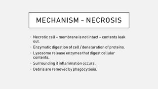


There is no inflammation in apoptosis because: - Apoptosis is an orderly and controlled process of programmed cell death that does not release intracellular contents into the surrounding tissue. - In apoptosis, the cell shrinks and its contents are packaged into apoptotic bodies which are then phagocytosed by neighboring cells or macrophages in a non-inflammatory manner. - There is no disruption of the cell membrane or release of cytoplasmic enzymes and other inflammatory mediators that would trigger an inflammatory response, as occurs in necrosis. The orderly packaging and removal of dead cells prevents collateral tissue damage and inflammation.


