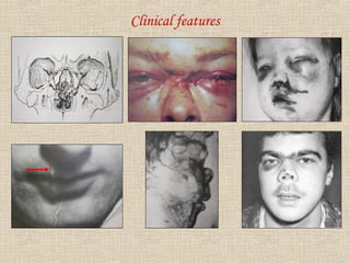
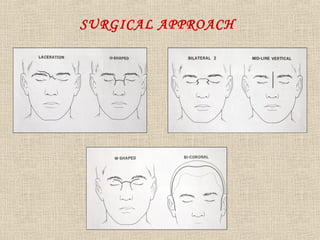
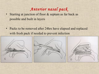
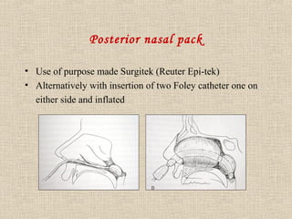
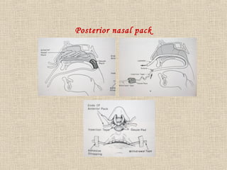
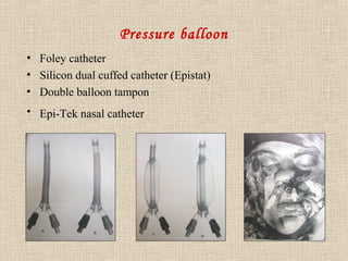
NASO ETHMOIDAL COMPLEX 
• Dr V.RAMKUMAR 
• CONSULTANT 
DENTAL&FACIOM 
AXILLARY 
SURGEON 
• REG NO: 4118- 
TAMILNADU-INDIA( 
ASIA)
Introduction 
• Middle third fracture of the face often involves 
Nasoethmoid complex. The skeletal foundation of the 
Nasoethmoid complex consists of a strong triangular 
shaped frame. Fractures in this region are almost invariably 
comminuted and involve numerous bones. 
• The skeletal structures situated in front of and behind 
this frame are relatively fragile and force sufficient to 
fracture the frame usually results in severe comminution 
and displacement of these thin bones.
Nasoethmoid complex
Classification 
( Nasoethmoid complex ) 
• Isolated nasoethmoidal injury 
– Bilateral 
– Unilateral 
• Combined Nasoethmoid injury + midface fractures 
– Bilateral 
– Unilateral
Classification: 
I: isolated naso- ethmoid and frontal region injury without 
other fractures of midface 
II: combined naso- ethmoid and frontal region injury with 
other fractures of midface
Clinical features 
• Nasal deformity 
• Frontal bone depression 
• Cerebrospinal fluid Rhinorrhoea 
• Haemorrhage (anterior or posterior branch of ethmoid artery) 
• Traumatic Telecanthus 
• Diplopia
Clinical features
Radiograph 
• Occipito -frontal view 
• Occipito -mental 
• Lateral view 
• CT
SURGICAL APPROACH
Nasoethmoid Fracture Emergencies 
• Cerebrospinal fluid leak 
• Unconsciousness 
• Skull fractures 
• Increasing intra cranial pressure 
• Meningitis 
• Persistent CSF leakage
CSF leakage 
Fracture of 
• Floor of anterior cranial fossa/base of skull 
Escape of CSF through 
• Ethmoidal sinus 
• Sphenoidal sinus 
• Cribriform plate 
• Frontal sinus 
Communication between 
• Meninges 
• Nose 
• Paranasal sinuses 
Dural laceration 
• Later becomes epithelialised - Fistula
• CSF leakage should be noted immediately after trauma 
• Blood clot of brain tissue may obstruct fluid passage 
• After lysis of clot or Increased intracranial pressure 
leakage is seen 
• Mobile midface fractures often creates pumping action-cause 
increased CSF leak
In Maxillofacial Injuries 
• Higher the level of fractures more chances for CSF 
Rhinorrhoea, Otorrhoea 
• Frequently missed 
– Reclining position 
– Blood stained 
• Later clean watery discharge
How to detect? 
• Pt position - Sitting and leans forward (drips from nose) 
• Salty / Metallic taste 
• “Tram line” 
• “Double halo”- when dropped on gauze sponge 
• Classical “bull’s eye ” ring will develop 
• “Clinistix ” (but colour change can occur with Lacrimal secretions )
• “Glucose test” (>30mg/dl) 
• Location of leak-CT scan 
• Intrathecal injection and assaying in different locations 
• Fluorescein & radioactive tracers (Indium)
Tram line
Precaution 
• Pre-nasal route intubation – avoided (but in maxillofacial injury 
it is safest & effective) 
• Pt in – semi recumbent position 
• Should not 
– Strain 
– Sneeze 
– Blow the nose 
• Avoid packing nose or ear- prevent retrograde infection 
• Infection- Meningitis 
• Antibiotics
Persistent/ recurrent leak 
• Duration 4-5 days (with or without fracture reduction) 
• Persistent leaks beyond 3 weeks- need formal dural repair 
• Surgical repair failure – Lumbo peritoneal shunting 
• Meningitis may occur
Treatment 
• Fracture reduction 
• Dural repair 
• Lumbo peritoneal shunting 
• Antibiotics
Hemorrhage management 
• Nasoethmoid fracture with midface bleeding manifest itself as 
Epistaxis 
• To locate the site of bleeding good visualization is needed 
• Mucous membrane should be shrunken & anesthetized with 
Phenylephrine hydrochloride (Neo-synephrine) or 4% cocaine 
solution or both 
• Cotton pledget soaked with vasoconstrictor 
• Cauterization with silver nitrate solution
• Merocel sponge 
• Anterior nasal pack 
• Posterior nasal pack 
• Pressure balloon 
If bleeding not stopped 
• Arteriogram 
• Embolization or Ligation
Airway obstruction 
• Patient head is positioned forward to prevent drainage of 
blood into pharynx which cause airway embarrassment 
• Blood from nose or retropharyngeal hematoma is a 
warning of potential airway obstruction
Anterior nasal pack 
• Starting at junction of floor & septum as far back as 
possible and built in layers 
• Packs to be removed after 24hrs have elapsed and replaced 
with fresh pack if needed to prevent infection
Posterior nasal pack 
• Use of purpose made Surgitek (Reuter Epi-tek) 
• Alternatively with insertion of two Foley catheter one on 
either side and inflated
Posterior nasal pack
Pressure balloon 
• Foley catheter 
• Silicon dual cuffed catheter (Epistat) 
• Double balloon tampon 
• Epi-Tek nasal catheter
THANK YOU

. nasoethmoid complex fractures

  • 2.
    NASO ETHMOIDAL COMPLEX • Dr V.RAMKUMAR • CONSULTANT DENTAL&FACIOM AXILLARY SURGEON • REG NO: 4118- TAMILNADU-INDIA( ASIA)
  • 3.
    Introduction • Middlethird fracture of the face often involves Nasoethmoid complex. The skeletal foundation of the Nasoethmoid complex consists of a strong triangular shaped frame. Fractures in this region are almost invariably comminuted and involve numerous bones. • The skeletal structures situated in front of and behind this frame are relatively fragile and force sufficient to fracture the frame usually results in severe comminution and displacement of these thin bones.
  • 4.
  • 5.
    Classification ( Nasoethmoidcomplex ) • Isolated nasoethmoidal injury – Bilateral – Unilateral • Combined Nasoethmoid injury + midface fractures – Bilateral – Unilateral
  • 6.
    Classification: I: isolatednaso- ethmoid and frontal region injury without other fractures of midface II: combined naso- ethmoid and frontal region injury with other fractures of midface
  • 7.
    Clinical features •Nasal deformity • Frontal bone depression • Cerebrospinal fluid Rhinorrhoea • Haemorrhage (anterior or posterior branch of ethmoid artery) • Traumatic Telecanthus • Diplopia
  • 8.
  • 9.
    Radiograph • Occipito-frontal view • Occipito -mental • Lateral view • CT
  • 10.
  • 11.
    Nasoethmoid Fracture Emergencies • Cerebrospinal fluid leak • Unconsciousness • Skull fractures • Increasing intra cranial pressure • Meningitis • Persistent CSF leakage
  • 12.
    CSF leakage Fractureof • Floor of anterior cranial fossa/base of skull Escape of CSF through • Ethmoidal sinus • Sphenoidal sinus • Cribriform plate • Frontal sinus Communication between • Meninges • Nose • Paranasal sinuses Dural laceration • Later becomes epithelialised - Fistula
  • 13.
    • CSF leakageshould be noted immediately after trauma • Blood clot of brain tissue may obstruct fluid passage • After lysis of clot or Increased intracranial pressure leakage is seen • Mobile midface fractures often creates pumping action-cause increased CSF leak
  • 14.
    In Maxillofacial Injuries • Higher the level of fractures more chances for CSF Rhinorrhoea, Otorrhoea • Frequently missed – Reclining position – Blood stained • Later clean watery discharge
  • 15.
    How to detect? • Pt position - Sitting and leans forward (drips from nose) • Salty / Metallic taste • “Tram line” • “Double halo”- when dropped on gauze sponge • Classical “bull’s eye ” ring will develop • “Clinistix ” (but colour change can occur with Lacrimal secretions )
  • 16.
    • “Glucose test”(>30mg/dl) • Location of leak-CT scan • Intrathecal injection and assaying in different locations • Fluorescein & radioactive tracers (Indium)
  • 17.
  • 18.
    Precaution • Pre-nasalroute intubation – avoided (but in maxillofacial injury it is safest & effective) • Pt in – semi recumbent position • Should not – Strain – Sneeze – Blow the nose • Avoid packing nose or ear- prevent retrograde infection • Infection- Meningitis • Antibiotics
  • 19.
    Persistent/ recurrent leak • Duration 4-5 days (with or without fracture reduction) • Persistent leaks beyond 3 weeks- need formal dural repair • Surgical repair failure – Lumbo peritoneal shunting • Meningitis may occur
  • 20.
    Treatment • Fracturereduction • Dural repair • Lumbo peritoneal shunting • Antibiotics
  • 21.
    Hemorrhage management •Nasoethmoid fracture with midface bleeding manifest itself as Epistaxis • To locate the site of bleeding good visualization is needed • Mucous membrane should be shrunken & anesthetized with Phenylephrine hydrochloride (Neo-synephrine) or 4% cocaine solution or both • Cotton pledget soaked with vasoconstrictor • Cauterization with silver nitrate solution
  • 22.
    • Merocel sponge • Anterior nasal pack • Posterior nasal pack • Pressure balloon If bleeding not stopped • Arteriogram • Embolization or Ligation
  • 23.
    Airway obstruction •Patient head is positioned forward to prevent drainage of blood into pharynx which cause airway embarrassment • Blood from nose or retropharyngeal hematoma is a warning of potential airway obstruction
  • 24.
    Anterior nasal pack • Starting at junction of floor & septum as far back as possible and built in layers • Packs to be removed after 24hrs have elapsed and replaced with fresh pack if needed to prevent infection
  • 25.
    Posterior nasal pack • Use of purpose made Surgitek (Reuter Epi-tek) • Alternatively with insertion of two Foley catheter one on either side and inflated
  • 26.
  • 27.
    Pressure balloon •Foley catheter • Silicon dual cuffed catheter (Epistat) • Double balloon tampon • Epi-Tek nasal catheter
  • 28.