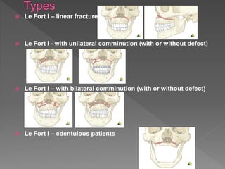
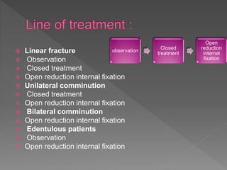
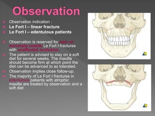
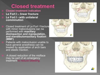
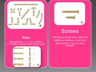
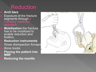
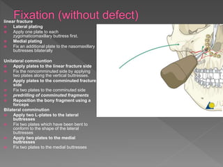
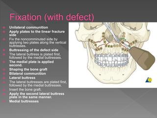
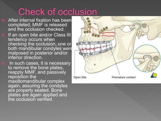
 The fracture extend from :
1. Lateral margin of anterior nasal aperture
2. Canine fossa
3. Below zygomatic buttress
4. Along the lateral wall of anturm
5. Posteriorly across the pterfgomaxillary
fissure
6. To fracture the lower 1/3 of pterygiod
laminae
1. Lateral margin of anterior nasal
aperture
2. Canine fossa
3. Below zygomatic buttress
4. Along the lateral wall of anturm
5. Posteriorly across the
fissure
6. To fracture the lower 1/3 of
pterygiod laminae
 Le Fort I – linear fracture
 Le Fort I - with unilateral comminution (with or without defect)
 Le Fort I – with bilateral comminution (with or without defect)
 Le Fort I – edentulous patients
 Linear fracture
 Observation
 Closed treatment
 Open reduction internal fixation
 Unilateral comminution
 Closed treatment
 Open reduction internal fixation
 Bilateral comminution
 Open reduction internal fixation
 Edentulous patients
 Observation
 Open reduction internal fixation
observation
Closed
treatment
Open
reduction
internal
fixation
 Observation indication :
 Le Fort I – linear fracture
 Le Fort I – edentulous patients
 Observation is reserved for non- or
minimally mobile Le Fort I fractures
with unaffected occlusion.
 The patient is advised to stay on a soft
diet for several weeks. The maxilla
should become firm at which point the
diet can be advanced to as tolerated.
 Observation implies close follow-up.
 The majority of Le Fort I fractures in
edentulous patients with atrophic
maxilla are treated by observation and a
soft diet
 Closed treatment indication
 Le Fort I – linear fracture
 Le Fort I - with unilateral
comminution
 Closed treatment of Le Fort I fractures
with minor malocclusions can be
performed with maxillary
disimpaction and manipulation,
and mandibulomaxillary fixation
(MMF)
 Patients with malocclusion unable to
have general anesthesia can be
treated by application of arch bars
and elastic traction.
 A closed reduction of the midface
may be part of an emergency
treatment to reduce bleeding.
 Open reduction internal fixation indication :
 Le Fort I – linear fracture
 Le Fort I - with unilateral comminution
(with or without defect)
 Le Fort I – with bilateral comminution
(with or without defect)
 Le Fort I – edentulous patients
 Principles
 Decision on whether to use bone graft
 Choice of implant
 Reduction
 Fixation (without bone graft)
 Fixation (with bone graft)
 Check of occlusion
 Reestablish the midfacial vertical buttresses.
 Reestablish the premorbid dental occlusion
 Class III tendency often occurs in the
postoperative due to?? To overcome??
 The goal is to achieve an anatomical correct
repositioning by means of 3-D reconstruction.
 Le Fort I fractures with bilateral comminution
display loss of the correct facial height
 Bone graft is used to fill defects and the buttresses are
restored using two plates.
 Indications (bone grafting):
 Loss of bone volume and loss of buttressing
 Contraindications (bone grafting):
 Inability to stabilize bone graft and maxilla
 Inability to obtain soft-tissue closure over graft
 Advantages (bone grafting):
 Support for facial soft tissues
 Restoration of bony buttresses
 Prevention of loss of facial height
 Disadvantages (bone grafting):
 Donor site required for bone graft harvesting
 Principles
 A plate that is placed for the fixation of the
fracture at the zygomaticomaxillary buttress
is generally a larger plate because it is the
point that will provide most stability to the Le
Fort I fracture. The highest forces of
mastication would be in this area.
 Another plate can also be applied at the
piriform rim.
Plate
Miniplate has 1, 1.3 ,1.5 ,2 system.
L,T,Y-plate or straight plate may by used.
Screws
Self-tapping (mostly uesd - pedrilling)
Selfdrilling (nodrilling –more force)
Plate fixed by 2 screw in each
fragment
 Arch bars
 Exposure of the fracture
segments through a
maxillary vestibular
approach
 Mobilization the fracture
has to be mobilized to
enable reduction and
fixation.
 Reduction instruments
 Rowe disimpaction forceps
 Bone hooks
 Placing the patient into
MMF
 Reducing the maxilla
linear fracture
 Lateral plating
 Apply one plate to each
zygomaticomaxillary buttress first.
 Medial plating
 Fix an additional plate to the nasomaxillary
buttresses bilaterally
Unilateral commiuntion
 Apply plates to the linear fracture side
 Fix the noncomminuted side by applying
two plates along the vertical buttresses.
 Apply plates to the comminuted fracture
side
 Fix two plates to the comminuted side
 predrilling of comminuted fragments
 Reposition the bony fragment using a
forceps
Bilateral comminution
 Apply two L-plates to the lateral
buttresses
 Fix two plates which have been bent to
conform to the shape of the lateral
buttresses
 Apply two plates to the medial
buttresses
 Fix two plates to the medial buttresses
 Unilateral communition
 Apply plates to the linear fracture
side
 Fix the noncomminuted side by
applying two plates along the vertical
buttresses.
 Buttressing of the defect side
 The lateral buttress is plated first,
followed by the medial buttresses.
 The medial plate is applied
second.
 Shaping the bone graft
 Bilateral communition
 Lateral buttress
 The lateral buttresses are plated first,
followed by the medial buttresses.
 Insert the bone graft.
 Apply the second lateral buttress
plate in the same manner.
 Medial buttresses
 After internal fixation has been
completed, MMF is released
and the occlusion checked.
 If an open bite and/or Class III
tendency occurs when
checking the occlusion, one or
both mandibular condyles were
malposed in posterior and/or
inferior direction.
 In such cases, it is necessary
to remove the bone plates,
reapply MMF, and passively
reposition the
maxillomandibular complex
again, assuring the condyles
are properly seated. Bone
plates are again applied and
the occlusion verified.
Le fort 1

Le fort 1

  • 2.
     The fractureextend from : 1. Lateral margin of anterior nasal aperture 2. Canine fossa 3. Below zygomatic buttress 4. Along the lateral wall of anturm 5. Posteriorly across the pterfgomaxillary fissure 6. To fracture the lower 1/3 of pterygiod laminae
  • 3.
    1. Lateral marginof anterior nasal aperture 2. Canine fossa 3. Below zygomatic buttress 4. Along the lateral wall of anturm 5. Posteriorly across the fissure 6. To fracture the lower 1/3 of pterygiod laminae
  • 4.
     Le FortI – linear fracture  Le Fort I - with unilateral comminution (with or without defect)  Le Fort I – with bilateral comminution (with or without defect)  Le Fort I – edentulous patients
  • 5.
     Linear fracture Observation  Closed treatment  Open reduction internal fixation  Unilateral comminution  Closed treatment  Open reduction internal fixation  Bilateral comminution  Open reduction internal fixation  Edentulous patients  Observation  Open reduction internal fixation observation Closed treatment Open reduction internal fixation
  • 6.
     Observation indication:  Le Fort I – linear fracture  Le Fort I – edentulous patients  Observation is reserved for non- or minimally mobile Le Fort I fractures with unaffected occlusion.  The patient is advised to stay on a soft diet for several weeks. The maxilla should become firm at which point the diet can be advanced to as tolerated.  Observation implies close follow-up.  The majority of Le Fort I fractures in edentulous patients with atrophic maxilla are treated by observation and a soft diet
  • 7.
     Closed treatmentindication  Le Fort I – linear fracture  Le Fort I - with unilateral comminution  Closed treatment of Le Fort I fractures with minor malocclusions can be performed with maxillary disimpaction and manipulation, and mandibulomaxillary fixation (MMF)  Patients with malocclusion unable to have general anesthesia can be treated by application of arch bars and elastic traction.  A closed reduction of the midface may be part of an emergency treatment to reduce bleeding.
  • 8.
     Open reductioninternal fixation indication :  Le Fort I – linear fracture  Le Fort I - with unilateral comminution (with or without defect)  Le Fort I – with bilateral comminution (with or without defect)  Le Fort I – edentulous patients
  • 9.
     Principles  Decisionon whether to use bone graft  Choice of implant  Reduction  Fixation (without bone graft)  Fixation (with bone graft)  Check of occlusion
  • 10.
     Reestablish themidfacial vertical buttresses.  Reestablish the premorbid dental occlusion  Class III tendency often occurs in the postoperative due to?? To overcome??  The goal is to achieve an anatomical correct repositioning by means of 3-D reconstruction.  Le Fort I fractures with bilateral comminution display loss of the correct facial height
  • 11.
     Bone graftis used to fill defects and the buttresses are restored using two plates.  Indications (bone grafting):  Loss of bone volume and loss of buttressing  Contraindications (bone grafting):  Inability to stabilize bone graft and maxilla  Inability to obtain soft-tissue closure over graft  Advantages (bone grafting):  Support for facial soft tissues  Restoration of bony buttresses  Prevention of loss of facial height  Disadvantages (bone grafting):  Donor site required for bone graft harvesting
  • 12.
     Principles  Aplate that is placed for the fixation of the fracture at the zygomaticomaxillary buttress is generally a larger plate because it is the point that will provide most stability to the Le Fort I fracture. The highest forces of mastication would be in this area.  Another plate can also be applied at the piriform rim.
  • 13.
    Plate Miniplate has 1,1.3 ,1.5 ,2 system. L,T,Y-plate or straight plate may by used. Screws Self-tapping (mostly uesd - pedrilling) Selfdrilling (nodrilling –more force) Plate fixed by 2 screw in each fragment
  • 14.
     Arch bars Exposure of the fracture segments through a maxillary vestibular approach  Mobilization the fracture has to be mobilized to enable reduction and fixation.  Reduction instruments  Rowe disimpaction forceps  Bone hooks  Placing the patient into MMF  Reducing the maxilla
  • 15.
    linear fracture  Lateralplating  Apply one plate to each zygomaticomaxillary buttress first.  Medial plating  Fix an additional plate to the nasomaxillary buttresses bilaterally Unilateral commiuntion  Apply plates to the linear fracture side  Fix the noncomminuted side by applying two plates along the vertical buttresses.  Apply plates to the comminuted fracture side  Fix two plates to the comminuted side  predrilling of comminuted fragments  Reposition the bony fragment using a forceps Bilateral comminution  Apply two L-plates to the lateral buttresses  Fix two plates which have been bent to conform to the shape of the lateral buttresses  Apply two plates to the medial buttresses  Fix two plates to the medial buttresses
  • 16.
     Unilateral communition Apply plates to the linear fracture side  Fix the noncomminuted side by applying two plates along the vertical buttresses.  Buttressing of the defect side  The lateral buttress is plated first, followed by the medial buttresses.  The medial plate is applied second.  Shaping the bone graft  Bilateral communition  Lateral buttress  The lateral buttresses are plated first, followed by the medial buttresses.  Insert the bone graft.  Apply the second lateral buttress plate in the same manner.  Medial buttresses
  • 17.
     After internalfixation has been completed, MMF is released and the occlusion checked.  If an open bite and/or Class III tendency occurs when checking the occlusion, one or both mandibular condyles were malposed in posterior and/or inferior direction.  In such cases, it is necessary to remove the bone plates, reapply MMF, and passively reposition the maxillomandibular complex again, assuring the condyles are properly seated. Bone plates are again applied and the occlusion verified.