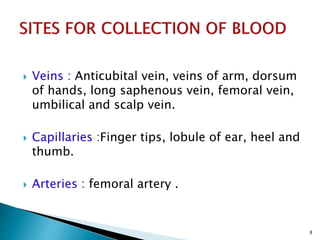
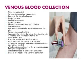
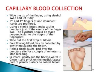
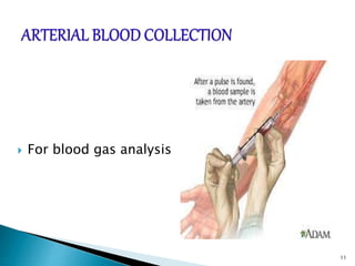
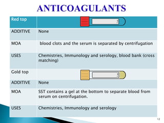
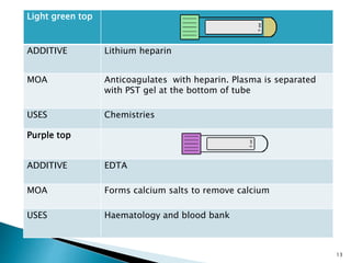
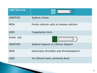
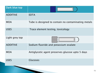
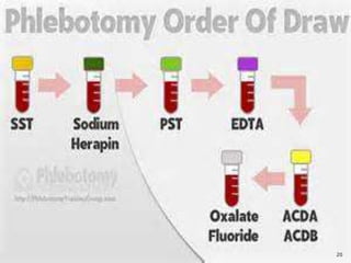
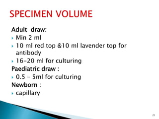
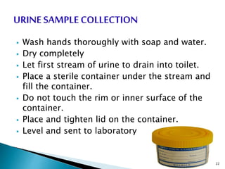
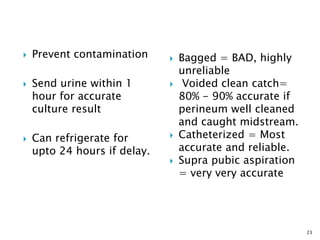
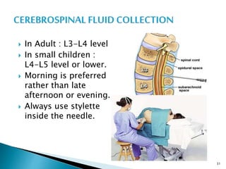
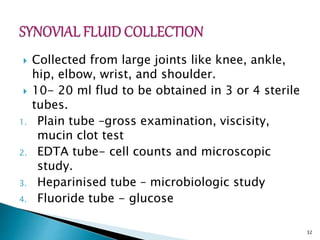
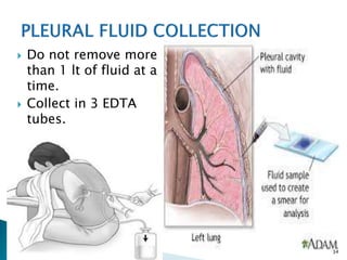
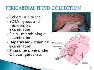
This document provides an overview of laboratory procedures for collecting and handling various clinical specimens. It discusses the appropriate collection containers, requirements, and procedures for collecting blood, urine, stool, sputum, cerebrospinal fluid, and other specimens. Specific topics covered include blood collection by venipuncture and fingerstick, urine collection and testing parameters, stool collection for culture and ova/parasite examination, and safety practices for handling hazardous materials in the laboratory.
The document discusses procedures for collecting small specimen amounts from patients for diagnostic testing purposes. It covers urine collection from infants, stool collection, and blood collection. Proper cleaning and labeling of specimens is emphasized to avoid contamination and ensure accurate test results. Maintaining sterile technique and recording all relevant information is important when obtaining and handling specimens.
Clinical pathology is the laboratory analysis of bodily fluids and tissues to diagnose disease. It involves collecting samples such as blood, urine, and tissues and analyzing them via techniques including virology, bacteriology, clinical chemistry, serology, and histology. Blood can be collected via arterial sampling, venipuncture, or fingerstick while urine is usually collected via clean-catch, catheterization, or suprapubic aspiration. Samples must be properly labeled and either immediately transported or preserved/refrigerated to maintain integrity for laboratory analysis and diagnosis.
The document outlines principles for proper collection and submission of specimens to the microbiology lab in order to obtain accurate test results. It discusses 9 key principles: 1) collecting specimens with minimal contamination close to site of infection, 2) collecting at optimal times, 3) obtaining sufficient quantity, 4) using appropriate collection devices and containers, 5) collecting before antibiotics, 6) proper labeling for transport, 7) minimizing transport time, 8) following special handling instructions, and 9) collecting the proper specimen for the ordered test. Specific guidelines are provided for various specimen types like blood, urine, sputum etc. to ensure specimens are of high quality for microbiological analysis.
This document provides guidelines for safely collecting urine and blood specimens. It outlines the personal protective equipment that should be worn to prevent transmission of pathogens when handling potentially infectious materials. Proper techniques are described for midstream urine collection, catheterized urine collection, blood collection via venipuncture, and transporting specimens in appropriate media. Adhering to safety, technique, and transport methods helps protect healthcare workers and ensures specimen viability for testing.
Dr. Rajju Tiwari discusses sample collection and processing procedures. Standard precautions include using personal protective equipment, proper hand hygiene, and handling all blood as potentially infectious. Phlebotomy involves making an incision in a vein with a needle to collect blood samples. Proper sample collection requires checking patient identity, sample type, and special requirements. Common sample types are blood, urine, and fluids. Centrifugation is used to separate plasma or serum from whole blood for processing. Samples must be properly labeled, transported, stored, and processed within specified timeframes for accurate testing and results.
This document provides guidance on collecting various specimen types, including blood, urine, deep specimens, feces, sputum, and genital specimens. It emphasizes the importance of collecting specimens correctly and transporting them to the laboratory promptly to ensure quality results. Key steps include using the proper collection container, obtaining informed consent, maintaining aseptic technique, and documenting and labeling the specimens accurately.
The document discusses the proper procedures for collecting, transporting, and processing microbiological specimens to accurately identify infectious agents, noting that specimens must be representative of the infection, collected and transported aseptically, and processed promptly in the laboratory to identify causative organisms and guide treatment. Close communication between clinicians and the microbiology lab is important to select the appropriate tests and properly interpret results.
The document provides guidance on proper specimen collection and transport procedures. It discusses appropriate containers, labeling, storage, and handling of spillages. Specimen containers must be sealed securely and labeled with patient details. Storage should be in a refrigerator if delivery to the lab is delayed. Only clinically-indicated specimens should be collected to avoid inappropriate antibiotic prescribing. Proper collection methods are outlined for various sample types like urine, sputum, and feces.
Methods of Blood Collection and AnticoagulantsShreya D Prabhu
This document discusses methods of blood collection and anticoagulants. It covers precollection variables that can affect test results, various tests affected by factors like diurnal variation and posture. It then discusses blood collection sites like venous, capillary and arterial. The proper techniques, equipment and safety procedures for venipuncture and arterial blood collection are explained. Finally, it covers anticoagulants, their uses in preventing coagulation during storage and transportation of blood samples, and the coagulation pathways they act on.
The document provides guidance on proper sample collection techniques in clinical microbiology, including using aseptic technique and collecting samples from appropriate sites and times, as well as guidelines for collecting and transporting various sample types such as respiratory, urine, and gastrointestinal samples to accurately diagnose infections. Proper collection and handling of samples is emphasized to obtain quality specimens and avoid contamination.
This document discusses the order of draw for blood collection tubes. It begins by explaining that the order of draw is important to prevent contamination between tubes. The standard order of draw is then listed as: 1) sterile tube for blood cultures, 2) blue top for coagulation tests, 3) serum tube, 4) heparin tube, 5) EDTA tube, 6) fluoride/oxalate tube. The rationale for this order is then explained for each tube type to minimize interference between tests. Proper procedure and potential issues caused by incorrect order of draw are also summarized.
The document provides guidelines for blood sample collection procedures. It discusses introducing oneself to the patient, checking identification, selecting a vein, using the proper order and techniques for drawing blood into tubes, labeling the tubes, and applying pressure after withdrawing the needle. Precautions are outlined such as universal safety protocols, proper disposal of sharps, and cleaning up spills. The document also provides information on collecting samples from infants and the indications for rejecting specimens.
Collecting blood samples and other biological specimens is crucial to the understanding, prevention, and treatment of disease. However, from the patient’s perspective, it can also be painful, unnerving, frightening, and inconvenient.
Specimen containers and transport bags must be used appropriately to safely transport specimens from patients. Specimens should only be collected when there are clinical signs of infection to avoid inappropriate antibiotic use. Proper labelling, storage, and transportation of specimens is required to prevent misdiagnosis and ensure specimens are not compromised. Spillages and leaks must be properly disinfected and recollected or decanted according to standard precautions to minimize infection risk.
Skin punctures like fingersticks and heel sticks are commonly used to obtain small blood samples from patients. Heel sticks are preferred for infants less than 1 year old because the heel has more tissue and no nerves, bones, or tendons nearby.
Earlobe punctures are mainly used to measure bleeding time by introducing a small wound and timing how long it takes to stop bleeding. Normal bleeding time is 1-3 minutes.
When performing punctures, it is important to clean the site, use the proper lancet or needle size for the patient, and control blood flow to obtain an adequate sample for testing while minimizing patient discomfort.
This document provides guidelines for the proper collection, transportation, preparation, processing, and storage of laboratory samples for disease diagnosis and monitoring. It discusses defining samples and specimens, examples of laboratory samples, requirements for sample collection, potential errors from improper collection, and objectives of collection. Specific guidance is given for collection and handling of various sample types, including blood, urine, cerebrospinal fluid, stool, and others. Proper labeling, transport, separation, preservation, and storage methods are also outlined to ensure sample quality and integrity.
This document discusses sample collection in clinical biochemistry. It outlines the essential steps in sample collection including patient identification, sample collection and processing, and storage and transport. It describes the proper procedures, equipment, and additives used for collecting different sample types like blood, urine, cerebrospinal fluid, and arterial blood gas. Potential errors are discussed along with ways to avoid errors like hemolysis, clotting, and contamination. Maintaining proper procedures is important for obtaining accurate laboratory test results.
The document discusses common laboratory procedures and related nursing responsibilities. It covers blood tests like complete blood counts and electrolyte panels. It also discusses urine tests, stool exams, sputum analysis, and various visualization procedures for the gastrointestinal and urinary systems. Nursing responsibilities include obtaining consent, preparing patients, collecting specimens, monitoring patients during tests, and following up after tests.
This document provides guidelines for routine venipuncture and specimen handling. It describes the venipuncture procedure, including patient preparation, vein selection, order of draw, labeling samples, and troubleshooting incomplete collections. It also lists the additive, function, and uses of various colored collection tube types, such as red top for chemistries, gold top for serum separation, and purple top for complete blood counts. Complications like hematomas are addressed.
This document discusses various laboratory tests that are important for hematopoietic stem cell transplantation (HSCT) and monitoring patients who have received HSCT. It describes tests in several categories: hematology, biochemistry, virology, coagulation, culture, flow cytometry, HLA typing, cytogenetics, and chimerism studies. It provides details on sample collection procedures, specific tests that should be performed at different intervals to monitor patients receiving immunosuppressive drugs or experiencing complications, and the services provided by different specialized laboratories like stem cell, flow cytometry, and HLA laboratories.
This document outlines policies and procedures for blood specimen collection and disposal of sharp objects at KFCH. It defines venipuncture and different types of collection tubes used, as well as nursing responsibilities for blood collection and labeling of samples. The standard operating procedure for disposal of sharp objects specifies using puncture-proof containers labeled as biohazardous waste and proper handling, transport, and disposal of filled sharp containers. Recommendations include strictly following policies to ensure staff and patient safety during phlebotomy and waste disposal.
The document discusses urinary diversion procedures which surgically reroute urine flow out of the body when the normal flow is blocked, including temporary procedures like urinary catheterization and nephrostomy tubes which drain urine until the blockage is treated, as well as permanent diversions that require creating a stoma or internal reservoir to reroute urine to an external pouch. It also provides instructions for caring for urinary diversions by checking the dressing, skin, and urine color and flow through the tubing.
The document discusses proper procedures for sample collection, handling, and transportation for effective microbial testing. It emphasizes that the pre-analytical stage, which involves collection and handling, is critical. Key points include using appropriate containers and transport media to preserve samples, maintaining sterile technique, proper labeling, and timely transportation while refrigerated. Following standard operating procedures at each stage helps ensure accurate diagnosis.
1. The document outlines various protocols for collecting and handling specimens for diagnostic testing in pathology. It discusses appropriate samples, containers, transport conditions, and preservation methods for urine, CSF, fluids, sputum, stool, semen, and blood samples.
2. Guidelines are provided for venous blood collection including equipment, anticoagulants used, and proper labelling and transport of samples.
3. An overview is also given of histopathology specimen handling, blood banking techniques including blood donation and components, and cross matching procedures.
1. Nurses are often responsible for collecting various specimen types including urine, stool, sputum, wound drainage and blood.
2. Proper specimen collection involves ensuring patient comfort, privacy, consent and clear instructions while following standard precautions including hand hygiene, labeling and timely delivery to the lab.
3. Urine specimens may include random, clean-catch midstream, catheterized or timed collections for testing or culture and sensitivity. Proper technique is required to ensure specimen integrity and accuracy of results.
This document provides guidelines on standard operating procedures for microbiology laboratory diagnosis. It discusses the proper collection, transport, and processing of various clinical specimens like blood, cerebrospinal fluid, sputum, urine, stool, and others. Key points emphasized include collecting specimens before antimicrobial administration, using appropriate containers, minimizing delays in transport and storage, and preventing contamination. The document also outlines procedures for staining techniques, sterilization, waste disposal, and quality control. Following standardized protocols helps ensure accurate laboratory diagnosis of infectious diseases.
2. Sample collection ,transport and acceptance and rejection.pptxMohanSinghDhakad1
This document provides guidelines for collecting, transporting, and testing various clinical specimens for microbiological analysis. It discusses sample types including body fluids, cerebrospinal fluid, blood, urine, respiratory samples, gastrointestinal samples, ear swabs, eye swabs, and abscess or wound samples. For each sample type, it provides information on containers, transport times and temperatures, and appropriate microbiological tests. The document aims to ensure proper specimen collection and handling to optimize pathogen recovery and identification.
This journal club article summarizes a study examining the clinicopathological significance of the cribriform pattern and intraductal carcinoma of the prostate (IDC-P) in prostate biopsy specimens. The study found that both were associated with higher Gleason grade, lymph node metastasis, extraprostatic extension, and lymphovascular invasion. Their presence, regardless of percentage or number of cores, provided prognostic information. The cribriform pattern and IDC-P were independent risk factors for each other and high Gleason grade. Their identification, even in a small portion of cores, should be reported to help predict patient outcomes.
This document describes techniques for preparing and preserving pathological specimens for museum collections. It discusses receiving specimens from hospitals and laboratories, preparing them by washing in saline and fixing in formalin-based solutions, restoring color using alcohol, and long-term preservation by mounting in glycerin-based solutions. Special techniques are described for hollow organs, maceration of bone specimens, and labeling and cataloging finished museum pieces. The goal is to preserve tissue in a life-like state for teaching and research over long periods of time.
This document presents a case of a 57-year-old male with a raised blackish lesion on his face for 2 years. Microscopic examination of a biopsy shows a tumor infiltrating the dermis and subcutis composed of basaloid cells arranged in islands and cords with peripheral palisading and minimal atypia. Differential diagnoses discussed include trichoepithelioma, trichoblastoma, basaloid follicular hamartoma, and infundibulocystic basal cell carcinoma. Trichoepithelioma cannot be ruled out as the lesion is solitary, adult-onset, <2cm in size, well-circumscribed, and composed of basaloid cells with peripheral palis
This document summarizes a slide seminar on lung biopsy findings for a 34-year-old female patient being evaluated for diffuse parenchymal lung disease. High-resolution CT showed numerous tiny calcified nodules throughout both lungs suggestive of pulmonary alveolar microlithiasis. Lung biopsy microscopic findings showed dilated alveoli filled with basophilic concentric calcific deposits, supporting a provisional diagnosis of pulmonary alveolar microlithiasis. This diagnosis was further supported by clinical features such as autosomal recessive inheritance pattern and characteristic radiologic and histopathologic findings of calcified microliths filling the alveolar air spaces. Differential diagnoses including pulmonary blue bodies and metastatic calc
The document provides guidelines for proper specimen collection and transport. It discusses general guidelines including aseptic technique, adequate volume, and proper timing and containers. It then describes appropriate collection and transport methods for various specimen types including blood, urine, stool, respiratory samples and more. Proper labeling, packaging and timely transport of specimens to the laboratory are emphasized.
Postmortem changes occur immediately, early, and late after death. Immediate changes include cessation of brain, circulatory, and respiratory functions. Early changes are facial pallor, skin changes, eye changes, cooling of the body, lividity, and rigor mortis. Late changes include decomposition, adipocere formation, and mummification. Determining postmortem changes aids in estimating time since death and investigating causes of death.
1) Duodenal biopsy of a 21-year old male showed Giardia lamblia trophozoites attached to the duodenal mucosa without invasion. Multiple Brunner's glands were seen in the submucosa.
2) Giardia lamblia, or Giardia intestinalis, is a common intestinal parasite spread through contaminated food or water. It attaches to the small intestine and can cause intermittent diarrhea but does not invade the mucosa.
3) The biopsy findings were consistent with giardiasis based on the morphology and location of the organisms seen as well as the patient's clinical history of abdominal pain and diarrhea.
This document provides information about blood collection and processing in a clinical laboratory. It discusses the different types of samples that can be collected including blood, urine, stool, sputum, and various body fluids. It outlines the collection procedures, types of tubes used, and additives in the tubes for different tests. Potential hazards in the laboratory are identified along with first aid measures for exposures or injuries. Safety practices are emphasized including proper chemical storage, use of personal protective equipment, and handling of biohazardous materials.
Specimen collection and transport are critical for accurate laboratory results. Proper guidelines include using appropriate containers and transport media, adequate labeling, and timely delivery. Key points are minimizing contamination, ensuring sufficient sample quantity and quality, and following instructions for different specimen types like blood, urine, stool and respiratory samples. Adherence to protocols helps produce reliable diagnostic test results.
This document discusses diseases of immunity and immune system disorders. It defines innate and adaptive immunity and their key cells. It describes the major histocompatibility complex and its role in self-recognition. It then discusses four types of hypersensitivity reactions, common autoimmune diseases like lupus and rheumatoid arthritis, and primary and secondary immunodeficiencies. It also provides summaries of AIDS, amyloidosis, and other conditions.
The document describes different features seen in normal and abnormal blood cells under a microscope. It notes that red blood cells normally have a pale center and white blood cells include granulocytes. Iron-deficiency anemia is shown by paler, smaller red blood cells. Sickle cell anemia features sickled red blood cells. Chronic myelogenous leukemia features an increased number of neutrophils. Hairy cell leukemia cells have hairy projections and Hodgkin lymphoma features large Reed-Sternberg cells.
I) Type II hypersensitivity reactions, also known as cytotoxic reactions, involve antibody-mediated destruction of cells through two main mechanisms:
1) Activation of the complement system leads to pore formation and lysis of the target cell.
2) Antibody-dependent cell-mediated cytotoxicity (ADCC) occurs when antibodies bind to target cells and recruit natural killer cells or macrophages to destroy the target cell.
Some examples of type II hypersensitivity reactions include hemolytic anemia caused by antibodies against blood cells, transfusion reactions due to ABO incompatibility, and hemolytic disease of the newborn from Rh incompatibility.
Type I, II, III, and IV hypersensitivity reactions are immune responses that are harmful or inappropriate. Type I is an immediate, antibody-mediated reaction like an allergy. Type II involves antibody-mediated cell destruction. Type III is immune complex-mediated responses causing issues like serum sickness. Type IV is a delayed, cell-mediated response like a tuberculin skin test or contact dermatitis. These hypersensitivities can cause issues in various tissues and organs.
The diagnosis of lymphoma involves an integrated process using clinical information, histology, immunophenotyping, cytogenetics, and molecular studies. A tissue sample suspected of lymphoma is tested with microscopy, immunohistochemistry, flow cytometry, cytogenetic analysis, fluorescence in situ hybridization, and molecular genetic analysis. The morphological analysis of lymph nodes is the cornerstone for diagnosis, involving examination of cell size, patterns, chromatin, and more under low and high power microscopy regardless of additional tests. Cell size can guide the grade of many lymphomas.
This document appears to be an outline for a SWOT analysis presentation slide. It includes the slide title "SWOT" and headings for strengths, weaknesses, opportunities, and threats but does not provide any actual content under these categories. The document seems to be missing the analysis part of the SWOT framework and only contains the template structure.
Postmortem changes occur immediately, early, and late after death. Immediate changes include cessation of brain, circulatory, and respiratory functions. Early changes are facial pallor, skin changes, eye changes, cooling of the body, lividity, and rigor mortis. Rigor mortis is caused by a chemical change in muscles and typically lasts 1-3 days. Late changes include decomposition through autolysis and bacterial action, resulting in discoloration, bloating, and maggot activity.
Hodgkin's lymphoma is a cancer of the lymphatic system that is characterized by the presence of Reed-Sternberg cells. There are two main types: classical Hodgkin's lymphoma and nodular lymphocyte predominant Hodgkin's lymphoma. Classical Hodgkin's lymphoma is further divided into subtypes based on the cellular background and presence of fibrosis, including nodular sclerosis, mixed cellularity, lymphocyte rich, and lymphocyte depleted. The cause is unknown but Epstein-Barr virus infection plays a role in some cases. Diagnosis involves identifying Reed-Sternberg cells on biopsy.
This document describes the case of a 22-year-old male with a painless swelling in his lower back that was found to be a cystic lesion measuring 13.8 x 6mm in the sacral region. Scant aspirated material showed scattered cells with bland nuclei and eosinophilic cytoplasm within a myxoid background. Differential diagnoses included notochordal vestige, extraosseous benign notochordal cell tumor, incipient chordoma, and chondroid syringoma. Features mostly supported a diagnosis of notochordal vestige given the location, cytomorphology, lack of atypia, and no bony destruction on imaging.
Blood parasites are microorganisms that infect blood cells. Common types include Plasmodium, which causes malaria, and Trypanosomes, which cause sleeping sickness. Plasmodium is transmitted between humans by mosquitoes and infects liver and blood cells, causing fever and anemia. Trypanosomes are transmitted by tsetse flies and infect the central nervous system, leading to fever, headache, and confusion. Both diseases are diagnosed through blood tests and treated with antimalarial or antiparasitic drugs.
- A 71-year-old male presented with a mass in the anal verge. Biopsy showed fragments of moderately differentiated adenocarcinoma.
- The WHO classification includes several types of benign and malignant epithelial tumors of the anal canal, including squamous cell carcinoma and adenocarcinoma.
- This case report describes a biopsy showing fragments of moderately differentiated adenocarcinoma invading the lamina propria in a 71-year-old male who presented with a mass in the anal verge.
8 Surprising Reasons To Meditate 40 Minutes A Day That Can Change Your Life.pptxHolistified Wellness
We’re talking about Vedic Meditation, a form of meditation that has been around for at least 5,000 years. Back then, the people who lived in the Indus Valley, now known as India and Pakistan, practised meditation as a fundamental part of daily life. This knowledge that has given us yoga and Ayurveda, was known as Veda, hence the name Vedic. And though there are some written records, the practice has been passed down verbally from generation to generation.
Does Over-Masturbation Contribute to Chronic Prostatitis.pptxwalterHu5
In some case, your chronic prostatitis may be related to over-masturbation. Generally, natural medicine Diuretic and Anti-inflammatory Pill can help mee get a cure.
Osteoporosis - Definition , Evaluation and Management .pdfJim Jacob Roy
Osteoporosis is an increasing cause of morbidity among the elderly.
In this document , a brief outline of osteoporosis is given , including the risk factors of osteoporosis fractures , the indications for testing bone mineral density and the management of osteoporosis
Here is the updated list of Top Best Ayurvedic medicine for Gas and Indigestion and those are Gas-O-Go Syp for Dyspepsia | Lavizyme Syrup for Acidity | Yumzyme Hepatoprotective Capsules etc
TEST BANK For Basic and Clinical Pharmacology, 14th Edition by Bertram G. Kat...rightmanforbloodline
TEST BANK For Basic and Clinical Pharmacology, 14th Edition by Bertram G. Katzung, Verified Chapters 1 - 66, Complete Newest Version.
Muktapishti is a traditional Ayurvedic preparation made from Shoditha Mukta (Purified Pearl), is believed to help regulate thyroid function and reduce symptoms of hyperthyroidism due to its cooling and balancing properties. Clinical evidence on its efficacy remains limited, necessitating further research to validate its therapeutic benefits.
TEST BANK For An Introduction to Brain and Behavior, 7th Edition by Bryan Kol...rightmanforbloodline
TEST BANK For An Introduction to Brain and Behavior, 7th Edition by Bryan Kolb, Ian Q. Whishaw, Verified Chapters 1 - 16, Complete Newest Versio
TEST BANK For An Introduction to Brain and Behavior, 7th Edition by Bryan Kolb, Ian Q. Whishaw, Verified Chapters 1 - 16, Complete Newest Version
Local Advanced Lung Cancer: Artificial Intelligence, Synergetics, Complex Sys...Oleg Kshivets
Overall life span (LS) was 1671.7±1721.6 days and cumulative 5YS reached 62.4%, 10 years – 50.4%, 20 years – 44.6%. 94 LCP lived more than 5 years without cancer (LS=2958.6±1723.6 days), 22 – more than 10 years (LS=5571±1841.8 days). 67 LCP died because of LC (LS=471.9±344 days). AT significantly improved 5YS (68% vs. 53.7%) (P=0.028 by log-rank test). Cox modeling displayed that 5YS of LCP significantly depended on: N0-N12, T3-4, blood cell circuit, cell ratio factors (ratio between cancer cells-CC and blood cells subpopulations), LC cell dynamics, recalcification time, heparin tolerance, prothrombin index, protein, AT, procedure type (P=0.000-0.031). Neural networks, genetic algorithm selection and bootstrap simulation revealed relationships between 5YS and N0-12 (rank=1), thrombocytes/CC (rank=2), segmented neutrophils/CC (3), eosinophils/CC (4), erythrocytes/CC (5), healthy cells/CC (6), lymphocytes/CC (7), stick neutrophils/CC (8), leucocytes/CC (9), monocytes/CC (10). Correct prediction of 5YS was 100% by neural networks computing (error=0.000; area under ROC curve=1.0).
2. Presented by:
Dr. Priyanka Buragohain
Guided by :
Dr Swapan Kr Chakraborty.
Prof. & HOD
Dr Anup Baishya
Associate Prof.
Dr. Hemen Kalita
Assistant Prof.
Dr. Mrinal Baishya
Lecturer
2
3. To study different investigative parameters
3
4. Blood
Urine
Stool
Sputum
Semen
Cerebrospinal fluid
Body fluids
Gastric washings
Ear/Eye/Nose/
Pernasal/Wound/
Throat/ Vaginal
swabs
Nasopharyngeal
aspirate
Fungal samples of
hair, nail & skin etc.
Biopsy material
4
5. Haematological study : Blood R/E, Hb%, CBC
count,PBS study, coagulation profile study,
genetic study etc.
Biochemical study : Blood sugar, LFT, Bl. Urea, Sr.
creatinine, Sr. uric acid etc.
Serology : CRP, VDRL, HIV, RA factor, ASO titre,
widal test, etc.
Bacteriological culture
Immunological study : detection of antibodies
and antigens, etc.
Blood transfusion services : ABO grouping, cross
matching of blood, etc.
5
6. For fasting BS- empty stomach with a
preferable fasting of 8-10 hours
For PPBS- a full meal or 75 gm of glucose
intake
For lipid profile- a fasting sample of not less
than 12 hours, no fatty foods in previous
meal
No alcohol before sample collection
6
8. Veins : Anticubital vein, veins of arm, dorsum
of hands, long saphenous vein, femoral vein,
umbilical and scalp vein.
Capillaries :Finger tips, lobule of ear, heel and
thumb.
Arteries : femoral artery .
8
9. Make the patient sit
Arrange the required equipments
Examine the site of collection
Locate the site
Apply the torniquet
Locate the vein
Cleanse the area with an alcohol wipe
Dry with gauze
Feel and fix the vein by pressing down on the
vein
Remove the needle shield
Approach the vein in the same direction the vein
is running with the needle 15* to the
participant’s arm
Push the needle with bevel facing up
Make sure that the needle is in the vein
Loosen the torniquet and allow the needle to fill
as much as needed
Withdraw the needle out of the arm, press gauze
firmly on the puncture
Collect the blood in approprite container.
Discard the needle into a sharps container.
9
10. Wipe the tip of the finger, using alcohol
swab and let it dry.
3rd and 4th fingers of non dominant
hands are preffered.
Using a sterile lancet, make a skin
puncture just of the centre of the finger
pad. The puncture should be made
perpendicular to the ridges of the
fingerprint.
Wipe out the first drop of blood.
Free flowing blood may be collected by
gently massaging the finger.
Hold a small gauze pad over the
puncture site for a couple of minutes to
stop bleeding.
In infant, gently rub the heel to warm it.
Clean it and prick on the medial/lateral
part of plantar surface to collect blood.
10
12. Red top
ADDITIVE None
MOA blood clots and the serum is separated by centrifugation
USES Chemistries, Immunology and serology, blood bank (cross
matching)
Gold top
ADDITIVE None
MOA SST contains a gel at the bottom to separate blood from
serum on centrifugation.
USES Chemistries, Immunology and serology
12
13. Light green top
ADDITIVE Lithium heparin
MOA Anticoagulates with heparin. Plasma is separated
with PST gel at the bottom of tube
USES Chemistries
Purple top
ADDITIVE EDTA
MOA Forms calcium salts to remove calcium
USES Haematology and blood bank
13
14. Light blue top
ADDITIVE Sodium citrate
MOA Forms calcium salts to remove calcium
USES Coagulation tests
Green top
ADDITIVE Sodium heparin or Lithium heparin
MOA Inactivates thrombin and thromboplastin
USES For lithium level, ammonia level
14
15. Dark blue top
ADDITIVE EDTA
MOA Tube is designed to contain no contaminating metals
USES Trace element testing, toxicology
Light grey top
ADDITIVE Sodium fluoride and potassium oxalate
MOA Antiglycotic agent preserves glucose upto 5 days
USES Glucoses
15
16. Yellow top
ADDITIVE ACD (acid citrate dextrose)
MOA Complement inactivation
USES HLA tissue typing, paternity testing, DNA studies
Yellow black top
ADDITIVE Broth mixture
MOA Preserves viability of microorganisms
USES microbiology- aerobes, anaerobes and fungi
16
17. Black top
ADDITIVE Sodium citrate
MOA Forms calcium salts to remove calcium
USES ESR
Orange top
ADDITIVE Thrombin
MOA Quickly clots blood
USES STAT serum chemistries
17
18. Light brown top
ADDITIVE Sodium heparin
MOA Inactivates thrombin and thromboplastin contains
no lead
USES Serum lead determination
Pink top
ADDITIVE Potassium EDTA
MOA Forms calcium salts
USES Immunohaematology
18
21. Adult draw:
Min 2 ml
10 ml red top &10 ml lavender top for
antibody
16-20 ml for culturing
Paediatric draw :
0.5 – 5ml for culturing
Newborn :
capillary
21
22. Wash hands thoroughly with soap and water.
Dry completely
Let first stream of urine to drain into toilet.
Place a sterile container under the stream and
fill the container.
Do not touch the rim or inner surface of the
container.
Place and tighten lid on the container.
Level and sent to laboratory.
22
23. Prevent contamination
Send urine within 1
hour for accurate
culture result
Can refrigerate for
upto 24 hours if delay.
Bagged = BAD, highly
unreliable
Voided clean catch=
80% - 90% accurate if
perineum well cleaned
and caught midstream.
Catheterized = Most
accurate and reliable.
Supra pubic aspiration
= very very accurate
23
24. Urine specimens for culture should be
collected in C&S preservative tubes.
Fill the tube upto the minimum fill line.
Mix tube 8-10 times immediately after filling.
Morning specimen is preffered.
Maintain normal daily fluid intake, avoid
alcohol.
In case of 24 hr urine collection do not void
on the collection container.
In case of creatinine clearance 1st hydrate the
pt. By administering a minimum of 600 ml of
water before the collection.
Avoid coffee, tea and drugs
24
25. Position sample collection paper across the
rim of toilet bowl.
Make a bowel movement onto the collection
paper.
Avoid mixing with urine or water from toilet.
Poke onto stool at six different sites.
Collect in a neat, clean, wide mouthed jar.
Do not clump, scoop, or fill the tube.
Screw it tightly and level it.
Store between 2-8 degree or room
temperature
25
27. Transport within 1 hour of collection or
refrigerate upto 24 hours.
Warm stools are best for detecting ova and
parasites.
Not recommended on patients who have
been hospitalisd for >3 days & not admitted
with a diagnosis of gastroenteritis.
Specimen should be collected before
antibiotic therapy is initiated.
Diarrhoeal stool will always give good results.
27
28. Mouth should be pernished before
sample collection.
For fungal culture – 3 consecutively
collected early morning specimens are
recommended.
For AFB – Collect 3 early morning
specimens from a deep cough or 3
consecutively collected specimens, each
collectad in 8-24 hour intervals with at
least one being an early morning
specimen.
Sample should be collected in a sterile
disposable, impermeable container.
28
31. In Adult : L3-L4 level
In small children :
L4-L5 level or lower.
Morning is preferred
rather than late
afternoon or evening.
Always use stylette
inside the needle.
31
32. Collected from large joints like knee, ankle,
hip, elbow, wrist, and shoulder.
10- 20 ml flud to be obtained in 3 or 4 sterile
tubes.
1. Plain tube –gross examination, viscisity,
mucin clot test
2. EDTA tube- cell counts and microscopic
study.
3. Heparinised tube – microbiologic study
4. Fluoride tube - glucose
32
34. Do not remove more
than 1 lt of fluid at a
time.
Collect in 3 EDTA
tubes.
34
35. Collect in 3 tubes
EDTA- gross and
microscopic
examination
Plain- microbiologic
examination
Heparinised- chemical
examination.
Should be done under
CT scan guidance.
35
36. Urinary bladder to be emptied
Patient to lie in supine or semi
reclined position.
Large bore I.V. needle is
introduced in the midway
between symphysis pubis and
umbillicus.
Needle is connected with a
rubber tubing hich drains the
fluid into the container.
20-50 ml fluid is sufficient for
diagnostic procedures.
36
37. Lie the patient in supine
position.
Determine the position of
foetus and pocket of
amniotic fluid with
ultrasound.
Prepare the area (mother’s
abdomen)
Anaesthesize the area locally
Spinal needle of 20 or 22
gauge is inserted into uterine
cavity.
10-20 ml fluid is aspirated.
37
40. A fire extinguisher should always be handy.
Keep sand bucket in the laboratory.
Take measures to prevent electrical short
circuiting.
No smoking in the working zone of the
laboratory.
Breakable items should be kept in proper racks
and never at the edge of the working table.
Do not suck anything with the mouth, use rubber
teets and bulbs for sucking.
Do not place eatables on the working bench.
Keep finger nails short
40
41. At the end of the day clean all working
benches with a disinfectant. See that
nothing except the required electrical
appliance is on.
Dispose all infected material properly.
The glasswares should be disinfected with a
suitable disinfectant and be cleaned
thoroughly with running water.
Use rubber gloves and a nose mask while
working.
Wear a laboratory gown or uniform.
41
43. Examples Storage Safe use
Flammable
chemicals
Ether, xylene,
ethanol,
methanol,
Romanowsky
stain, acid
alcohol etc.
• Cool storage
• In fireproof
metal box
• At ground
level
• Less quantity
• Never heat
over flame
• Use water
bath or
electric hot
plate
• Flame should
be at least 10
feet away
Corrosive
chemicals
Strong acids like
conc. Sulphuric
acid, nitric acid
etc
Strong alkalis
like sodium
hydroxide,potas
sium hydroxide
Store them at
low levels
• Never mouth
pipette
• Pour at below
eye level
• Always add
corrosive
substance to
water slowly
43
44. Examples Storage Safe use
Toxic,
harmful &
irritating
chemicals
Potassium
cyanide,
iodine,
formaldehyde,
chloroform,
methanol, etc
Store in locked
cupboard
• Wear protective gloves
• Wash hands after
using them
• Never mouth pipette
Oxidising
chemicals
Chlorates,
perchlorate,
strong
peroxide,
chromic acid,
etc
• Keep away
from organic
materials and
reducing
agents
• Keep away
from
flammable
chemicals
Handle with utmost care
44
45. Examples Storage Safe use
Explosive
chemicals
Picric acid Store under
water
• Do not allow to
dry
• Never expose
to flame
Carcinogens Benzidine,
nitrosamines,
nitrosophenols,
o-tolidine, o-
dianisidine
• Level them as
carcinogenic
• Handle with
special
precautions
• Wear protective
gloves, mask,
eye shields
• Do not allow
contact with
skin
• After use wash
well with cold
water
45
46. ACIDS
ALKALIS
TOXIC SUBSTANCES
HEAT
BROKEN GLASS
ELECTRIC SHOCK
CONTAMINATION BY
INFECTED MATERIAL
46
47. Splashes on the skin : bathe the
area with 5% aqueous sodium
carbonate.
Splashes in the eye : put 4 drops
of 2% aqueous sodium
bicarbonate into the eye.
Swallowing of acid :
Drink 5% soap solution
immediately
Gurgle with soap solution
Give him 2 whites of egg mixed
with 500 ml of milk or water
3 glasses of water
Rinse the mouth and lips with 2%
aqueous sodium bicarbonate
47
48. Splashes on the skin : bathe the
area with 5% acetic acid/ vinegar
Splashes in the eye : apply drops
of saturated solution of boric
acid
Swallowing of alkalis :
Make him drink 5% solution of
acetic acid/lemon juice/diluted
vinegar
Gurgle with the same
3 glasses of water
Rinse the mouth and lips with 5%
acetic acid
48
49. Send for a physician or qualified nurse
Place the victim in the open air
49
50. Severe burns :
If splashed with burning flammable
solvent;roll him in a blanket or
overall
Lay the victim on t5he ground
Cover him if he is cold
Inform the physician
Minor burns :
Plunge the area with cold water or
ice water.
Apply mercurochrome or
acriflavine ointment
Apply dry gauze dressing
If infected inform the physician.
50
51. Wash the wound and remove
the glass pieces.
Apply mercurochrome or
acriflavin ointment on the
wound
Cover with gauze and
adhesive tape.
If it bleeds profusely try to
stop bleeding by pressing
down on it and refer the
patient to a physician.
51
52. Wash the wound.
If it is not bleeding squeeze hard to make it
bleed
Bathe the area with antiseptic lotion.
Wash thoroughly with soapy water.
Bathe again with antiseptic lotion
Refer the patient to the physician.
If swallowed wash thoroughly with water and
diluted antiseptic lotion
52
53. It is rare
Put off the main switch
Send for a physician
Begin mouth to mouth
respiration
immediately
53