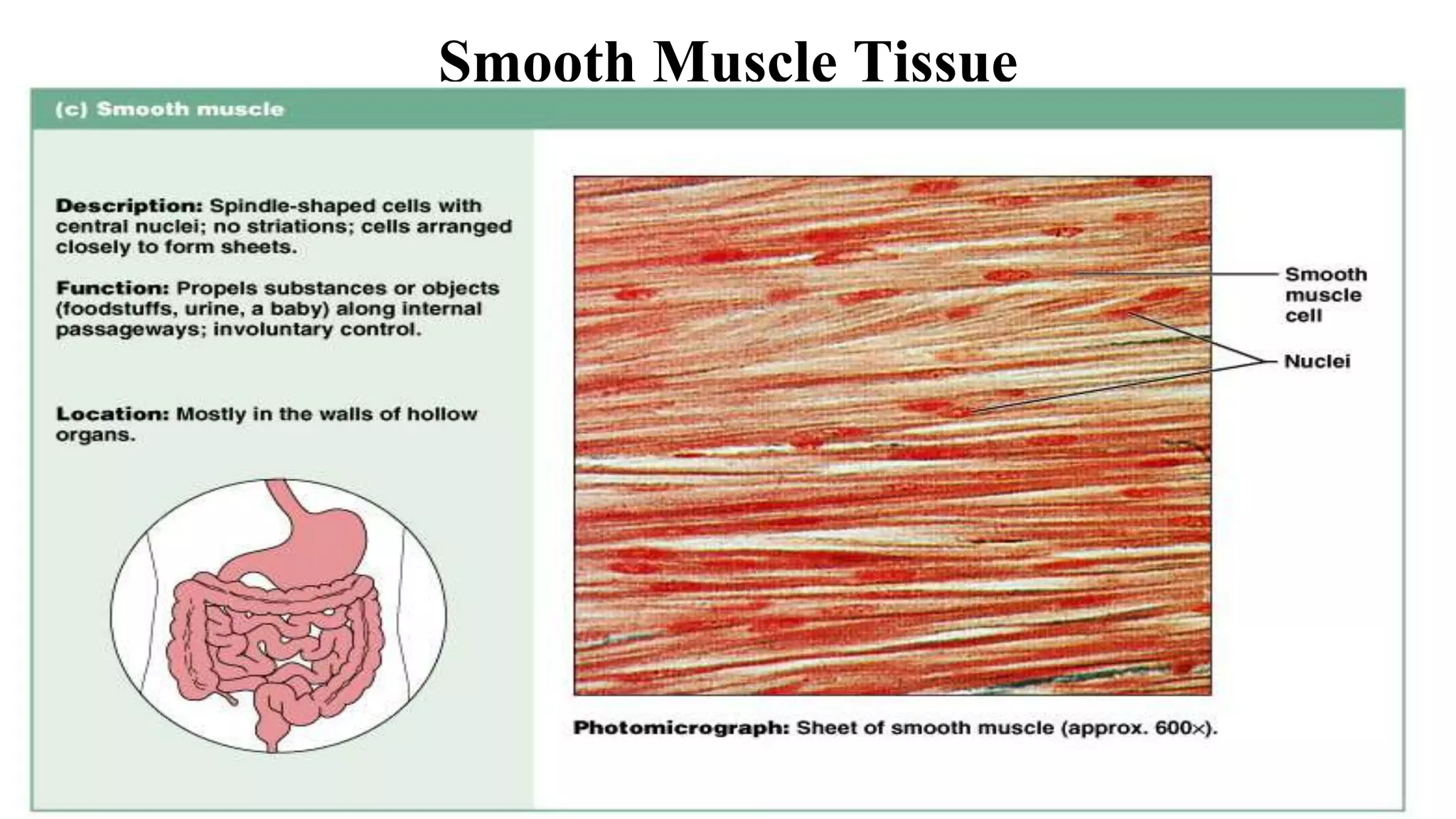
MUSCULAR
TISSUES
MUSCLE TISSUE
• Abundant, dense tissue
– 40-50% of body weight
• High metabolic activity
– high energy use/output;
– O2 consumption;
– CO2 production
MUSCLE TISSUE
• Consist of cells & intercellular substance.
• Cells are called fibres b/c they are elongated.
• Contractile property is due to myofibrils.
• Plasma lemma is called sarcolemma.
• Cytoplasm is called sarcoplasm.
Characteristics of Muscle Tissue
• Contractility
– Muscle tissue shortens producing force.
• Excitability
– Responds to nerve impulses by contracting.
• Extensibility
– Ability of a muscle to extend to a predetermined endpoint.
• Elasticity
– Recoils after being stretched.
Functions of Muscle Tissue
• Produce movement by contracting
– Locomotion & manipulation
– Movement of blood, food, urine, etc. through hollow organs
• Maintenance of posture
• Stabilize joints
– so that other muscles can produce movement or maintain posture
• Heat production
– Maintenance of body temperature.
Three Kinds of Muscle Tissue
• Skeletal muscle:
– Striated, voluntary, attached to skeleton
• Cardiac muscle:
– Striated, involuntary, walls of heart
• Smooth muscle:
– Non-striated, involuntary, wrapping of hollow organs
– E.g.: Stomach, intestines, arteries, bladder, vas deferens, uterus, etc.
Skeletal Muscle Tissue
Figure 4.14a
• Long, cylindrical cells,
• Multinucleate (nuclei peripheral),
• Cytoplasm = sarcoplasm
• ER = sarcoplasmic reticulum
• Covered by plasma membrane (sarcolemma).
Muscle sheath
• Epimysium
– Dense connective tissue that surrounds entire muscle
• Perimysium
– Thin connective tissue layer that surrounds fiber bundles (fascicles)
• Endomysium
– Surrounds each muscle fiber (cell)
– Mainly reticular fibers and basal lamina
Architecture of a skeletal
muscle fiber
• Muscle fibers are composed of myofibrils
• Myofibrils are composed of repeating units called sarcomeres
• Sarcomeres are the smallest functional unit of skeletal muscle
• Sarcomeres are composed of overlapping thick and thin
filaments
• Thin filaments are composed of actin and associated proteins
• Thick filaments are composed of myosin
Myofibrils
• Made of three types of filaments (or
myofilaments):
– Thick (myosin II)
– Thin (actin)
– Elastic (titin)
Thick myofilament:
• Three main proteins: the actin monomer, troponin
complex (comprising three protein components:- troponin-
I, troponin-C and troponin-T) and two strands of
tropomyosin molecules.
• Composed of 2 heavy chains & 2 pair of light chains.
• Heavy chain has a shape of golf club, has 2 binding sites
i.e. for ATP & actin.
• Light chain are of 2 types: one pair of essential light chain
& one of regulatory light chain.
Thin myofilament:
• Composed mainly of actin but tropomyosin & troponin is also
present.
• Actin exist as long filamentous polymer called F-actin polymer,
w/c composed of two strands of globular sub units of G-actin (has
an active site that can bind to the head of a myosin molecule).
• Tropomyosin, the protein that blocks the active sites of the thin
filaments when the muscle is relaxed.
• Each tropomyosin molecule has a smaller calcium-binding protein
called troponin bound to it.
The Sarcolemma
• The cell membrane of a muscle cell
• Surrounds the sarcoplasm (cytoplasm of muscle fiber)
• A change in transmembrane potential begins contractions
• All regions of the cell must contract simultaneously
Muscle Striations
• A striped or striated pattern within myofibrils:
– alternating dark, thick filaments (A bands) and light, thin filaments (I
bands)
• M line:
– the center of the A band
– at midline of sarcomere
• Z lines:
– the centers of the I bands
– at 2 ends of sarcomere
H Zone
• The area around the M line
• Has thick filaments but no thin filaments
Titin
• Are strands of protein
• Reach from tips of thick filaments to the Z line
• Stabilize the filaments
Sarcomeres
-The contractile units of muscle.
-Structural units of myofibrils.
-Form visible patterns within
myofibrils.
Sarcomere regions
• I Band, isotropic- similar polarization characteristics
throughout
• A Band, anisotropic- different polarization characteristics
throughout
• H Band- ‘heller’ or bright
• Z line, between
• M line, ‘mittel’ or middle
T-tubules
• Invaginations of sarcolemma that surround myofibril at
every sarcomere in the region of the A-I band
• Adjacent to each T-tubule is a sarcoplasmic reticulum,
which stores calcium.
• Action potential travels along sarcolemma, down T-tubule
• This causes calcium to be released from sarcoplasmic
reticulum
The triad
• Formed at A-I junction.
• the triad is a diagnostic feature of skeletal muscle
• It is composed of two terminal cisternae on either side of a T-
tubule.
• It is not found in cardiac or smooth muscle.
Cisternae
• Concentrate Ca2+
(via ion pumps)
• Release Ca2+
into sarcomeres to begin muscle contraction
Cardiac Muscle Tissue
Figure 4.14b
• Bundles form thick myocardium
• Cardiac muscle cells are single cells (not called fibers)
• Cells branch with 1-2 nuclei in center.
• Cells join at intercalated discs (intercellular junction)
• Here “fiber” = long row of joined cardiac muscle cells
• Inherent rhythmicity: each cell! (muscle cells beat separately
without any stimulation)
• Cells are small, have short, wide T tubules but no triads
• have SR with no terminal cisternae
• are aerobic (high in myoglobin, mitochondria)
Intercalated Discs
• Are specialized contact points between
cardiocytes
• Join cell membranes of adjacent cardiocytes (gap
junctions, desmosomes & fasciae adherent)
Functions of Intercalated Discs
• Maintain structure
• Enhance molecular and electrical connections
• Conduct action potentials
Smooth Muscle Tissue
Figure 4.14c
Characteristics of Smooth Muscle Cells
1. Long, slender, and spindle shaped
2. Have a single, central nucleus
3. Have no T tubules, myofibrils, or sarcomeres
4. Have no tendons or aponeuroses.
5. Contractions are slow, sustained and resistant to fatigue
6. Does not always require a nervous signal: can be stimulated by
stretching or hormones
7. Contain contractile apparatus, w/c enables shortening.
8. Have scattered myosin fibers, have more heads per thick filament
9. Have thin filaments attached to dense bodies, w/c transmit
contractions from cell to cell.
Dense bodies
• Round, amorphous bodies scattered through the cytoplasm of
smooth muscle fibers.
• Intermediate filament & actin are inserted into dense bodies.
• They contain α-actinin (actin binding protein)
• Actin filaments within the cells are either anchored to the internal
surface of the plasma membrane, or onto irregularly placed dense
bodies (rather than z-lines).
• Contractile units are situated diagonally across the cell (rather than
running parallel as in skeletal muscle)
• 6 major locations:
1. Inside the eye
2. Walls of vessels
3. Respiratory tubes
4. Digestive tubes
5. Urinary organs
6. Reproductive organs
Thank You

Muscular tissue.ppt

  • 1.
  • 2.
    MUSCLE TISSUE • Abundant,dense tissue – 40-50% of body weight • High metabolic activity – high energy use/output; – O2 consumption; – CO2 production
  • 3.
    MUSCLE TISSUE • Consistof cells & intercellular substance. • Cells are called fibres b/c they are elongated. • Contractile property is due to myofibrils. • Plasma lemma is called sarcolemma. • Cytoplasm is called sarcoplasm.
  • 4.
    Characteristics of MuscleTissue • Contractility – Muscle tissue shortens producing force. • Excitability – Responds to nerve impulses by contracting. • Extensibility – Ability of a muscle to extend to a predetermined endpoint. • Elasticity – Recoils after being stretched.
  • 5.
    Functions of MuscleTissue • Produce movement by contracting – Locomotion & manipulation – Movement of blood, food, urine, etc. through hollow organs • Maintenance of posture • Stabilize joints – so that other muscles can produce movement or maintain posture • Heat production – Maintenance of body temperature.
  • 6.
    Three Kinds ofMuscle Tissue • Skeletal muscle: – Striated, voluntary, attached to skeleton • Cardiac muscle: – Striated, involuntary, walls of heart • Smooth muscle: – Non-striated, involuntary, wrapping of hollow organs – E.g.: Stomach, intestines, arteries, bladder, vas deferens, uterus, etc.
  • 7.
  • 8.
    • Long, cylindricalcells, • Multinucleate (nuclei peripheral), • Cytoplasm = sarcoplasm • ER = sarcoplasmic reticulum • Covered by plasma membrane (sarcolemma).
  • 9.
    Muscle sheath • Epimysium –Dense connective tissue that surrounds entire muscle • Perimysium – Thin connective tissue layer that surrounds fiber bundles (fascicles) • Endomysium – Surrounds each muscle fiber (cell) – Mainly reticular fibers and basal lamina
  • 11.
    Architecture of askeletal muscle fiber • Muscle fibers are composed of myofibrils • Myofibrils are composed of repeating units called sarcomeres • Sarcomeres are the smallest functional unit of skeletal muscle • Sarcomeres are composed of overlapping thick and thin filaments • Thin filaments are composed of actin and associated proteins • Thick filaments are composed of myosin
  • 13.
    Myofibrils • Made ofthree types of filaments (or myofilaments): – Thick (myosin II) – Thin (actin) – Elastic (titin)
  • 14.
    Thick myofilament: • Threemain proteins: the actin monomer, troponin complex (comprising three protein components:- troponin- I, troponin-C and troponin-T) and two strands of tropomyosin molecules. • Composed of 2 heavy chains & 2 pair of light chains. • Heavy chain has a shape of golf club, has 2 binding sites i.e. for ATP & actin. • Light chain are of 2 types: one pair of essential light chain & one of regulatory light chain.
  • 15.
    Thin myofilament: • Composedmainly of actin but tropomyosin & troponin is also present. • Actin exist as long filamentous polymer called F-actin polymer, w/c composed of two strands of globular sub units of G-actin (has an active site that can bind to the head of a myosin molecule). • Tropomyosin, the protein that blocks the active sites of the thin filaments when the muscle is relaxed. • Each tropomyosin molecule has a smaller calcium-binding protein called troponin bound to it.
  • 16.
    The Sarcolemma • Thecell membrane of a muscle cell • Surrounds the sarcoplasm (cytoplasm of muscle fiber) • A change in transmembrane potential begins contractions • All regions of the cell must contract simultaneously
  • 17.
    Muscle Striations • Astriped or striated pattern within myofibrils: – alternating dark, thick filaments (A bands) and light, thin filaments (I bands) • M line: – the center of the A band – at midline of sarcomere • Z lines: – the centers of the I bands – at 2 ends of sarcomere
  • 18.
    H Zone • Thearea around the M line • Has thick filaments but no thin filaments Titin • Are strands of protein • Reach from tips of thick filaments to the Z line • Stabilize the filaments
  • 19.
    Sarcomeres -The contractile unitsof muscle. -Structural units of myofibrils. -Form visible patterns within myofibrils.
  • 20.
    Sarcomere regions • IBand, isotropic- similar polarization characteristics throughout • A Band, anisotropic- different polarization characteristics throughout • H Band- ‘heller’ or bright • Z line, between • M line, ‘mittel’ or middle
  • 21.
    T-tubules • Invaginations ofsarcolemma that surround myofibril at every sarcomere in the region of the A-I band • Adjacent to each T-tubule is a sarcoplasmic reticulum, which stores calcium. • Action potential travels along sarcolemma, down T-tubule • This causes calcium to be released from sarcoplasmic reticulum
  • 22.
    The triad • Formedat A-I junction. • the triad is a diagnostic feature of skeletal muscle • It is composed of two terminal cisternae on either side of a T- tubule. • It is not found in cardiac or smooth muscle. Cisternae • Concentrate Ca2+ (via ion pumps) • Release Ca2+ into sarcomeres to begin muscle contraction
  • 24.
  • 25.
    • Bundles formthick myocardium • Cardiac muscle cells are single cells (not called fibers) • Cells branch with 1-2 nuclei in center. • Cells join at intercalated discs (intercellular junction) • Here “fiber” = long row of joined cardiac muscle cells • Inherent rhythmicity: each cell! (muscle cells beat separately without any stimulation) • Cells are small, have short, wide T tubules but no triads • have SR with no terminal cisternae • are aerobic (high in myoglobin, mitochondria)
  • 26.
    Intercalated Discs • Arespecialized contact points between cardiocytes • Join cell membranes of adjacent cardiocytes (gap junctions, desmosomes & fasciae adherent) Functions of Intercalated Discs • Maintain structure • Enhance molecular and electrical connections • Conduct action potentials
  • 27.
  • 29.
    Characteristics of SmoothMuscle Cells 1. Long, slender, and spindle shaped 2. Have a single, central nucleus 3. Have no T tubules, myofibrils, or sarcomeres 4. Have no tendons or aponeuroses. 5. Contractions are slow, sustained and resistant to fatigue 6. Does not always require a nervous signal: can be stimulated by stretching or hormones
  • 30.
    7. Contain contractileapparatus, w/c enables shortening. 8. Have scattered myosin fibers, have more heads per thick filament 9. Have thin filaments attached to dense bodies, w/c transmit contractions from cell to cell.
  • 31.
    Dense bodies • Round,amorphous bodies scattered through the cytoplasm of smooth muscle fibers. • Intermediate filament & actin are inserted into dense bodies. • They contain α-actinin (actin binding protein) • Actin filaments within the cells are either anchored to the internal surface of the plasma membrane, or onto irregularly placed dense bodies (rather than z-lines). • Contractile units are situated diagonally across the cell (rather than running parallel as in skeletal muscle)
  • 32.
    • 6 majorlocations: 1. Inside the eye 2. Walls of vessels 3. Respiratory tubes 4. Digestive tubes 5. Urinary organs 6. Reproductive organs
  • 33.