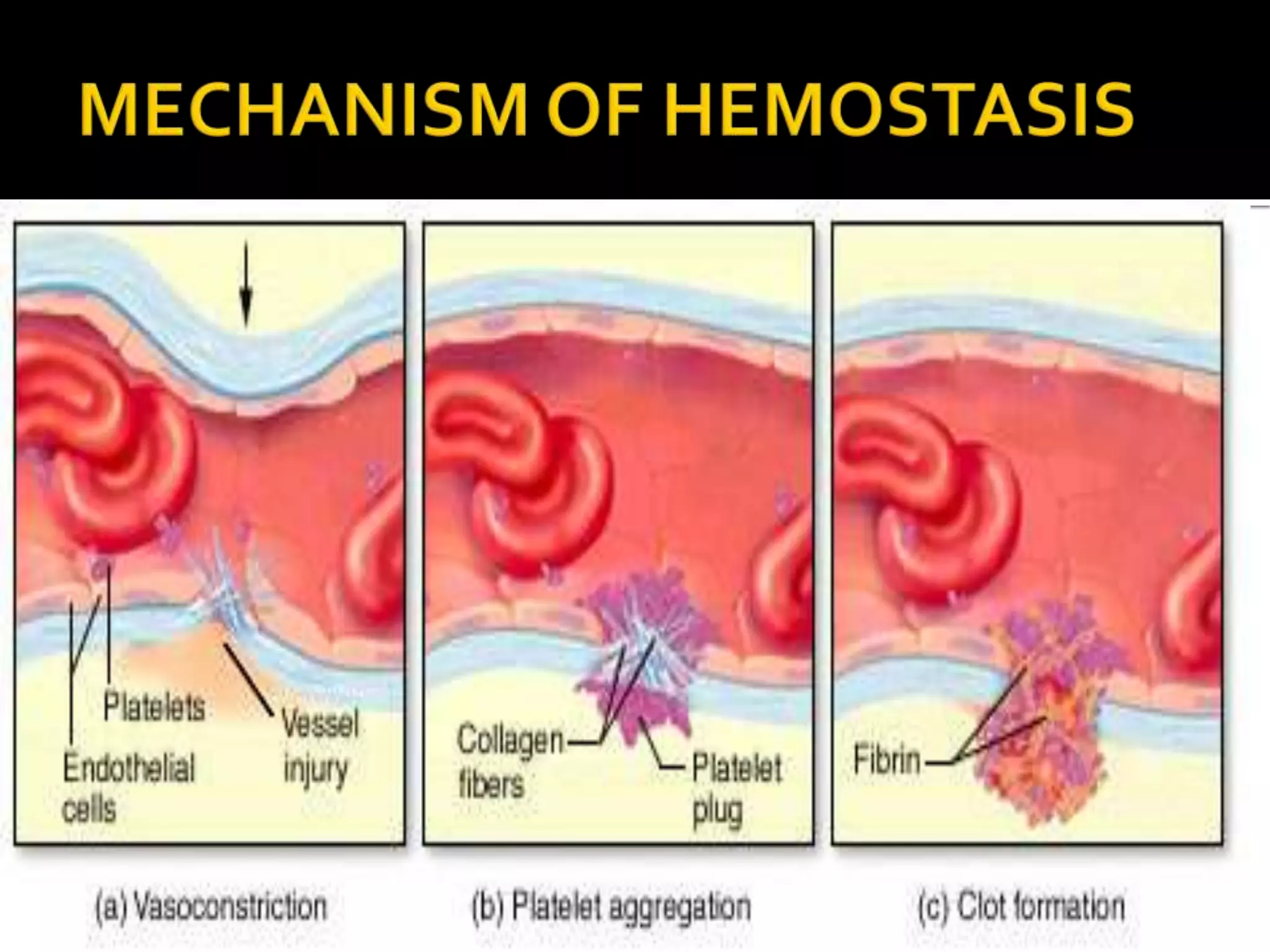
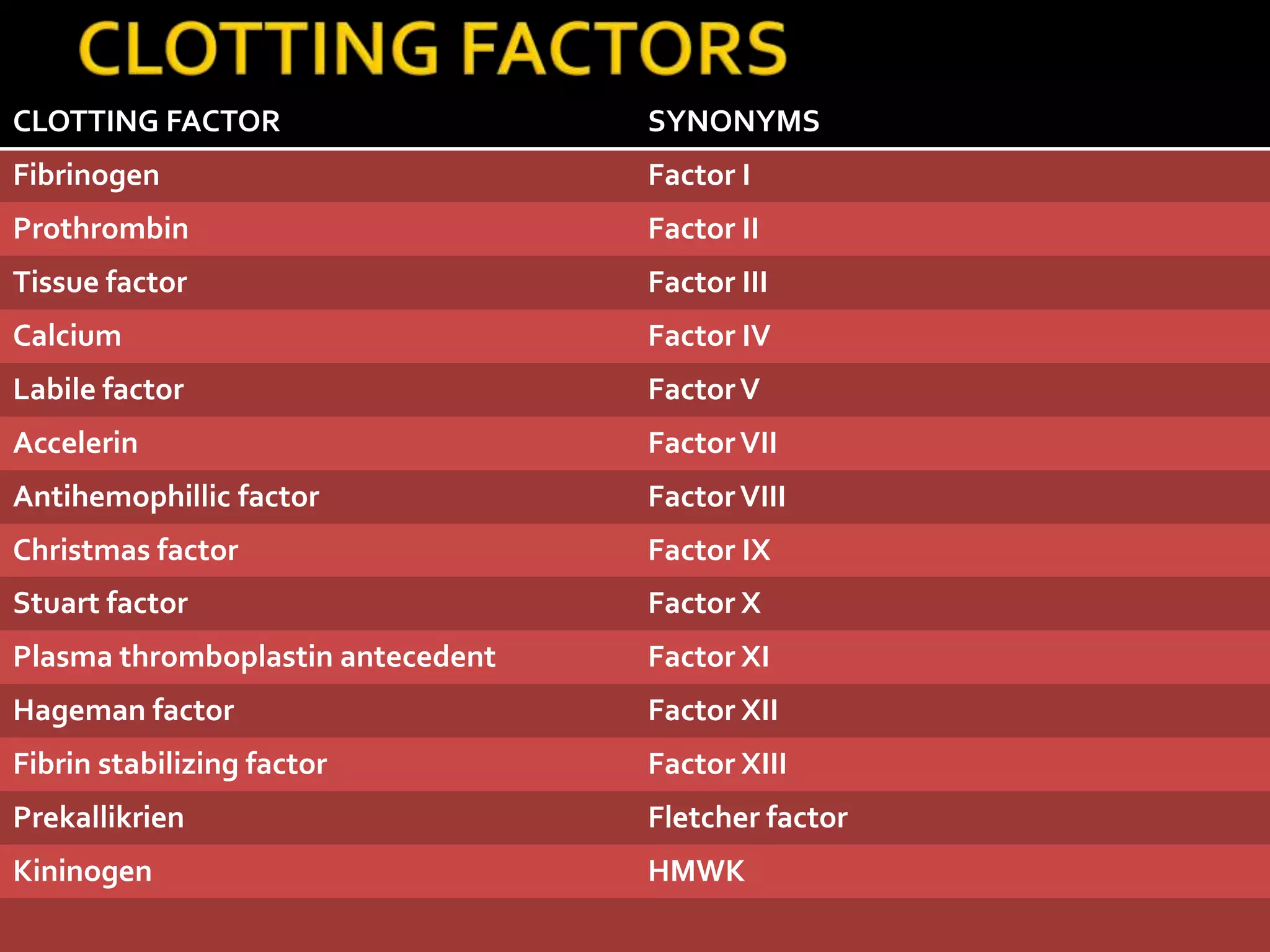
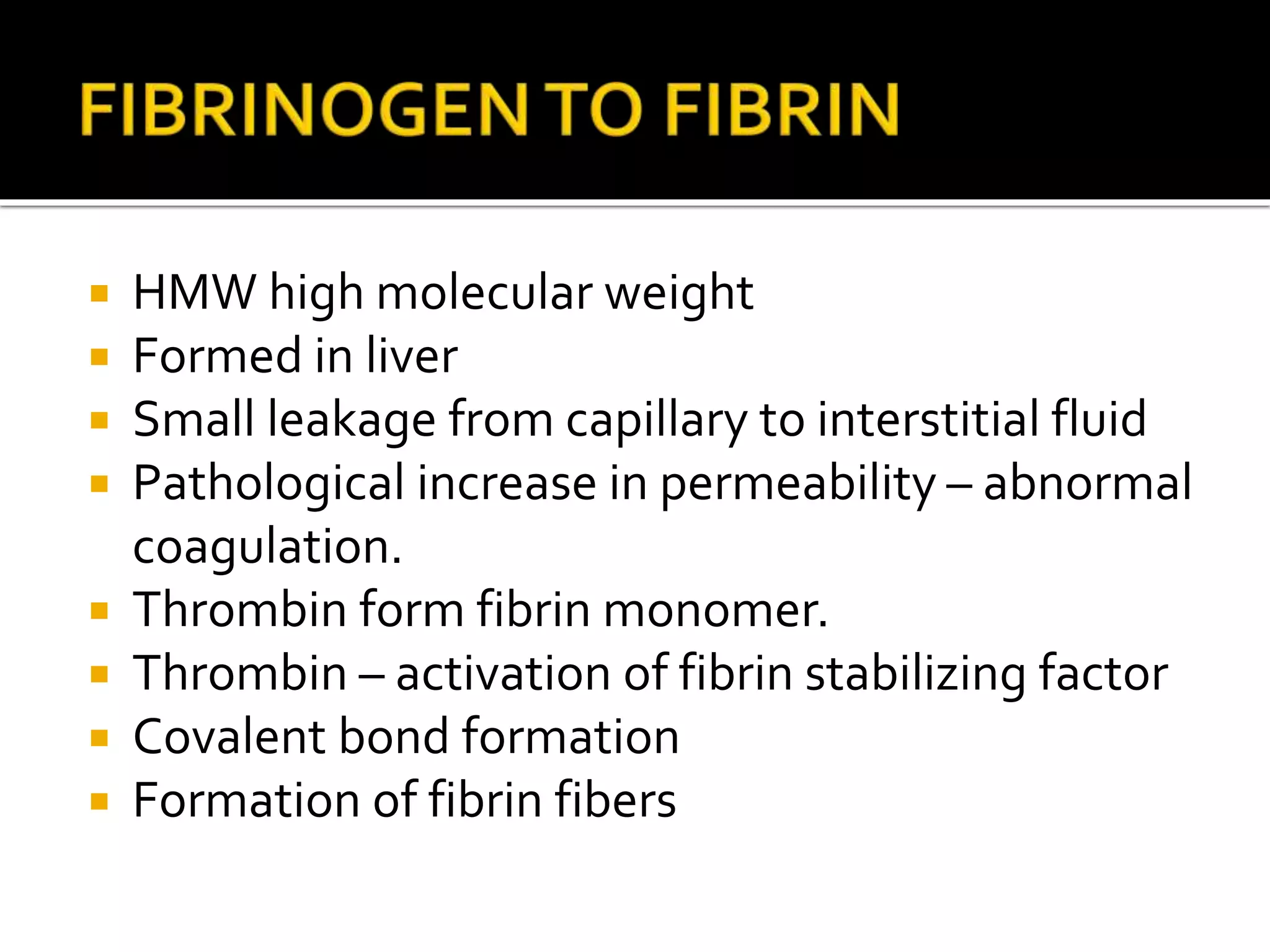
“Prevention of blood loss”
 Vascular constriction
 Formation of platelet plug
 Blood clot formation
 Fibrous tissue organization to close the hole
of veins.
 Smooth muscle contraction to reduce blood
flow after an injury or cut.
 3 factors responsible for contraction.
 Nerve reflexes
 Autocoids from platelets or traumatized tissue
 Myogenic spasm
 Thrombocytes
 Formed from megakaryocytes
 Anucleated
 1,50000 – 300,000 / µl
 Actin myosin molecules & thrombosthenin
 Residual ER & golgi apparatus
 Enzyme system to synthesizeATP from ADP
 Enzyme system to synthesize PGs.
 Fibrin stabilizing factor – form stable
fibrin threads
 Growth factor
 Glycoprotein on surface
 Phospholipids
 Adenyl cyclase  cAMP for further
activation of platelets
 Platelets in contact with collagen in damaged
vessels
 Stick to collagen fibres
 Secretion of ADP & thromboxane A2
 Increased number of activated platelets
gathered to form platelet plug.
 Close small dents in vessel wall
 Fibrin threads
 Clot – when blood looses its fluidity.
 Clot forms – 15 – 20 sec
 Activator substances – adheres to
traumatized vessel
 Clot formed
CLOTTING FACTOR SYNONYMS
Fibrinogen Factor I
Prothrombin Factor II
Tissue factor Factor III
Calcium Factor IV
Labile factor FactorV
Accelerin FactorVII
Antihemophillic factor FactorVIII
Christmas factor Factor IX
Stuart factor Factor X
Plasma thromboplastin antecedent Factor XI
Hageman factor Factor XII
Fibrin stabilizing factor Factor XIII
Prekallikrien Fletcher factor
Kininogen HMWK
 Clot Follows 2 ways.
 Invaded by fibroblasts
 Can be dissolved
 Procoagulants
 Anticoagulants
 Formation of prothrombin activator
 Conversion of prothrombin to thrombin
 Conversion of fibrinogen to fibrin
 Rate limiting step - formation of prothrombin
activator.
 Calcium and prothrombin activator – convert
prothrombin to thrombin
 Prothrombin – unstable protein
 Produced by liver
 Vitamin K – formation of prothrombin.
 HMW high molecular weight
 Formed in liver
 Small leakage from capillary to interstitial fluid
 Pathological increase in permeability – abnormal
coagulation.
 Thrombin form fibrin monomer.
 Thrombin – activation of fibrin stabilizing factor
 Covalent bond formation
 Formation of fibrin fibers
 Extrinsic pathway
 Intrinsic pathway
 Traumatized vessel release tissue factor .
 Factor III with factorVII  act on factor X 
Factor Xa
 Factor Xa complexes with phospholipids &
factorV .
 Prothrombin activator formed.
 Trauma to blood or damage s platelets due to
adherence to collagen.
 Activates factor XII.
 XIIa in presence of HMW kinogens &
prekallikrien  XI
 XIa  IXa
 IXa +VIII  Xa
 Xa + phospholipids +V  prothrombin activator
 Extraction of fluid after coagulation i.e.
serum
 Disseminated intravascular coagulation (DIC)
 Hemophilia
 Thrombocytopenia
 Purpura
 Thrombo embolic condition
 HemophiliaA
(VIII small component def.)
 Von willebrands disease
(VIII large component def.)
 Hemophilia B
(IX def)
 Hemophilia C
(XI def.)
BLOOD COAGULATION & HEMOSTASIS.pptx

BLOOD COAGULATION & HEMOSTASIS.pptx

  • 2.
  • 3.
     Vascular constriction Formation of platelet plug  Blood clot formation  Fibrous tissue organization to close the hole of veins.
  • 4.
     Smooth musclecontraction to reduce blood flow after an injury or cut.  3 factors responsible for contraction.  Nerve reflexes  Autocoids from platelets or traumatized tissue  Myogenic spasm
  • 5.
     Thrombocytes  Formedfrom megakaryocytes  Anucleated  1,50000 – 300,000 / µl  Actin myosin molecules & thrombosthenin  Residual ER & golgi apparatus  Enzyme system to synthesizeATP from ADP
  • 6.
     Enzyme systemto synthesize PGs.  Fibrin stabilizing factor – form stable fibrin threads  Growth factor  Glycoprotein on surface  Phospholipids  Adenyl cyclase  cAMP for further activation of platelets
  • 7.
     Platelets incontact with collagen in damaged vessels  Stick to collagen fibres  Secretion of ADP & thromboxane A2  Increased number of activated platelets gathered to form platelet plug.  Close small dents in vessel wall  Fibrin threads
  • 8.
     Clot –when blood looses its fluidity.  Clot forms – 15 – 20 sec  Activator substances – adheres to traumatized vessel  Clot formed
  • 11.
    CLOTTING FACTOR SYNONYMS FibrinogenFactor I Prothrombin Factor II Tissue factor Factor III Calcium Factor IV Labile factor FactorV Accelerin FactorVII Antihemophillic factor FactorVIII Christmas factor Factor IX Stuart factor Factor X Plasma thromboplastin antecedent Factor XI Hageman factor Factor XII Fibrin stabilizing factor Factor XIII Prekallikrien Fletcher factor Kininogen HMWK
  • 12.
     Clot Follows2 ways.  Invaded by fibroblasts  Can be dissolved
  • 13.
     Procoagulants  Anticoagulants Formation of prothrombin activator  Conversion of prothrombin to thrombin  Conversion of fibrinogen to fibrin
  • 14.
     Rate limitingstep - formation of prothrombin activator.  Calcium and prothrombin activator – convert prothrombin to thrombin  Prothrombin – unstable protein  Produced by liver  Vitamin K – formation of prothrombin.
  • 15.
     HMW highmolecular weight  Formed in liver  Small leakage from capillary to interstitial fluid  Pathological increase in permeability – abnormal coagulation.  Thrombin form fibrin monomer.  Thrombin – activation of fibrin stabilizing factor  Covalent bond formation  Formation of fibrin fibers
  • 17.
     Extrinsic pathway Intrinsic pathway
  • 18.
     Traumatized vesselrelease tissue factor .  Factor III with factorVII  act on factor X  Factor Xa  Factor Xa complexes with phospholipids & factorV .  Prothrombin activator formed.
  • 19.
     Trauma toblood or damage s platelets due to adherence to collagen.  Activates factor XII.  XIIa in presence of HMW kinogens & prekallikrien  XI  XIa  IXa  IXa +VIII  Xa  Xa + phospholipids +V  prothrombin activator
  • 21.
     Extraction offluid after coagulation i.e. serum
  • 22.
     Disseminated intravascularcoagulation (DIC)  Hemophilia  Thrombocytopenia  Purpura  Thrombo embolic condition
  • 23.
     HemophiliaA (VIII smallcomponent def.)  Von willebrands disease (VIII large component def.)  Hemophilia B (IX def)  Hemophilia C (XI def.)