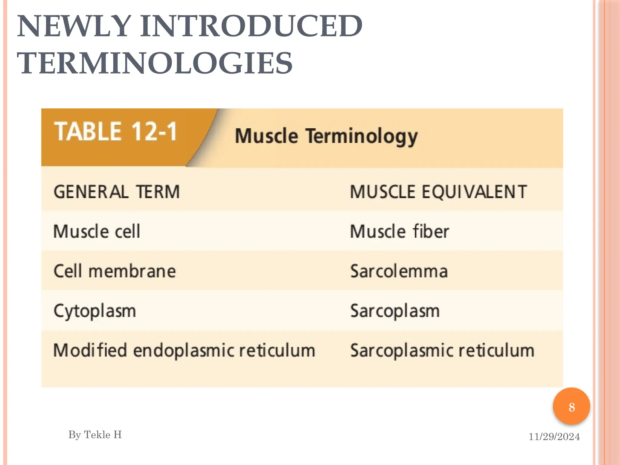
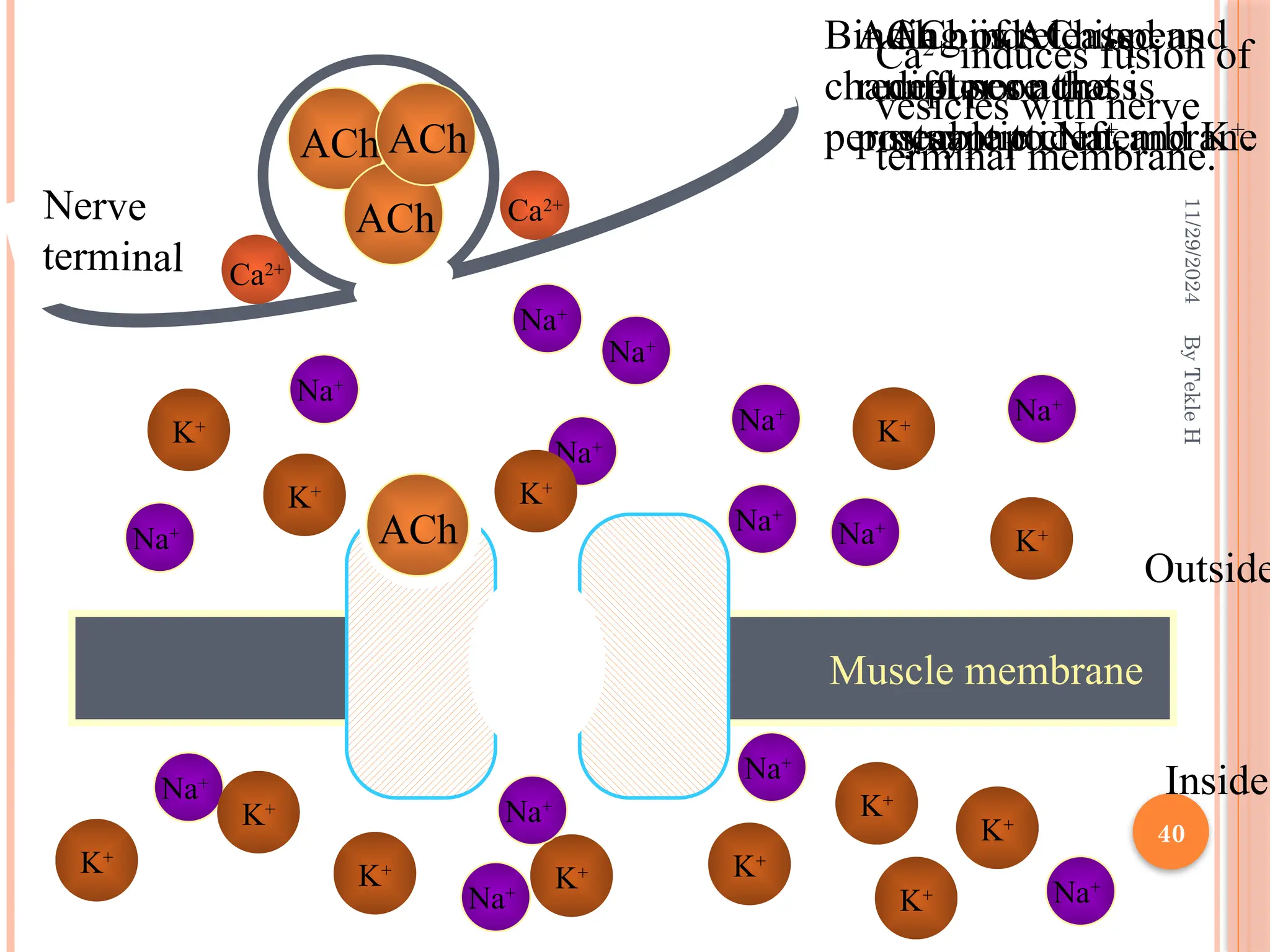
11/29/2024
By
Tekle
H
1
MUSCLE PHYSIOLOGY
By Tekle H.
11/29/2024
2
By
Tekle
H
CONTENTS
 Introduction
 Skeletal muscle
 Structure and organization
 Muscle fiber anatomy
 Mechanism of muscle contraction
 Neuromuscular junction
 Excitation contraction coupling
 Sliding filament mechanism
 Mechanism of muscle relaxation
 Cardiac muscles
 Smooth muscles
11/29/2024
3
By
Tekle
H
INTRODUCTION
Muscle:
 Is a fleshy, reddish colored tissue in the body
 Comprises the largest group of tissues in the body
 Muscles constitutes up to 50% of body weight
 About 40% of the body is skeletal muscle, and
 Another 10% is smooth and cardiac muscle
 Muscle cells are specialized to generate mechanical force from chemical
energy
 This force is used to regulate the internal environment, and used for movement
of the body in reference to the external environment
11/29/2024
4
By
Tekle
H
 There are three types of muscle cells and tissues
 Skeletal, cardiac, or smooth muscle cells and tissues
 Most skeletal muscles are attached to bone
 Their contraction is responsible for supporting and moving
the skeleton
 Contraction of skeletal muscle is initiated by action
potential from motor neurons of the somatic nervous
system
 That is why these are under voluntary control
11/29/2024
5
By
Tekle
H
 Smooth muscle surround various hollow organs and
tubular structure
 Including the digestive tract, urinary bladder and tracts,
uterus, blood vessels, and airways
 Contraction of smooth muscles decreases either the
diameter or the length of these structures and
 Propel the luminal contents through the hollow organs, or
 Regulate internal flow by changing the tube diameter
 For example, contraction of smooth muscle cells along the
esophagus helps “squeeze” swallowed food to the stomach
 Furthermore contraction of smooth muscle stand up the
hairs of the skin and change diameter of the pupil
11/29/2024
6
By
Tekle
H
 Smooth muscle contraction is involuntary
 It occurs under the control of autonomic nervous system,
hormones, autocrine or paracrine signals, and other local
chemical factors
 In some cases it occurs autonomously
 Cardiac muscle is the muscle found in the walls of
the heart
 Its contraction enables the heart to pump blood
 Like smooth muscle, this is also involuntary
 Regulated by the autonomic nervous system, hormones, and
other glandular signals
 It can undergo spontaneous contractions
7
COMMON PROPERTIES OF
MUSCLES
1. Excitability- ability to respond to stimulus which could be from motor
neuron or a hormone
2. Contractility- The ability to shorten forcibly when adequately
stimulated
3. Extensibility- The ability to be stretched or extended
4. Elasticity- The ability to recoil and resume original length after being
stretched or contracted
11/29/2024
By Tekle H
8
NEWLY INTRODUCED
TERMINOLOGIES
11/29/2024
By Tekle H
11/29/2024
9
By
Tekle
H
SKELETAL MUSCLE
Skeletal Muscles
 Are muscles that
causes the skeleton to
move at joints
 They are attached to
the skeleton by
tendons
 The tendons transmit
the muscle force to
the bone
10
Skeletal muscles are
 Long cylindrical cells
 Many nuclei per cell
 Voluntary
 Rapid contractions
 Are well supplied with nerves and blood vessels
 Are striated muscle type
 Due to the distinct series of alternating light and dark
bands
 Cardiac muscles have same feature
 But, the smooth muscle lacks striation and is named after
it
11/29/2024
By Tekle H
11/29/2024
11
By
Tekle
H
MUSCLE ORGANIZATION
EPIMYSIUM
Surrounds the entire muscle
PERIMYSIUM
Surrounds bundle of muscle
Fibers (Fascicle)
ENDOMYSIUM
Surrounds individual muscle
fibers
12
11/29/2024
By Tekle H
11/29/2024
13
By
Tekle
H
MUSCLE FIBER ANATOMY
 Sarcolemma - cell membrane
 Surrounds the sarcoplasm (cytoplasm of fiber)
 Contains many of the same organelles seen in other cells
 An abundance of the oxygen-binding protein myoglobin
 Myofibrils -cylindrical structures within muscle fiber
 Are bundles of protein filaments (myofilaments)
 Two types of myofilaments
1. Actin (thin) filaments
2. Myosin (thick) filaments
– Fills majority of the sarcoplasmic space
– At each end of the fiber, myofibrils are anchored to the inner
surface of the sarcolemma
 When myofibril shortens, the muscle contracts
14
11/29/2024
By
Tekle
H
11/29/2024
15
By
Tekle
H
SARCOPLASMIC RETICULUM (SR)
 SR refers to smooth endoplasmic reticulum
 Runs longitudinally and surrounds each myofibril
 SR forms a series of sleeve like segments around each myofibril
 At it’s end there are two enlarged regions which are called terminal
cisternae (also called lateral sacs)
 The terminal cisternae are connected to each other by a series of smaller
tubular elements
 SR stores Ca2+
 When stimulated, calcium is released into sarcoplasm
 SR membrane has Ca2+
pump that function to pump Ca2+
back into
the SR after contraction
16
11/29/2024
By
Tekle
H
17
 There is also separate tubular structure, the transverse
tubule (T-tubule), that lies between adjacent terminal
cisternae
 T-tubules and terminal cisternae surrounds the myofibrils at
the region of the sarcomeres where the A and I bands meet
 T-tubules are continuous with the sarcolemma
 Action potentials propagating along the surface membrane also travel
throughout the interior of the muscle fiber by way of the T-tubules
 The lumen of the T-tubule is continuous with the extracellular
fluid surrounding the muscle fiber
 Terminal cisternae together with T-tubules form “Triad”
11/29/2024
By
Tekle
H
18
11/29/2024
By
Tekle
H
19
11/29/2024
By
Tekle
H
11/29/2024
20
By
Tekle
H
Sarcomere - repeating functional units of a myofibril
 There around 10,000 sarcomeres per myofibril
 Each is about 2 µm long
 Extends between two successive Z lines (Z disks)
The Sarcomere contains two sets of
Myofilaments
(i) Actin filament
(ii) Myosin filament
11/29/2024
21
By
Tekle
H
22
 It contains different bands, zones, disc, line,….
 A bands: a dark band; full length of thick (myosin)
filament
 H zone:- narrow, light band in the center of the A
band
Which is the space between the opposing ends of the
two sets of thin filaments in each sarcomere
 M line - a narrow, dark band formed by proteins that
link the central region of adjacent thick filaments
11/29/2024
By
Tekle
H
23
11/29/2024
By
Tekle
H
11/29/2024
24
By
Tekle
H
 I bands: a light band that lies between the ends of the
A bands of two adjacent sarcomeres
 Formed by the portions of the thin filaments that do not
overlap with the thick filament
 Z disk: filamentous network of protein
 Serves as attachment for actin filaments
 The boundary between two sarcomeres
 Titin filaments
 Elastic protein extending from the Z line to the M line
(linked to both M Line proteins and the thick filaments)
 Keep thick and thin filaments in proper alignment in
the middle of each sarcomere
11/29/2024
25
By
Tekle
H
11/29/2024
26
By
Tekle
H
THICK FILAMENT
 Around 200 myosin molecules form the thick filament
 Forms the dark band (A band) of the alternating dark and light bands
of a muscle fiber
 Located in the centre of the sarcomere and attached to the Z-
line by Titin filaments
 Myosin is contractile protein composed of two heavy polypeptide
chains and four light chains
 The polypeptides combine to form
 Two globular heads (each containing 01 folded heavy and 02 light
chains)
 Light chains help control the function of the head
 A long tail formed by the two intertwined heavy chains
11/29/2024
27
By
Tekle
H
 The tail of each myosin molecule lies along the axis of the
thick filament
 The two globular heads extend out to the sides of the
filament forming cross-bridges
 This is the part that makes contact with the thin
filament and exert force during muscle contraction
 Each globular head contains two binding sites, one for
attaching to the thin filament and one for ATP
 The ATP binding site also serves as an enzyme (ATPase)
that hydrolyzes the bound ATP, producing its energy for
contraction
11/29/2024
28
By
Tekle
H
11/29/2024
29
By
Tekle
H
THIN FILAMENT
 Composed of three proteins, Actin, Troponin and
Tropomyosin
 One end of the thin filaments is connected to the Z-line
while the other end is directed to the center
 These are about half of the diameter of the thick filaments
 An actin monomers form a polymer made up of two helical
chains (the core of a thin filament)
 Each actin molecule contains a binding site for myosin
11/29/2024
30
By
Tekle
H
ACTIN:
Double-stranded helix formed from actin monomers
Having the binding site for myosin
TROPOMYOSIN:
 Two polypeptide chains coiled around each other
 Covers myosin binding sites of actin
11/29/2024
31
TROPONIN:
Globular units located at intervals along the
tropomyosin molecule
Has 3 components
 Troponin C- contain binding site for calcium
 Troponin T – binds the troponin component to
tropomyosin
 Troponin I –inhibit the interaction of myosin with actin.
By
Tekle
H
11/29/2024
32
By
Tekle
H
11/29/2024
33
By
Tekle
H
MECHANISMS OF SKELETAL MUSCLE
CONTRACTION
 Muscle contraction refers to the activation of the
force-generating sites within muscle fibers (cross-
bridges)
 But it does not always mean shortening of the muscle
 Following contraction, the mechanisms that
generate force are turned off and
 Allowing relaxation of muscle fibers
 Skeletal muscles always need neuronal stimulation
to initiate an action potential
11/29/2024
34
By
Tekle
H
 Motor neurons (somatic neurons) are responsible for
that stimulation
 A single motor neuron innervates many muscle fibers
 Each muscle fiber is controlled by a branch from only
one motor neuron
 A motor neuron plus the muscle fibers it innervates
is called a motor unit
 So, an action potential from the motor neuron
stimulates the muscle fibers in its motor unit
11/29/2024
35
By
Tekle
H
THE NEUROMUSCULAR JUNCTION (NMJ)
 This is the area where the axon terminal of a motor
neuron and the muscle membrane meet
 The region of the muscle fiber plasma membrane
that lies directly under the axon terminal is known
as the motor end plate.
 The space in between is called synaptic cleft
 The axon terminals of a motor neuron contain vesicles
that contain the neurotransmitter acetylcholine (ACh)
11/29/2024
36
By
Tekle
H
11/29/2024
37
By
Tekle
H

Nerve impulse reaches
nerve terminal
 Opening of voltage gated
calcium channels
 Calcium diffuses from
the ECF into the nerve
terminal
 Release of Ach from the
synaptic vesicles into the
synaptic cleft by process
of Exocytosis
11/29/2024
38
By
Tekle
H
Binding of Ach with
receptor, formation of
Ach- receptor complex
 Opening of Ligand gated
sodium channels
 Entry of sodium ions
into the ICF
 Development of end
plate potential ( EPP )
Stimulation of nearby
membrane & action
potential development
11/29/2024
By
Tekle
H
39
Neuromuscular
Transmission:
Step by Step
Nerve action
potential invades
axon terminal
-
+
-
-
-
-
-
-
+
+
+
+
+
+
+
-
-
-
+ +
Depolarization
of terminal
opens Ca channels
+ +
11/29/2024
By
Tekle
H
40
K+
Outside
Inside
Na+
Na+
Na+
Na+
Na+
Na+
Na+ Na+
Na+
Na+
Na+
Na+
K+ K+
K+
K+
K+
K+
K+
K+
K+
K+
K+
ACh
ACh
ACh
Ca2+
induces fusion of
vesicles with nerve
terminal membrane.
ACh is released and
diffuses across
synaptic cleft.
ACh
ACh binds to its
receptor on the
postsynaptic membrane
Binding of ACh opens
channel pore that is
permeable to Na+
and K+
.
Na+
Na+
K+
Muscle membrane
Nerve
terminal Ca2+
Ca2+
11/29/2024
41
By
Tekle
H
Resting membrane potential of skeletal muscle
membrane is -90mV.
When end plate potential reaches a threshold of
potential, it depolarizes the surface membrane of
the muscle and results in the generation of action
potential.
Once it reaches the muscle fibers then the muscles
give mechanical response by contraction.
11/29/2024
42
By
Tekle
H
 After Ach acts on
the receptors, it is
hydrolyzed by the
enzyme cholinesterase
into Acetate and
Choline
 Choline is actively
reabsorbed into the
nerve terminal to be
used again to form Ach
 The whole process of
Ach release, action and
destruction takes
about 5 –10 ms
11/29/2024
43
By
Tekle
H
EXCITATION - CONTRACTION
COUPLING
 Refers to the sequence of events by which an action
potential in the plasma membrane activates the force-
generating mechanisms
 To do so, the action potential in the membrane should
result in an increased cytosolic Ca2+
concentration
 First action potential propagates through the T tubules and
causes shape change in the dihydropyridine (DHP) receptors
 This in turn causes conformational change in ryanodine
receptors in the sarcoplasmic reticulum membrane
 Then Ca2+
channels are opened and Ca2+
will be released in to
cytosol
11/29/2024
44
By
Tekle
H
 When there is adequate Ca2+
in the sarcoplasm, it will bind with
troponin C
 This relaxes its inhibitory grip and displaces tropomyosin and
exposes myosin binding sites
 Then there will be cross-bridge formation and sliding of the
filaments
 Conversely, the removal of Ca2+
from troponin reverses the
process, turning off contractile activity
 Achieved by lowering the Ca2+
concentration in the cytosol back to its
prerelease level (pumped by Ca2+
-ATPases back to sarcoplasmic
reticulum)
 So, the main source of Ca2+
in skeletal muscle contraction is the
sarcoplasmic reticulum within the muscle fiber
11/29/2024
45
By
Tekle
H
SLIDING-FILAMENT MECHANISM
 Myosin head is always in activated state
 So when Ca2+
binds with troponin C, there will be
displacement of tropomyosin and exposure of myosin
binding sites
 Myosin head will bind with actin and pulls the actin
filament to the center of the sarcomere
 The filaments are sliding over each other resulting in
muscle contraction
 Therefore sliding of the thin and thick filaments is the
basis for muscle contraction
11/29/2024
46
By
Tekle
H
11/29/2024
By
Tekle
H
47
11/29/2024
By
Tekle
H
48
11/29/2024 By Tekle H 49
 cross-bridges attach to thin filament pull
towards center detach attach further down
pull it again,….. This process continues
11/29/2024
50
By
Tekle
H
 Generally ATP has four importance in skeletal muscle
contraction
 Source of energy in Na+
/K+
ATPase
 Source of energy in Ca2+
ATPase
 Source of energy in forming energized state of myosin
 ATP binding (not hydrolysis) to myosin breaks the link
formed between actin and myosin during the cycle
 Rigor mortis – failure of the detachment of actin and
myosin after death due to the absence of energy (ATP)
11/29/2024
By
Tekle
H
51
11/29/2024
52
By
Tekle
H
MECHANISM OF MUSCLE RELAXATION
 Increased sarcoplasmic calcium level during muscle contraction
 To decrease it and bring muscle relaxation
 Breakdown of Ach by acetyl cholinesterase
 Activation of Ca2+
ATPase
 Ca2+
pumped back into the SR
 Decreased sarcoplasmic Ca2+
level
 Detachment of Ca2+
from troponin
 Cessation of actin myosin interaction
 Muscle relaxation
11/29/2024
53
By
Tekle
H
11/29/2024
54
By
Tekle
H
11/29/2024
By
Tekle
H
55
PHYSIOLOGY OF CARDIAC
MUSCLE
56
PHYSIOLOGIC ANATOMY OF CARDIAC MUSCLE
 Two forms of muscles fibers in the heart
 Contractile muscle fibers
 Muscle fibers found in the walls of the
heart chambers that generate force used
to pump blood
 Excitatory and conductive muscle fibers
 Specialized muscle fibers mainly involved
in either
o Automatic rhythmical electrical
discharge OR
o Conduction of the action potentials
 The muscle fibers are arranged in a latticework
fashion
o Fibers divide, rejoin, and then divide
o The muscle fibers are striated similar to that of
skeletal muscle fiber and there is almost similar
o Arrangement of actin and myosin filaments
o Sliding during contraction
11/29/2024 By Tekle H
57
 Cardiac muscle cells form Syncytium
 Due to intercalated discs and branching, so fusion of
cytoplasm
 Intercalated disc have gap-junctions that allow rapid diffusion
of ions
 As a result ions (action potential) can move easily between two
adjacent cells
 So, when a given cell becomes excited, the action potential
rapidly spreads to all of the other muscle cells
 The heart have two syncytium
 The atrial syncytium
 The ventricular syncytium
 Fibrous connective tissue separates the atria and
ventricles preventing direct transmission of potentials
between the two syncytium
 AV bundles are the only means of potential transmission
between the two syncytium
 Any physiologic importance of this nature????
 This is important to have alternate excitation and contraction
of the atria and ventricles
11/29/2024 By Tekle H
58
ACTION POTENTIALS IN CARDIAC
MUSCLE
11/29/2024 By Tekle H
The membrane potential of ventricular muscle
fiber can reach up to +20 millivolts during
depolarization
The membrane remains depolarized for about 0.2
second (plateau),
This potential plateau is important to have long
lasting (15X that of skeletal muscle) ventricular
contraction
Two main reasons are responsible for this plateau
formation in cardiac muscle cells
1. Depolarization is happening as a result of influx of both
Na+
(fast opening and closure of ion channels) and Ca2+
(slow opening and closure of ion channels)
2. Decreased permeability of cardiac muscle membrane for
potassium ions (5x decreases)
59
EXCITATION-CONTRACTION COUPLING
 During muscle excitation, the mechanism of Ca2+
release in
skeletal muscles also applies
 But there is also additional mechanism of release
 So when there is excitation there is opening of voltage gated Ca2+
channels in the muscle membrane
 Then influx of Ca2+
in to the sarcoplasm
 That Ca2+
will stimulate the ryanodine receptors and additional
Ca2+
will be released from sarcoplasmic reticulum
 The Ca2+
diffuse into the myofibrils and initiate sliding of the
actin and myosin filaments - then muscle contraction
11/29/2024 By Tekle H
60
 Cardiac muscle’s sarcoplasmic reticulum is less developed and it
does not store enough calcium
 Because, calcium supply in cardiac muscles is mainly dependent
from external sources
 As a result these muscles have wider T-tubules with high
mucopolysaccharides (negatively charged molecules)
 Bind more Ca2+
and let it to be ready for diffusion to sarcoplasm when
needed
 Therefore, the concentration of Ca2+
in the ECF highly determine
the strength of cardiac muscle contraction
 Finally, muscle relaxation occurs as a result of Ca2+
pumping either
to SR (Ca2+
ATPase) or to the ECF (sodium-calcium exchanger)
11/29/2024 By Tekle H
61
11/29/2024 By Tekle H
11/29/2024
By
Tekle
H
62
SMOOTH MUSCLE
11/29/2024
63
By
Tekle
H
Grouped into sheets in walls of hollow visceral organs
• Longitudinal layer – muscle fibers run parallel to organ’s long axis
• Circular layer – muscle fibers run around circumference of the
organ
Surround:
• Blood vessels
• Digestive tract
• Organs (stomach, bladder; uterus)
11/29/2024
64
By
Tekle
H
Generally found in:-
 Cardiovascular system
 Respiratory system
 Digestive system
 Renal system
 Reproductive system
11/29/2024
65
By
Tekle
H
General features
 Involuntary muscle – controlled by ANS, Hormones, chemicals, …
 Unstriated muscle (lack visible cross striations)- plain muscle.
 No Sarcomere
Actin and Myosin not arranged as symmetrically as in skeletal muscle, thus
NO Sarcomere and striations
 More actin than myosin, Thin filaments lack Troponin
 Smaller, spindle shaped with varying dimensions.
a) Digestive system- 30-40 µm long and 5 µm diameter.
b) Blood vessels- 15-20 µm long and 2-3 µm in diameter.
c) Uterus 300 µm long and 10 µm diameter.
 Receive dual nerve supply from two divisions of autonomic
nervous system.
11/29/2024
66
By
Tekle
H
11/29/2024
67
By
Tekle
H
Types of smooth muscles
11/29/2024
68
By
Tekle
H
Visceral or single unit smooth muscle.
Multiunit smooth muscle.
11/29/2024
69
Visceral or single unit smooth muscle.
• The fibers are arranged as large sheets
• Exhibit unstable RMP. This is responsible for their
spontaneous activity.
• Muscle fibers are connected by tight junctions and gap
junctions. Electrical activity is conducted by ionic
movements = Syncytial fashion
By
Tekle
H
11/29/2024
70
By
Tekle
H
11/29/2024
71
By
Tekle
H
 Common sites: walls of the hollow viscera- GIT, bile duct,
bronchi, uterus, ureters, urinary bladder and in some blood
vessels
 The muscles are characterized by their spontaneous activity
in certain areas due to the pacemakers
 Receives nerve supply from autonomic nervous system.
But Independent of their innervations.
Nerves only modify the activity.
11/29/2024
72
By
Tekle
H
Multiunit smooth muscle.
11/29/2024
73
By
Tekle
H
 Made up of individual units without interconnecting
bridges- Non Syncytial.
 Common sites: Ciliary muscles of eye, pilomotor muscles of
skin, muscles of blood vessels.
 Richly innervated and each muscle fiber has its own nerve
supply
 These muscles only contact in response to the stimulus
through their nerves.
 Muscles do not respond to stretch
11/29/2024
74
By
Tekle
H
11/29/2024
75
By
Tekle
H
Smooth Muscle Regulation
 Innervated by autonomic nervous system
 Neurotransmitter like acetylcholine
 Hormones like epinephrine and oxytocin
 Cold, stretch stimulate contraction
 Hypoxia, hypercapnia- relaxation of smooth
muscle.
11/29/2024
76
By
Tekle
H
Mechanism of smooth muscle contraction:
• Activated by opening calcium channels on the cells
surface, there is influx of extra-cellular calcium.
11/29/2024
77
By
Tekle
H
11/29/2024
78
By
Tekle
H
11/29/2024
79
By
Tekle
H
Mechanism of smooth muscle relaxation:
11/29/2024
80
By
Tekle
H
11/29/2024
81
By
Tekle
H
11/29/2024
82
By
Tekle
H
THANK
YOU!!!

Muscle physiology power point pres-4.pptx

  • 1.
  • 2.
    11/29/2024 2 By Tekle H CONTENTS  Introduction  Skeletalmuscle  Structure and organization  Muscle fiber anatomy  Mechanism of muscle contraction  Neuromuscular junction  Excitation contraction coupling  Sliding filament mechanism  Mechanism of muscle relaxation  Cardiac muscles  Smooth muscles
  • 3.
    11/29/2024 3 By Tekle H INTRODUCTION Muscle:  Is afleshy, reddish colored tissue in the body  Comprises the largest group of tissues in the body  Muscles constitutes up to 50% of body weight  About 40% of the body is skeletal muscle, and  Another 10% is smooth and cardiac muscle  Muscle cells are specialized to generate mechanical force from chemical energy  This force is used to regulate the internal environment, and used for movement of the body in reference to the external environment
  • 4.
    11/29/2024 4 By Tekle H  There arethree types of muscle cells and tissues  Skeletal, cardiac, or smooth muscle cells and tissues  Most skeletal muscles are attached to bone  Their contraction is responsible for supporting and moving the skeleton  Contraction of skeletal muscle is initiated by action potential from motor neurons of the somatic nervous system  That is why these are under voluntary control
  • 5.
    11/29/2024 5 By Tekle H  Smooth musclesurround various hollow organs and tubular structure  Including the digestive tract, urinary bladder and tracts, uterus, blood vessels, and airways  Contraction of smooth muscles decreases either the diameter or the length of these structures and  Propel the luminal contents through the hollow organs, or  Regulate internal flow by changing the tube diameter  For example, contraction of smooth muscle cells along the esophagus helps “squeeze” swallowed food to the stomach  Furthermore contraction of smooth muscle stand up the hairs of the skin and change diameter of the pupil
  • 6.
    11/29/2024 6 By Tekle H  Smooth musclecontraction is involuntary  It occurs under the control of autonomic nervous system, hormones, autocrine or paracrine signals, and other local chemical factors  In some cases it occurs autonomously  Cardiac muscle is the muscle found in the walls of the heart  Its contraction enables the heart to pump blood  Like smooth muscle, this is also involuntary  Regulated by the autonomic nervous system, hormones, and other glandular signals  It can undergo spontaneous contractions
  • 7.
    7 COMMON PROPERTIES OF MUSCLES 1.Excitability- ability to respond to stimulus which could be from motor neuron or a hormone 2. Contractility- The ability to shorten forcibly when adequately stimulated 3. Extensibility- The ability to be stretched or extended 4. Elasticity- The ability to recoil and resume original length after being stretched or contracted 11/29/2024 By Tekle H
  • 8.
  • 9.
    11/29/2024 9 By Tekle H SKELETAL MUSCLE Skeletal Muscles Are muscles that causes the skeleton to move at joints  They are attached to the skeleton by tendons  The tendons transmit the muscle force to the bone
  • 10.
    10 Skeletal muscles are Long cylindrical cells  Many nuclei per cell  Voluntary  Rapid contractions  Are well supplied with nerves and blood vessels  Are striated muscle type  Due to the distinct series of alternating light and dark bands  Cardiac muscles have same feature  But, the smooth muscle lacks striation and is named after it 11/29/2024 By Tekle H
  • 11.
    11/29/2024 11 By Tekle H MUSCLE ORGANIZATION EPIMYSIUM Surrounds theentire muscle PERIMYSIUM Surrounds bundle of muscle Fibers (Fascicle) ENDOMYSIUM Surrounds individual muscle fibers
  • 12.
  • 13.
    11/29/2024 13 By Tekle H MUSCLE FIBER ANATOMY Sarcolemma - cell membrane  Surrounds the sarcoplasm (cytoplasm of fiber)  Contains many of the same organelles seen in other cells  An abundance of the oxygen-binding protein myoglobin  Myofibrils -cylindrical structures within muscle fiber  Are bundles of protein filaments (myofilaments)  Two types of myofilaments 1. Actin (thin) filaments 2. Myosin (thick) filaments – Fills majority of the sarcoplasmic space – At each end of the fiber, myofibrils are anchored to the inner surface of the sarcolemma  When myofibril shortens, the muscle contracts
  • 14.
  • 15.
    11/29/2024 15 By Tekle H SARCOPLASMIC RETICULUM (SR) SR refers to smooth endoplasmic reticulum  Runs longitudinally and surrounds each myofibril  SR forms a series of sleeve like segments around each myofibril  At it’s end there are two enlarged regions which are called terminal cisternae (also called lateral sacs)  The terminal cisternae are connected to each other by a series of smaller tubular elements  SR stores Ca2+  When stimulated, calcium is released into sarcoplasm  SR membrane has Ca2+ pump that function to pump Ca2+ back into the SR after contraction
  • 16.
  • 17.
    17  There isalso separate tubular structure, the transverse tubule (T-tubule), that lies between adjacent terminal cisternae  T-tubules and terminal cisternae surrounds the myofibrils at the region of the sarcomeres where the A and I bands meet  T-tubules are continuous with the sarcolemma  Action potentials propagating along the surface membrane also travel throughout the interior of the muscle fiber by way of the T-tubules  The lumen of the T-tubule is continuous with the extracellular fluid surrounding the muscle fiber  Terminal cisternae together with T-tubules form “Triad” 11/29/2024 By Tekle H
  • 18.
  • 19.
  • 20.
    11/29/2024 20 By Tekle H Sarcomere - repeatingfunctional units of a myofibril  There around 10,000 sarcomeres per myofibril  Each is about 2 µm long  Extends between two successive Z lines (Z disks) The Sarcomere contains two sets of Myofilaments (i) Actin filament (ii) Myosin filament
  • 21.
  • 22.
    22  It containsdifferent bands, zones, disc, line,….  A bands: a dark band; full length of thick (myosin) filament  H zone:- narrow, light band in the center of the A band Which is the space between the opposing ends of the two sets of thin filaments in each sarcomere  M line - a narrow, dark band formed by proteins that link the central region of adjacent thick filaments 11/29/2024 By Tekle H
  • 23.
  • 24.
    11/29/2024 24 By Tekle H  I bands:a light band that lies between the ends of the A bands of two adjacent sarcomeres  Formed by the portions of the thin filaments that do not overlap with the thick filament  Z disk: filamentous network of protein  Serves as attachment for actin filaments  The boundary between two sarcomeres  Titin filaments  Elastic protein extending from the Z line to the M line (linked to both M Line proteins and the thick filaments)  Keep thick and thin filaments in proper alignment in the middle of each sarcomere
  • 25.
  • 26.
    11/29/2024 26 By Tekle H THICK FILAMENT  Around200 myosin molecules form the thick filament  Forms the dark band (A band) of the alternating dark and light bands of a muscle fiber  Located in the centre of the sarcomere and attached to the Z- line by Titin filaments  Myosin is contractile protein composed of two heavy polypeptide chains and four light chains  The polypeptides combine to form  Two globular heads (each containing 01 folded heavy and 02 light chains)  Light chains help control the function of the head  A long tail formed by the two intertwined heavy chains
  • 27.
    11/29/2024 27 By Tekle H  The tailof each myosin molecule lies along the axis of the thick filament  The two globular heads extend out to the sides of the filament forming cross-bridges  This is the part that makes contact with the thin filament and exert force during muscle contraction  Each globular head contains two binding sites, one for attaching to the thin filament and one for ATP  The ATP binding site also serves as an enzyme (ATPase) that hydrolyzes the bound ATP, producing its energy for contraction
  • 28.
  • 29.
    11/29/2024 29 By Tekle H THIN FILAMENT  Composedof three proteins, Actin, Troponin and Tropomyosin  One end of the thin filaments is connected to the Z-line while the other end is directed to the center  These are about half of the diameter of the thick filaments  An actin monomers form a polymer made up of two helical chains (the core of a thin filament)  Each actin molecule contains a binding site for myosin
  • 30.
    11/29/2024 30 By Tekle H ACTIN: Double-stranded helix formedfrom actin monomers Having the binding site for myosin TROPOMYOSIN:  Two polypeptide chains coiled around each other  Covers myosin binding sites of actin
  • 31.
    11/29/2024 31 TROPONIN: Globular units locatedat intervals along the tropomyosin molecule Has 3 components  Troponin C- contain binding site for calcium  Troponin T – binds the troponin component to tropomyosin  Troponin I –inhibit the interaction of myosin with actin. By Tekle H
  • 32.
  • 33.
    11/29/2024 33 By Tekle H MECHANISMS OF SKELETALMUSCLE CONTRACTION  Muscle contraction refers to the activation of the force-generating sites within muscle fibers (cross- bridges)  But it does not always mean shortening of the muscle  Following contraction, the mechanisms that generate force are turned off and  Allowing relaxation of muscle fibers  Skeletal muscles always need neuronal stimulation to initiate an action potential
  • 34.
    11/29/2024 34 By Tekle H  Motor neurons(somatic neurons) are responsible for that stimulation  A single motor neuron innervates many muscle fibers  Each muscle fiber is controlled by a branch from only one motor neuron  A motor neuron plus the muscle fibers it innervates is called a motor unit  So, an action potential from the motor neuron stimulates the muscle fibers in its motor unit
  • 35.
    11/29/2024 35 By Tekle H THE NEUROMUSCULAR JUNCTION(NMJ)  This is the area where the axon terminal of a motor neuron and the muscle membrane meet  The region of the muscle fiber plasma membrane that lies directly under the axon terminal is known as the motor end plate.  The space in between is called synaptic cleft  The axon terminals of a motor neuron contain vesicles that contain the neurotransmitter acetylcholine (ACh)
  • 36.
  • 37.
    11/29/2024 37 By Tekle H  Nerve impulse reaches nerveterminal  Opening of voltage gated calcium channels  Calcium diffuses from the ECF into the nerve terminal  Release of Ach from the synaptic vesicles into the synaptic cleft by process of Exocytosis
  • 38.
    11/29/2024 38 By Tekle H Binding of Achwith receptor, formation of Ach- receptor complex  Opening of Ligand gated sodium channels  Entry of sodium ions into the ICF  Development of end plate potential ( EPP ) Stimulation of nearby membrane & action potential development
  • 39.
    11/29/2024 By Tekle H 39 Neuromuscular Transmission: Step by Step Nerveaction potential invades axon terminal - + - - - - - - + + + + + + + - - - + + Depolarization of terminal opens Ca channels + +
  • 40.
    11/29/2024 By Tekle H 40 K+ Outside Inside Na+ Na+ Na+ Na+ Na+ Na+ Na+ Na+ Na+ Na+ Na+ Na+ K+ K+ K+ K+ K+ K+ K+ K+ K+ K+ K+ ACh ACh ACh Ca2+ inducesfusion of vesicles with nerve terminal membrane. ACh is released and diffuses across synaptic cleft. ACh ACh binds to its receptor on the postsynaptic membrane Binding of ACh opens channel pore that is permeable to Na+ and K+ . Na+ Na+ K+ Muscle membrane Nerve terminal Ca2+ Ca2+
  • 41.
    11/29/2024 41 By Tekle H Resting membrane potentialof skeletal muscle membrane is -90mV. When end plate potential reaches a threshold of potential, it depolarizes the surface membrane of the muscle and results in the generation of action potential. Once it reaches the muscle fibers then the muscles give mechanical response by contraction.
  • 42.
    11/29/2024 42 By Tekle H  After Achacts on the receptors, it is hydrolyzed by the enzyme cholinesterase into Acetate and Choline  Choline is actively reabsorbed into the nerve terminal to be used again to form Ach  The whole process of Ach release, action and destruction takes about 5 –10 ms
  • 43.
    11/29/2024 43 By Tekle H EXCITATION - CONTRACTION COUPLING Refers to the sequence of events by which an action potential in the plasma membrane activates the force- generating mechanisms  To do so, the action potential in the membrane should result in an increased cytosolic Ca2+ concentration  First action potential propagates through the T tubules and causes shape change in the dihydropyridine (DHP) receptors  This in turn causes conformational change in ryanodine receptors in the sarcoplasmic reticulum membrane  Then Ca2+ channels are opened and Ca2+ will be released in to cytosol
  • 44.
    11/29/2024 44 By Tekle H  When thereis adequate Ca2+ in the sarcoplasm, it will bind with troponin C  This relaxes its inhibitory grip and displaces tropomyosin and exposes myosin binding sites  Then there will be cross-bridge formation and sliding of the filaments  Conversely, the removal of Ca2+ from troponin reverses the process, turning off contractile activity  Achieved by lowering the Ca2+ concentration in the cytosol back to its prerelease level (pumped by Ca2+ -ATPases back to sarcoplasmic reticulum)  So, the main source of Ca2+ in skeletal muscle contraction is the sarcoplasmic reticulum within the muscle fiber
  • 45.
    11/29/2024 45 By Tekle H SLIDING-FILAMENT MECHANISM  Myosinhead is always in activated state  So when Ca2+ binds with troponin C, there will be displacement of tropomyosin and exposure of myosin binding sites  Myosin head will bind with actin and pulls the actin filament to the center of the sarcomere  The filaments are sliding over each other resulting in muscle contraction  Therefore sliding of the thin and thick filaments is the basis for muscle contraction
  • 46.
  • 47.
  • 48.
  • 49.
    11/29/2024 By TekleH 49  cross-bridges attach to thin filament pull towards center detach attach further down pull it again,….. This process continues
  • 50.
    11/29/2024 50 By Tekle H  Generally ATPhas four importance in skeletal muscle contraction  Source of energy in Na+ /K+ ATPase  Source of energy in Ca2+ ATPase  Source of energy in forming energized state of myosin  ATP binding (not hydrolysis) to myosin breaks the link formed between actin and myosin during the cycle  Rigor mortis – failure of the detachment of actin and myosin after death due to the absence of energy (ATP)
  • 51.
  • 52.
    11/29/2024 52 By Tekle H MECHANISM OF MUSCLERELAXATION  Increased sarcoplasmic calcium level during muscle contraction  To decrease it and bring muscle relaxation  Breakdown of Ach by acetyl cholinesterase  Activation of Ca2+ ATPase  Ca2+ pumped back into the SR  Decreased sarcoplasmic Ca2+ level  Detachment of Ca2+ from troponin  Cessation of actin myosin interaction  Muscle relaxation
  • 53.
  • 54.
  • 55.
  • 56.
    56 PHYSIOLOGIC ANATOMY OFCARDIAC MUSCLE  Two forms of muscles fibers in the heart  Contractile muscle fibers  Muscle fibers found in the walls of the heart chambers that generate force used to pump blood  Excitatory and conductive muscle fibers  Specialized muscle fibers mainly involved in either o Automatic rhythmical electrical discharge OR o Conduction of the action potentials  The muscle fibers are arranged in a latticework fashion o Fibers divide, rejoin, and then divide o The muscle fibers are striated similar to that of skeletal muscle fiber and there is almost similar o Arrangement of actin and myosin filaments o Sliding during contraction 11/29/2024 By Tekle H
  • 57.
    57  Cardiac musclecells form Syncytium  Due to intercalated discs and branching, so fusion of cytoplasm  Intercalated disc have gap-junctions that allow rapid diffusion of ions  As a result ions (action potential) can move easily between two adjacent cells  So, when a given cell becomes excited, the action potential rapidly spreads to all of the other muscle cells  The heart have two syncytium  The atrial syncytium  The ventricular syncytium  Fibrous connective tissue separates the atria and ventricles preventing direct transmission of potentials between the two syncytium  AV bundles are the only means of potential transmission between the two syncytium  Any physiologic importance of this nature????  This is important to have alternate excitation and contraction of the atria and ventricles 11/29/2024 By Tekle H
  • 58.
    58 ACTION POTENTIALS INCARDIAC MUSCLE 11/29/2024 By Tekle H The membrane potential of ventricular muscle fiber can reach up to +20 millivolts during depolarization The membrane remains depolarized for about 0.2 second (plateau), This potential plateau is important to have long lasting (15X that of skeletal muscle) ventricular contraction Two main reasons are responsible for this plateau formation in cardiac muscle cells 1. Depolarization is happening as a result of influx of both Na+ (fast opening and closure of ion channels) and Ca2+ (slow opening and closure of ion channels) 2. Decreased permeability of cardiac muscle membrane for potassium ions (5x decreases)
  • 59.
    59 EXCITATION-CONTRACTION COUPLING  Duringmuscle excitation, the mechanism of Ca2+ release in skeletal muscles also applies  But there is also additional mechanism of release  So when there is excitation there is opening of voltage gated Ca2+ channels in the muscle membrane  Then influx of Ca2+ in to the sarcoplasm  That Ca2+ will stimulate the ryanodine receptors and additional Ca2+ will be released from sarcoplasmic reticulum  The Ca2+ diffuse into the myofibrils and initiate sliding of the actin and myosin filaments - then muscle contraction 11/29/2024 By Tekle H
  • 60.
    60  Cardiac muscle’ssarcoplasmic reticulum is less developed and it does not store enough calcium  Because, calcium supply in cardiac muscles is mainly dependent from external sources  As a result these muscles have wider T-tubules with high mucopolysaccharides (negatively charged molecules)  Bind more Ca2+ and let it to be ready for diffusion to sarcoplasm when needed  Therefore, the concentration of Ca2+ in the ECF highly determine the strength of cardiac muscle contraction  Finally, muscle relaxation occurs as a result of Ca2+ pumping either to SR (Ca2+ ATPase) or to the ECF (sodium-calcium exchanger) 11/29/2024 By Tekle H
  • 61.
  • 62.
  • 63.
    11/29/2024 63 By Tekle H Grouped into sheetsin walls of hollow visceral organs • Longitudinal layer – muscle fibers run parallel to organ’s long axis • Circular layer – muscle fibers run around circumference of the organ Surround: • Blood vessels • Digestive tract • Organs (stomach, bladder; uterus)
  • 64.
    11/29/2024 64 By Tekle H Generally found in:- Cardiovascular system  Respiratory system  Digestive system  Renal system  Reproductive system
  • 65.
    11/29/2024 65 By Tekle H General features  Involuntarymuscle – controlled by ANS, Hormones, chemicals, …  Unstriated muscle (lack visible cross striations)- plain muscle.  No Sarcomere Actin and Myosin not arranged as symmetrically as in skeletal muscle, thus NO Sarcomere and striations  More actin than myosin, Thin filaments lack Troponin  Smaller, spindle shaped with varying dimensions. a) Digestive system- 30-40 µm long and 5 µm diameter. b) Blood vessels- 15-20 µm long and 2-3 µm in diameter. c) Uterus 300 µm long and 10 µm diameter.  Receive dual nerve supply from two divisions of autonomic nervous system.
  • 66.
  • 67.
  • 68.
    11/29/2024 68 By Tekle H Visceral or singleunit smooth muscle. Multiunit smooth muscle.
  • 69.
    11/29/2024 69 Visceral or singleunit smooth muscle. • The fibers are arranged as large sheets • Exhibit unstable RMP. This is responsible for their spontaneous activity. • Muscle fibers are connected by tight junctions and gap junctions. Electrical activity is conducted by ionic movements = Syncytial fashion By Tekle H
  • 70.
  • 71.
    11/29/2024 71 By Tekle H  Common sites:walls of the hollow viscera- GIT, bile duct, bronchi, uterus, ureters, urinary bladder and in some blood vessels  The muscles are characterized by their spontaneous activity in certain areas due to the pacemakers  Receives nerve supply from autonomic nervous system. But Independent of their innervations. Nerves only modify the activity.
  • 72.
  • 73.
    11/29/2024 73 By Tekle H  Made upof individual units without interconnecting bridges- Non Syncytial.  Common sites: Ciliary muscles of eye, pilomotor muscles of skin, muscles of blood vessels.  Richly innervated and each muscle fiber has its own nerve supply  These muscles only contact in response to the stimulus through their nerves.  Muscles do not respond to stretch
  • 74.
  • 75.
    11/29/2024 75 By Tekle H Smooth Muscle Regulation Innervated by autonomic nervous system  Neurotransmitter like acetylcholine  Hormones like epinephrine and oxytocin  Cold, stretch stimulate contraction  Hypoxia, hypercapnia- relaxation of smooth muscle.
  • 76.
    11/29/2024 76 By Tekle H Mechanism of smoothmuscle contraction: • Activated by opening calcium channels on the cells surface, there is influx of extra-cellular calcium.
  • 77.
  • 78.
  • 79.
  • 80.
  • 81.
  • 82.