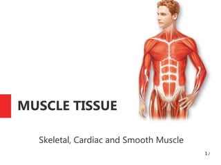
2. Muscle Tissue: General Protperties
One of 4 basic tissue types
along with epithelial,
connective, and nerve tissue
3 types of muscle:
Skeletal: mostly attached to
skeleton; vouluntary
Cardiac: in heart; involuntary
Smooth: in visceral organs;
involuntary
Made up specialised
elongated cells called
myofibres or muscle
fibres; capable of
contraction
Contraction results from
conversion of chemical
energy in ATP into
mechanical energy
2 /
3. Skeletal Muscle: Striated and Voluntary
Myofibres form long
multinucleated syncytium,
grouped in bundles
ensheathed in connective
tissue membranes
Extend from site of origin to
their insertion
Connective tissue sheaths
continuous with tendons;at
myotendinous junction
Connective tissue sheaths:
Epimysium: dense
connective tissue; entire
muscle
Perimysium: muscle
bundles (fascicles)
Endomysium: delicate
reticular fibres and matrix;
individual fibres
6. Skeletal Myofibres:
Characteristics
Formed from fusion of
embryonic myoblasts into
multinucleated myotube
Myotube matures into a
long muscle fibre (diameter,
10-100 µm; length, several
cm.)
Cell membrane called
sarcolemma surrounded by
basement membrane and
satellite cells
Sarcolemma forms tubular
invaginations into cell
cytoplasm (sarcoplasm);
transverse (T) tubules
T tubules associated with
sarcoplasmic reticulm
(SR); an intracellular Ca2+
store
Muscle action potential
spreads deep into cell along
T tubules, and cause
release of Ca2+ from SR
7. Skeletal Myofibres:
Characteristics(2)
80% sarcoplasm occupied
by myofibrils surrounded by
mitochondria; sarcosomes
Myofibrils contain thick and
thin myofilaments;
contractile proteins
Thick filaments (15 nm
width) contain myosin
Thin filaments (7 nm width)
contain actin
Thin filaments insert into Z-
disk and alternate with
thick filaments
Sarcomere: alternating
thick and thin filaments is
basic contractile unit of
striated muscle
Sarcomere extends
between 2 successive Z
disks
10. Skeletal Myofibres:
Characteristics(3)
Banding pattern observed
under microscope in cardiac
and skeletal muscle due to
alternating arrangement of
thin and thick filaments
(striated)
A (dark) band: region of
sarcomere with thick
filaments
I (light) band: region with
thin filaments
H band (zone): region of A
band with thick filaments
only; contains enzyme
creatine kinase
M line: line in H zone due to
bridges and filaments linking
thick filaments
Z disk: borders sarcomeres
and provide attachment for
filaments
11.
12. Banding in
Striated
Muscle
Z disc interval is
sarcomere
I band: thin
filaments
A band: thin and
thick filaments
H zone: area of
thick filaments
only
13. Composition of Thin and Thick
Myofilaments
Thin filaments:
(1) F-actin: double-
stranded actin
twisted in a spiral;
inserts into Z disk
(2) Tropomyosin:
lies in a groove
formed by actin helix
(3) Troponin:
complex of 3
proteins; T
(tropomyosin), I
(actin) and C
(Ca2+).
14.
15. Composition of Thin and Thick
Myofilaments(2)
Thick filaments:
Main component is
myosin: 4 light chains
and 2 heavy chains
Myosin is a motor
protein; uses ATPase
activity to generate
movement
Binds to F-actin in a
reversible manner
Attached to Z disks by
protein titin
16. Composition of Thin and Thick
Myofilaments(3)
Other molecules
present in
muscle:
Nebulin:
associated with
actin; stabilises
Desmin: links
and stabilises
myofibrils and
links them to
sarcolema via
interaction with
dystrophin-
associated
complex
17. Neuromuscular Junction (NMJ)
Junction b/n muscle
fibre and motor nerve
Made up of membrane
of muscle, membrane
of synaptic bouton
and nanometer cleft
Chemical released at
NMJ: acetylcholine
ACh
Motor unit: a motor
nerve and all muscle
fibre innervated
19. Mechanism of Muscle
Contraction
3 important facts about contraction:
1. Length of myofilaments does not change (e.g. A band length
does not change)
2. Muscle shortening occurs due to sliding of thin filaments
over thick filaments (I & H band width narrow): sliding
filament theory
3. Force of contraction is generated by ATP-powered
conformational changes of myosin proteins
Maintenance of steady ATP levels provided by creatine
phosphate
21. Contraction Mechanism
Excitation of muscle fibre at NMJ releases acetylcholine
(ACh)
ACh induces muscle action potential, which spreads along
sarcolemma and T tubules
Muscle action potential causes release of Ca2+ from SR
Ca2+ binds troponin, enabling displacement of tropomyosin
and exposure of myosin binding sites on actin
Myosin binds and undergoes changes that generates the
force for filament sliding
24. Cardiac Muscle (Cardiocytes)
Branched; 85-100 µm long, 15 µm wide
Single, centrally-located nucleus
Larger T tubules than in skeletal myocytes
SR not as extensive as skeletal myocytes
More abundant mitochondria
Cardiocytes joined end-to-end by junctional complexes;
intercalated disks
Intercalated disks contain gap junctions: enable signals to
spread through out large regions to contract simultaneously
27. Smooth Muscle
Found in viscera: wall of gut, bile ducts, ureters, bladder,
respiratory tract, uterus, blood vessels
Cells are spindle-shaped, tapering with central nucleus
Myofilaments not organised into sarcomeres; no striations
Have caveolae in place of T tubules, transmit signals to a
small SR
Linked to one another by gap junctions, enabling
synchronous contraction
Lack troponin, dependent on another Ca2+ sensor,
calmodulin