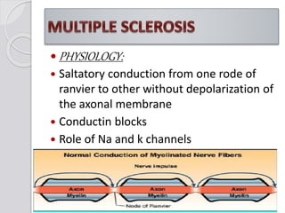
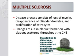
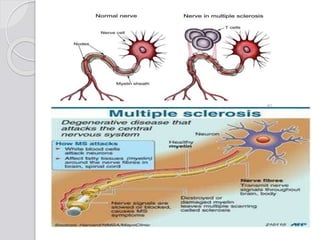
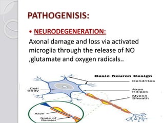
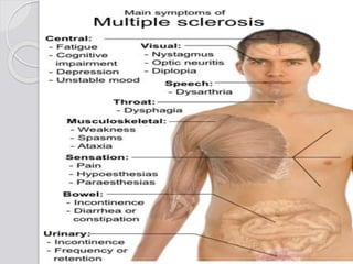
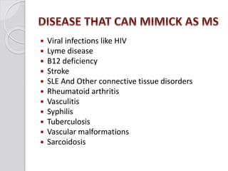
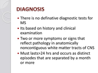
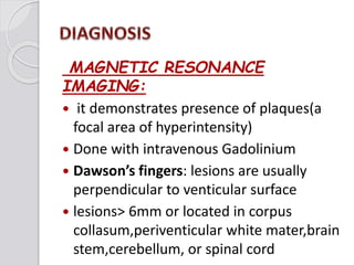
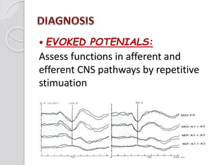
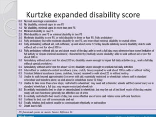
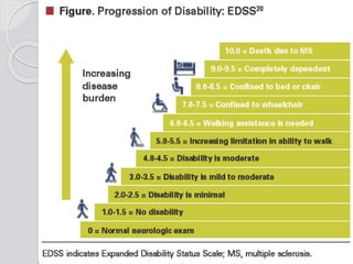
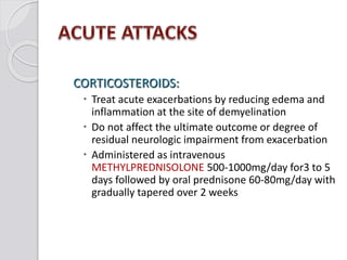
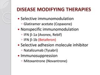
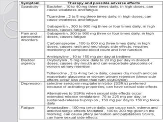
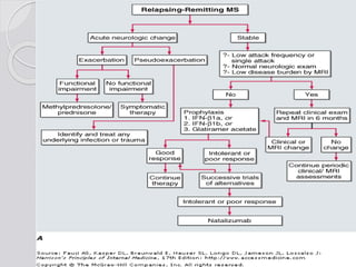
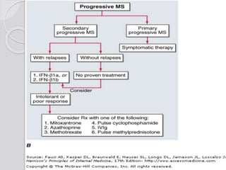
Dr Waqar Ali
Mayo Hospital Lahore
 Chronic, progressive, degenerative
disorder of the CENTRAL NERVOUS
SYSTEM characterized by
selective destruction of central nervous
system Myelin..
 The PERIPHERAL NERVOUS SYSTEM is
spared..
 It is chacterized by triad of
1. Inflammation
2. Demyelination
3. Gliosis(scarring)
Lesions of MS occurs at DIFFERENT TIMES
and in DIFFERENT LOCATIONS in CNS..
MEANS DISSEMINATED IN TIME AND
SPACE,
Manifestations of it vary from a benign
illness to a rapidly evolving and
incapacitating lifestyle adjustments
 PHYSIOLOGY:
 Saltatory conduction from one rode of
ranvier to other without depolarization of
the axonal membrane
 Conductin blocks
 Role of Na and k channels
 Disease process consists of loss of myelin,
disappearance of oligodendrocytes, and
proliferation of astrocytes
 Changes result in plaque formation with
plaques scattered throughout the CNS
 Perivenular cuffing with
 Inflammatory mononuclear cells
 Predominantly T cells and macrophages
 BBB is disrupted
 Myelin specific Autoantibodies
 Heterogenicity
 Shadow plaques
 Relative sparing of axons is typical
 Immunology:
 AUTOREACTIVE T LYMPHOCYTES:
Myelin basic protein(MBP) - a T cell antigen
 HUMORAL IMMUNITY:
Autoantibodies
oligoclonal antibodies
 CYTOKINES
IL 2,TNF alpha and gamma
Triggers:
Upper respiratory tract infections
 NEURODEGENERATION:
Axonal damage and loss via activated
microglia through the release of NO
,glutamate and oxygen radicals..
 EPIDEMOLOGY:
 Usually affects young to middle- aged
adults, with onset between 20 and 40
years of age
 Women affected three times more than
men
 2.5 million population is affected
worldwide
EPIDEMOLOGY:
Geographical distribution
 Unknown cause
 Related to infectious, immunologic, and
genetic factors
 Possible precipitating factors include
Infection like EBV
Physical injury
Emotional stress
Excessive fatigue
Pregnancy
Poor state of health
 GENETIC COSIDERATION:
 CAUSCASIONS more prone as compared
to asians or africans
 POLYGENIC
 MHC on chromosome 6 specifically DR2
• Relapsing-remitting MS (RRMS)
–Affects 85% of newly diagnosed
–Characterized by Discrete attacks
–Attacks followed by partial or complete
recovery
–Between attacks patients are
neurologically stable
• Secondary-progressive MS (SPMS)
 Always begins as RRMS
Occasional relapses but symptoms remain
constant, no remission
Progressive disability late in disease course
Steady deterioration in functions unassociated
with acute attacks
Seems to be a late stage of RRMS
Primary-progressive MS (PPMS)
◦ Affects approximately 15% of MS
population
◦ Slow onset but steady functional
deterioration from disease onset
◦ These usually do not experience attacks
 Progressive-relapsing MS (PRMS)
◦ Rarest form
◦ Affects approx. 5%
◦ Steady worsening of condition
at onset
 Abrupt or insidious
 Severe or trivial
 Depends upon location and severity
initial symptoms of MS
symptom Percent of cases symptom Percent of cases
Sensory loss 37 Lhermitte 3
Optic neuritis 36 Pain 3
Weakness 35 Dementia 2
Paresthesias 24 Visual loss 2
Diplopia 15 Facial palsy 1
Ataxia 11 Impotence 1
Vertigo 06 Myokymia 1
Paroxysmal
attacks
04 Epilepsy 1
Bladder 04 Falling 1
 Motor manifestations
◦ Weakness or paralysis of limbs, trunk, and head
are of upper motor neuron type
◦ Diplopia (double vision)
internuclear ophthalmoplagia
horizontal gaze palsy
Nystagmus
◦ Scanning speech
◦ Spasticity of muscles
 Sensory manifestations
◦ Numbness and tingling, formication “pins and
needles” or unpleasant sensations or a dead
feeling..
◦ Optic neuritis
◦ Blurred vision
◦ Vertigo and tinnitus
◦ Decreased hearing
◦ Chronic neuropathic pain
 Cerebellar manifestations
◦ Nystagmus
 Involuntary eye movements
◦ Ataxia
◦ Dysarthria(scanning speech):
 Lack of coordination in articulating
 speech
◦ Dysphagia
 Difficulty swallowing
 Emotional manifestations
◦ Anger
◦ Depression
◦ Euphoria
 Bladder dysfunction
detrusor hyper reflexia and
detrusor sphincter dyssynergia
 Constipation
 Sexual dysfunction
 Fatigue
 Facial weakness
 Vertigo
 Uhthoff’s symptoms:
unilateral visual blurring during a hot shower
or physical exercise
 Lhermitte’s symptoms:
electrical shock like sensations by flexion of
neck
 Paroxysmal symptoms:
Brief duration,high frequency,lack of ASOC,self
limited course
 Trigeminal neuralgia
 Facial myokymia
 Viral infections like HIV
 Lyme disease
 B12 deficiency
 Stroke
 SLE And Other connective tissue disorders
 Rheumatoid arthritis
 Vasculitis
 Syphilis
 Tuberculosis
 Vascular malformations
 Sarcoidosis
 There is no definative diagnostic tests for
MS
 Its based on history and clinical
examination
 Two or more symptoms or signs that
reflect pathology in anatomically
noncontiguous white matter tracts of CNS
 Must lasts>24 hrs and occurs as distinct
episodes that are separated by a month
or more
MAGNETIC RESONANCE
IMAGING:
 it demonstrates presence of plaques(a
focal area of hyperintensity)
 Done with intravenous Gadolinium
 Dawson’s fingers: lesions are usually
perpendicular to venticular surface
 lesions> 6mm or located in corpus
collasum,periventicular white mater,brain
stem,cerebellum, or spinal cord
 EVOKED POTENIALS:
Assess functions in afferent and
efferent CNS pathways by repetitive
stimuation
 CEREBROSPINAL FLUID:
increased levels of intrathecally
synthesized IgG
Total CSF protiens may be normal
IgG index: ratio of IgG to albumin in CSF to
same in serum
OLIGO CLONAL BANDING:
 Other tests
Like serum B12 levels,
ANA,ESR,trepoemal antibodies
 GOOD PROGNOSIS:
Optic neuritis or sensory symptoms at
onset
Fewer than 2 relapses in first year
Minimal impairement after 5 years
 Bad prognosis:
Truncal ataxia
Action tremors
Pyramidal symptoms
Progressive disease course
Kurtzke expanded disability score
1. Treatments of acute attacks as they
occur
2. Treatment with disease modifying
agents
3. Symptomatic therapy
 Acute attacks:
First think is it acute attack or
PSEUDOEXACERBATION?
Pseudoexacerbation are usually not treated
CORTICOSTEROIDS:
 Treat acute exacerbations by reducing edema and
inflammation at the site of demyelination
 Do not affect the ultimate outcome or degree of
residual neurologic impairment from exacerbation
 Administered as intravenous
METHYLPREDNISOLONE 500-1000mg/day for3 to 5
days followed by oral prednisone 60-80mg/day with
gradually tapered over 2 weeks
 PLASMA EXCHANGE:
seven exchanges 40-60ml/kg per exchange
every other day for 14 days
But evidence of efficacy is only preliminary
 Selective immunomodulation
◦ Glatiramer acetate (Copaxone)
 Nonspecific immunomodulation
◦ IFN b-1a (Avonex, Rebif)
◦ IFN b-1b (Betaferon)
 Selective adhesion molecule inhibitor
◦ Natalizumab (Tysabri)
 Immunosuppression
◦ Mitoxantrone (Novantrone)
 Others:
 Methotrexate
 Cyclophosphamide
 Intravenous immunoglobulins
 IFN b: (Avonex, Rebif,Betaseron)
 Induces an antiproliferative effect
 Blocks T cell activation
 Induces apoptosis of autoreactive T cells
 IFN- antagonistic
 Has antiviral effect
 Acts in periphery (ie, does not cross BBB)
 Indirect effects on CNS
 IFN b:
Reduces attack rates
Improves disease severity measures like
EDSS
Can be use in RRMS ,SPMS with relapses
INF b 1a 30ug by intramuscular injections
once every week
INF b 1b 250ug by subcutaneous
injections every other day
 :GLATIRAMER ACETATE (Copaxone)
 Blocks autoimmune T cells
 Binding to MHC molecule so displace MBP
 Induces anti-inflammatory TH2 cells
 Upregulates neuronal preservation
 Augmentation of processes of neurogenesis:
cell proliferation, migration, differentiation
 GLATIRAMER ACETATE:
 Reduce attack rates
 Used in only RRMS not in progressive
 20mg every day by subcutaneous
injections
 NATALIZUMAB: (Tysabri)
 Primary mechanism related to blockade
of
interaction between the a4b1-integrin
which is expressed on T lymphocytes
brain receptors so prevent there entry
through BBB..
 NATALIZUMAB:
Reduces attack rates
Significantly improves all measures of
disease severity
300ug by intravenous infusions every
month
 Mitoxantrone (Novantrone) :
◦ Antineoplastic drug
◦ Acting by interlacing into DNA and producing
strand breaks,iterfering with RNA synthesis
and inhibiting topoisomeras..
◦ Indicated in RRMS,SPMS and in worsening
RRMS
◦ 12mg/m2
 Side effects of disease modyfying agents:
 Local injection site irritation/reactions
 Flu like symptoms
 Rise in liver enzymes
 Decreased white cell count and platelets
 Opportunistic infections
 Depression
 Progressive multifocal leukoencephalopathy
(PML)
 Cardiotoxicity with mitoxantrone
Multiple sclerosis
Multiple sclerosis
Multiple sclerosis
Multiple sclerosis

Multiple sclerosis

  • 1.
    Dr Waqar Ali MayoHospital Lahore
  • 2.
     Chronic, progressive,degenerative disorder of the CENTRAL NERVOUS SYSTEM characterized by selective destruction of central nervous system Myelin..  The PERIPHERAL NERVOUS SYSTEM is spared..
  • 3.
     It ischacterized by triad of 1. Inflammation 2. Demyelination 3. Gliosis(scarring)
  • 4.
    Lesions of MSoccurs at DIFFERENT TIMES and in DIFFERENT LOCATIONS in CNS.. MEANS DISSEMINATED IN TIME AND SPACE, Manifestations of it vary from a benign illness to a rapidly evolving and incapacitating lifestyle adjustments
  • 5.
     PHYSIOLOGY:  Saltatoryconduction from one rode of ranvier to other without depolarization of the axonal membrane  Conductin blocks  Role of Na and k channels
  • 7.
     Disease processconsists of loss of myelin, disappearance of oligodendrocytes, and proliferation of astrocytes  Changes result in plaque formation with plaques scattered throughout the CNS
  • 8.
     Perivenular cuffingwith  Inflammatory mononuclear cells  Predominantly T cells and macrophages  BBB is disrupted  Myelin specific Autoantibodies  Heterogenicity  Shadow plaques  Relative sparing of axons is typical
  • 10.
     Immunology:  AUTOREACTIVET LYMPHOCYTES: Myelin basic protein(MBP) - a T cell antigen  HUMORAL IMMUNITY: Autoantibodies oligoclonal antibodies  CYTOKINES IL 2,TNF alpha and gamma Triggers: Upper respiratory tract infections
  • 11.
     NEURODEGENERATION: Axonal damageand loss via activated microglia through the release of NO ,glutamate and oxygen radicals..
  • 12.
     EPIDEMOLOGY:  Usuallyaffects young to middle- aged adults, with onset between 20 and 40 years of age  Women affected three times more than men  2.5 million population is affected worldwide
  • 13.
  • 14.
     Unknown cause Related to infectious, immunologic, and genetic factors
  • 15.
     Possible precipitatingfactors include Infection like EBV Physical injury Emotional stress Excessive fatigue Pregnancy Poor state of health
  • 16.
     GENETIC COSIDERATION: CAUSCASIONS more prone as compared to asians or africans  POLYGENIC  MHC on chromosome 6 specifically DR2
  • 17.
    • Relapsing-remitting MS(RRMS) –Affects 85% of newly diagnosed –Characterized by Discrete attacks –Attacks followed by partial or complete recovery –Between attacks patients are neurologically stable
  • 18.
    • Secondary-progressive MS(SPMS)  Always begins as RRMS Occasional relapses but symptoms remain constant, no remission Progressive disability late in disease course Steady deterioration in functions unassociated with acute attacks Seems to be a late stage of RRMS
  • 19.
    Primary-progressive MS (PPMS) ◦Affects approximately 15% of MS population ◦ Slow onset but steady functional deterioration from disease onset ◦ These usually do not experience attacks
  • 20.
     Progressive-relapsing MS(PRMS) ◦ Rarest form ◦ Affects approx. 5% ◦ Steady worsening of condition at onset
  • 23.
     Abrupt orinsidious  Severe or trivial  Depends upon location and severity
  • 24.
    initial symptoms ofMS symptom Percent of cases symptom Percent of cases Sensory loss 37 Lhermitte 3 Optic neuritis 36 Pain 3 Weakness 35 Dementia 2 Paresthesias 24 Visual loss 2 Diplopia 15 Facial palsy 1 Ataxia 11 Impotence 1 Vertigo 06 Myokymia 1 Paroxysmal attacks 04 Epilepsy 1 Bladder 04 Falling 1
  • 25.
     Motor manifestations ◦Weakness or paralysis of limbs, trunk, and head are of upper motor neuron type ◦ Diplopia (double vision) internuclear ophthalmoplagia horizontal gaze palsy Nystagmus ◦ Scanning speech ◦ Spasticity of muscles
  • 27.
     Sensory manifestations ◦Numbness and tingling, formication “pins and needles” or unpleasant sensations or a dead feeling.. ◦ Optic neuritis ◦ Blurred vision ◦ Vertigo and tinnitus ◦ Decreased hearing ◦ Chronic neuropathic pain
  • 28.
     Cerebellar manifestations ◦Nystagmus  Involuntary eye movements ◦ Ataxia ◦ Dysarthria(scanning speech):  Lack of coordination in articulating  speech ◦ Dysphagia  Difficulty swallowing
  • 29.
     Emotional manifestations ◦Anger ◦ Depression ◦ Euphoria
  • 30.
     Bladder dysfunction detrusorhyper reflexia and detrusor sphincter dyssynergia  Constipation  Sexual dysfunction  Fatigue  Facial weakness  Vertigo
  • 31.
     Uhthoff’s symptoms: unilateralvisual blurring during a hot shower or physical exercise  Lhermitte’s symptoms: electrical shock like sensations by flexion of neck  Paroxysmal symptoms: Brief duration,high frequency,lack of ASOC,self limited course  Trigeminal neuralgia  Facial myokymia
  • 33.
     Viral infectionslike HIV  Lyme disease  B12 deficiency  Stroke  SLE And Other connective tissue disorders  Rheumatoid arthritis  Vasculitis  Syphilis  Tuberculosis  Vascular malformations  Sarcoidosis
  • 34.
     There isno definative diagnostic tests for MS  Its based on history and clinical examination  Two or more symptoms or signs that reflect pathology in anatomically noncontiguous white matter tracts of CNS  Must lasts>24 hrs and occurs as distinct episodes that are separated by a month or more
  • 35.
    MAGNETIC RESONANCE IMAGING:  itdemonstrates presence of plaques(a focal area of hyperintensity)  Done with intravenous Gadolinium  Dawson’s fingers: lesions are usually perpendicular to venticular surface  lesions> 6mm or located in corpus collasum,periventicular white mater,brain stem,cerebellum, or spinal cord
  • 39.
     EVOKED POTENIALS: Assessfunctions in afferent and efferent CNS pathways by repetitive stimuation
  • 40.
     CEREBROSPINAL FLUID: increasedlevels of intrathecally synthesized IgG Total CSF protiens may be normal IgG index: ratio of IgG to albumin in CSF to same in serum OLIGO CLONAL BANDING:
  • 41.
     Other tests Likeserum B12 levels, ANA,ESR,trepoemal antibodies
  • 43.
     GOOD PROGNOSIS: Opticneuritis or sensory symptoms at onset Fewer than 2 relapses in first year Minimal impairement after 5 years
  • 44.
     Bad prognosis: Truncalataxia Action tremors Pyramidal symptoms Progressive disease course
  • 45.
  • 47.
    1. Treatments ofacute attacks as they occur 2. Treatment with disease modifying agents 3. Symptomatic therapy
  • 48.
     Acute attacks: Firstthink is it acute attack or PSEUDOEXACERBATION? Pseudoexacerbation are usually not treated
  • 49.
    CORTICOSTEROIDS:  Treat acuteexacerbations by reducing edema and inflammation at the site of demyelination  Do not affect the ultimate outcome or degree of residual neurologic impairment from exacerbation  Administered as intravenous METHYLPREDNISOLONE 500-1000mg/day for3 to 5 days followed by oral prednisone 60-80mg/day with gradually tapered over 2 weeks
  • 50.
     PLASMA EXCHANGE: sevenexchanges 40-60ml/kg per exchange every other day for 14 days But evidence of efficacy is only preliminary
  • 51.
     Selective immunomodulation ◦Glatiramer acetate (Copaxone)  Nonspecific immunomodulation ◦ IFN b-1a (Avonex, Rebif) ◦ IFN b-1b (Betaferon)  Selective adhesion molecule inhibitor ◦ Natalizumab (Tysabri)  Immunosuppression ◦ Mitoxantrone (Novantrone)
  • 52.
     Others:  Methotrexate Cyclophosphamide  Intravenous immunoglobulins
  • 53.
     IFN b:(Avonex, Rebif,Betaseron)  Induces an antiproliferative effect  Blocks T cell activation  Induces apoptosis of autoreactive T cells  IFN- antagonistic  Has antiviral effect  Acts in periphery (ie, does not cross BBB)  Indirect effects on CNS
  • 54.
     IFN b: Reducesattack rates Improves disease severity measures like EDSS Can be use in RRMS ,SPMS with relapses INF b 1a 30ug by intramuscular injections once every week INF b 1b 250ug by subcutaneous injections every other day
  • 55.
     :GLATIRAMER ACETATE(Copaxone)  Blocks autoimmune T cells  Binding to MHC molecule so displace MBP  Induces anti-inflammatory TH2 cells  Upregulates neuronal preservation  Augmentation of processes of neurogenesis: cell proliferation, migration, differentiation
  • 56.
     GLATIRAMER ACETATE: Reduce attack rates  Used in only RRMS not in progressive  20mg every day by subcutaneous injections
  • 57.
     NATALIZUMAB: (Tysabri) Primary mechanism related to blockade of interaction between the a4b1-integrin which is expressed on T lymphocytes brain receptors so prevent there entry through BBB..
  • 58.
     NATALIZUMAB: Reduces attackrates Significantly improves all measures of disease severity 300ug by intravenous infusions every month
  • 59.
     Mitoxantrone (Novantrone): ◦ Antineoplastic drug ◦ Acting by interlacing into DNA and producing strand breaks,iterfering with RNA synthesis and inhibiting topoisomeras.. ◦ Indicated in RRMS,SPMS and in worsening RRMS ◦ 12mg/m2
  • 61.
     Side effectsof disease modyfying agents:  Local injection site irritation/reactions  Flu like symptoms  Rise in liver enzymes  Decreased white cell count and platelets  Opportunistic infections  Depression  Progressive multifocal leukoencephalopathy (PML)  Cardiotoxicity with mitoxantrone