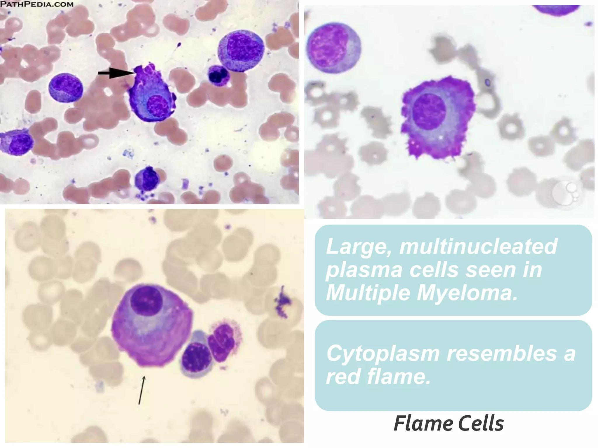
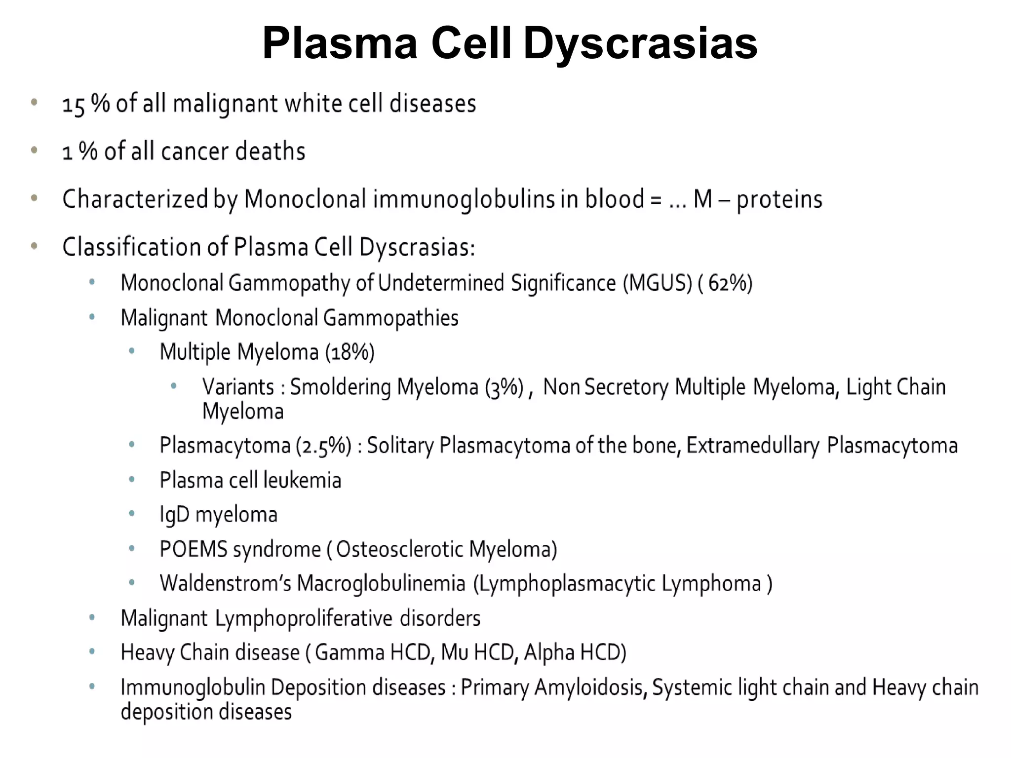
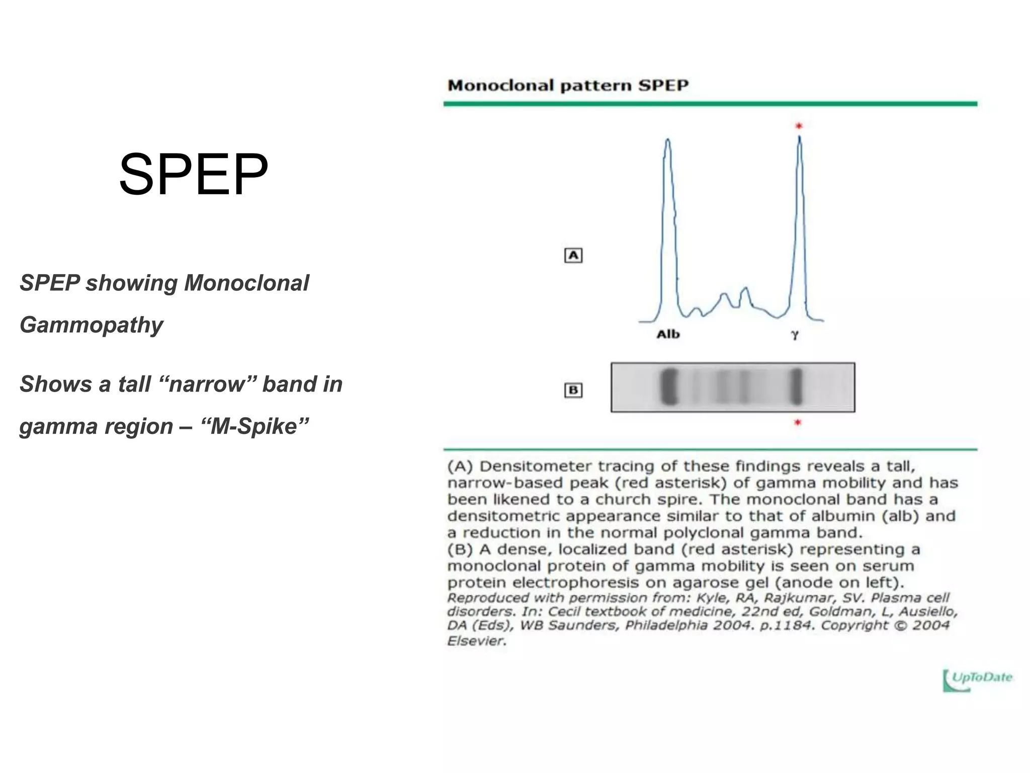
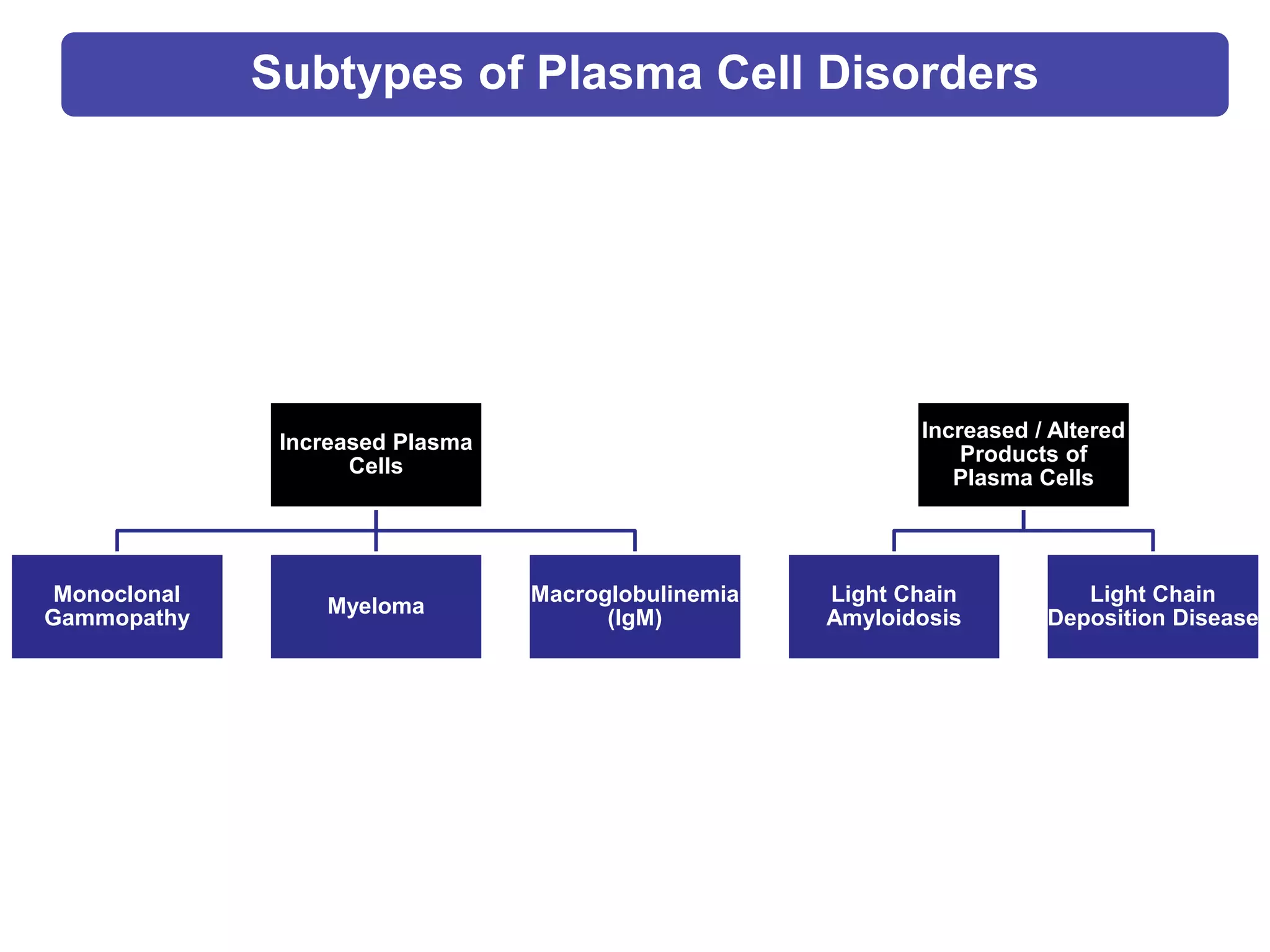
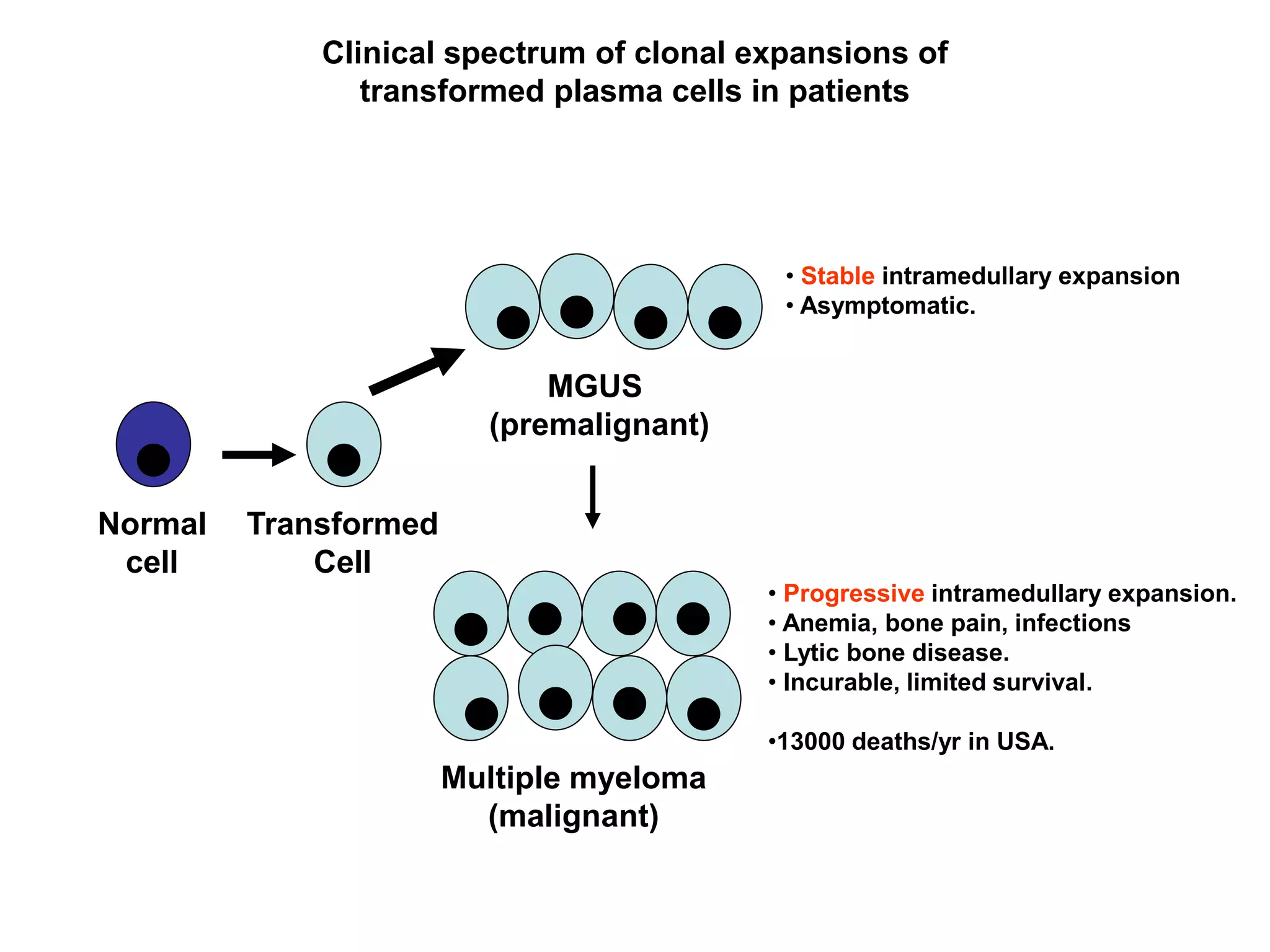
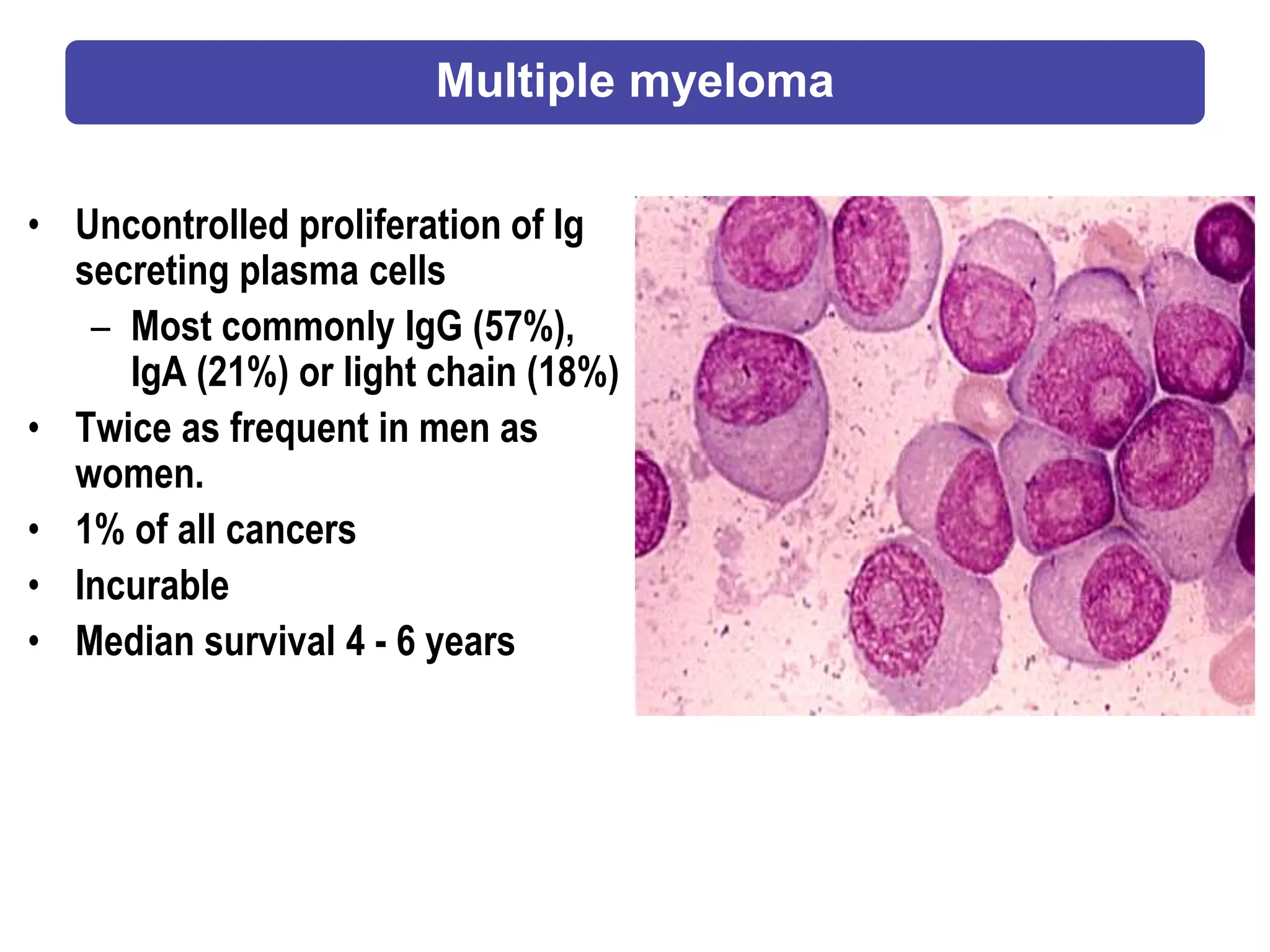
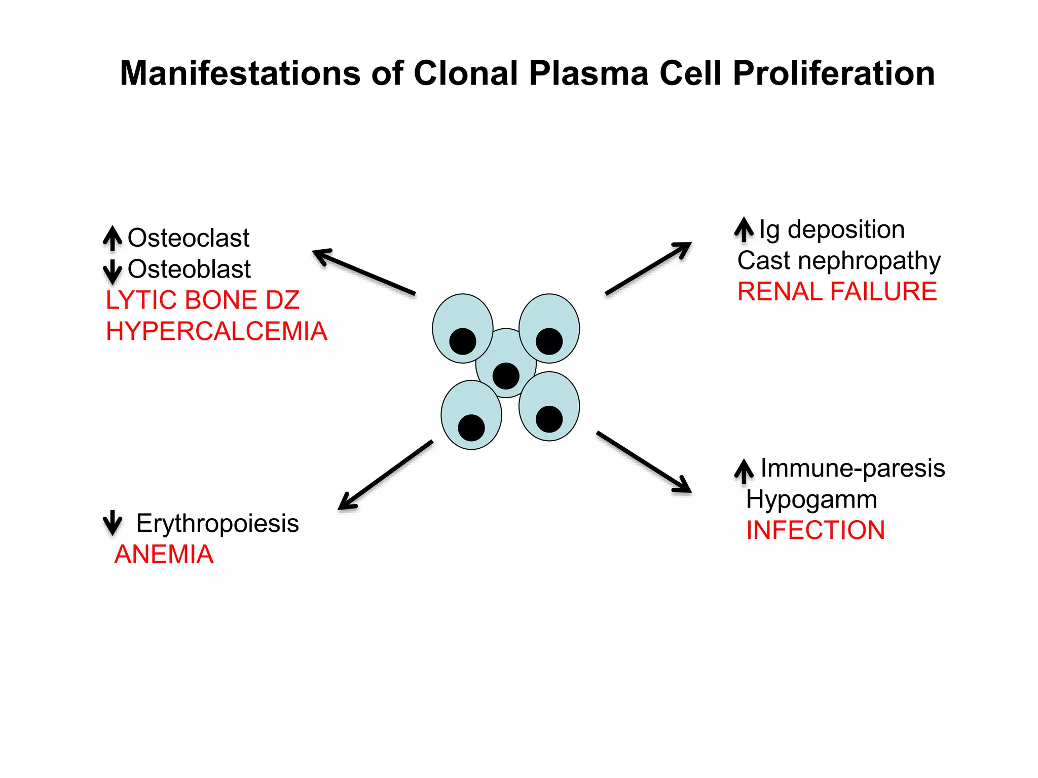
Plasma Cell Disorders
Dr. Indranil Bhattacharya
MD & WHO Fellow (Pathology)
HOD – Pathology & Laboratory Medicine
Jagjivan Ram Hospital
Mumbai
Definition:
Group of lymphoid neoplasm of
terminally differentiated B - cells that
have in common the expansion of a
single clone of immunoglobulin (Ig) -
secreting plasma cells and a resultant
increase in serum levels of a single
homogeneous (Monoclonal) Ig or it’s
fragments.
Plasma Cell Dyscrasias
Plasma Cell
• Terminally differentiated B-cells.
• Not normally found in peripheral blood.
• Less than 3.5% of nucleated cells in the bone
marrow.
• Oval cells with low N:C ratio. Cytoplasm is basophilic
blue. Nucleus (30-40% of the cell) is oval or round and
placed eccentrically of the cell.
• A clear, colorless area adjacent to the nucleus contains
Golgi apparatus.
• Russell bodies: Globules (2-3 μm) of accumulated
immunoglobulin in the cytoplasm of plasma cells.
Mott cells
• Plasma cells crowded with
Russell bodies.
• An obstruction blocks the
release of Golgi secretions.
• Can be found in case of
chronic plasmacytosis.
Flame Cells
Large, multinucleated
plasma cells seen in
Multiple Myeloma.
Cytoplasm resembles a
red flame.
Plasma Cell Dyscrasias: Synonyms
Gammopathy
Monoclonal Gammopathy
Dysproteinemia
Paraproteinemia
Plasma Cell Dyscrasias
Serum Protein Electrophoresis
• Serum is placed on special paper
treated with agarose gel and
exposed to an electric current. This
separates the serum protein
components into five classifications
by size and electrical charge: serum
albumin, alpha-1 globulins, alpha-2
globulins, beta globulins, and
gamma globulins.
• Immunoglobulins (IgG, IgM, IgA)
usually migrate to gamma region, may
sometimes extend to beta region.
• SPEP should always be performed in
combination with serum
immunofixation in order to determine
clonality
SPEP
SPEP showing Monoclonal
Gammopathy
Shows a tall “narrow” band in
gamma region – “M-Spike”
SPEP
• SPEP showing Polyclonal
Gammopathy
• Shows a broad based peak in
gamma region.
• Seen in chronic infections,
inflammation, connective tissue
disease, lymphoproliferative disease.
Immunofixation
• More sensitive than SPEP
• Immunofixation is performed when SPEP
shows a sharp “peak” or a plasma cell
disorder is suspected despite a normal
SPEP
• Immunofixation always done to confirm the
presence of M-Protein and to determine the
type (IgM or IgG etc and the light chain
restriction : k or λ)
• Why do both SPEP and IF ? Why not just IF
in initial diagnosis ?
• Unlike SPEP, immunofixation does not give
an estimate of the size of the M protein (ie,
its serum concentration), and thus should
be done in conjunction with
electrophoresis.
Subtypes of Plasma Cell Disorders
Increased Plasma
Cells
Monoclonal
Gammopathy
Myeloma
Macroglobulinemia
(IgM)
Increased / Altered
Products of
Plasma Cells
Light Chain
Amyloidosis
Light Chain
Deposition Disease
Case Presentation
• 78 year male
• Admitted with 2 wk history of fatigue and diffuse
bone pain.
• Evaluation:
– Increased serum creatinine 5.5 mg/dl (normal < 1.2 mg/dl)
– Increased serum calcium 12 mg/dl (normal < 10.5 mg/dl)
– Increased serum globulin 5.0 g/dl (normal < 3 g/dl)
Serum/ Urine Protein
Electrophoresis (S/UPEP)
Immunofixation
Electrophoresis (IFE)
Evaluation of Abnormal Serum Globulins
Serum
Urine
Serum M spike 4 g/dl; typed as IgG kappa monoclonal Ig
Bone Marrow Biopsy – Increased Plasma Cells
Skeletal survey — Lytic bone disease
Normal
cell
Transformed
Cell
MGUS
(premalignant)
Multiple myeloma
(malignant)
Clinical spectrum of clonal expansions of
transformed plasma cells in patients
• Stable intramedullary expansion
• Asymptomatic.
• Progressive intramedullary expansion.
• Anemia, bone pain, infections
• Lytic bone disease.
• Incurable, limited survival.
•13000 deaths/yr in USA.
Diagnostic Criteria in Monoclonal Gammopathies
MGUS
• < 10% bone marrow plasma
cells and M spike < 3 g/dl
• Monoclonal protein / clonal
plasma cell population
• No End organ damage
Myeloma
• > 10% marrow plasma cells
• End Organ Damage
Indolent / Smoldering
Myeloma
• > 10% marrow plasma cells
or M spike > 3 g/dl
• No End organ damage
Criteria for End-Organ Damage
in Monoclonal Gammopathies
• Calcium > 10 mg/dl above
ULN
• Renal Insufficiency (> 2 mg/dl)
• Anemia (< 10 g/dl)
• Bone Lesions (Lytic lesions or
Osteopenia)
CRAB
A Model for Pathogenesis of Myeloma
Monoclonal Gammopathy of Undetermined
Significance (MGUS)
• Common, age-related
• Prevalence: 3.2% in persons over 50 yrs old.
– ~5% in age >70
• Higher prevalence in African populations.
• ? Association with inflammatory states: Obesity, Gaucher’s disease
• Increased risk for thrombosis and fractures.
• Risk of progression in entire population: 1% /yr
• Risk factors for progression: %Plasma Cell, level M spike, Free light
chain, IgA protein.
Smoldering Myeloma (SMM)
• Patients with Plasma Cell > 10% or M spike > 3 g/dl, but lacking
CRAB symptoms.
• 10% per year progression to overt MM
• Most eventually require therapy.
• Current recommendation is observation until progressive disease.
Kyle et al. NEJM 356: 2582, 2007
Disease Progression in MGUS and SMM
Multiple myeloma
• Uncontrolled proliferation of Ig
secreting plasma cells
– Most commonly IgG (57%),
IgA (21%) or light chain (18%)
• Twice as frequent in men as
women.
• 1% of all cancers
• Incurable
• Median survival 4 - 6 years
Work-up in suspected myeloma
• Assessment of serum/urine protein
– SPEP/IF, 24 hr urine for UPEP/IF
– Free light chains (kappa, lambda)
• CBC, Creatinine, Calcium, Albumin, LDH,
• Serum beta 2 microglobulin (B2M)
• Skeletal survey
• Bone marrow aspirate and biopsy
– Cytogenetics (including FISH)
• Under investigation:
– MRI spine
– PET scans
– Bone densitometry, Urine n-telopeptide
Key clinical aspects of Myeloma
• Predominantly intra-medullary growth.
• Absence of clinical LN or spleen involvement.
• Low proliferative fraction.
• Long periods of stability in MGUS.
• Osteoclast activation, osteoblast inhibition, and bone
loss.
• Multi-focal growth of tumor cells.
Clinical presentation
Osteoclast
Osteoblast
LYTIC BONE DZ
HYPERCALCEMIA
Erythropoiesis
ANEMIA
Ig deposition
Cast nephropathy
RENAL FAILURE
Immune-paresis
Hypogamm
INFECTION
Manifestations of Clonal Plasma Cell Proliferation
Historical aside…
At age 39, developed fatigue and bone pain from several fractures. She died 4 years
later; autopsy showed that her marrow was replaced by a red, gelatinous substance
Multiple Myeloma: Skeletal Complications
Renal Pathology in MM
Light Chain Deposition Disease
Light Chain Cast Nephropathy AL Amyloid
Principles of Treatment
No evidence (yet)
that early treatment
prolongs survival
Wait for symptoms,
or evidence of
disease
progression, to
start treatment
• Drink plenty of fluids daily
• Treat infections promptly
• Prophylactic bisphosphonates reduce skeletal complications
in patients with osteopenia and lytic bone disease
• Anemia often responds to erythropoietin.
Supportive
measures are
critically important
“Myeloma treatment is a marathon, not a sprint”
Major drugs in myeloma
• Alkylators - 1962
– Melphalan, cyclophosphamide
– High dose melphalan and ASCT
• Glucocorticoids - 1966
– Prednisone, dexamethasone
• IMiDs - 1999
– Thalidomide
– Revlimid
– Pomalidomide
• Proteasome Inhibitors - 2001
– Bortezomib
– Carfilzomib
Treatment course
Asymptomatic
MGUS
Stable MM
Symptomatic Acute
Pancytopenia
Plasma cell leukemia
Years Months Days
Treatments
M protein
Factors Associated with Increased Disease Risk in MM
• Gene expression profile (GEP) 70 (or GEP15) high risk signature
• FISH:
– t(4:14); t(14:16)
– Del 17p
– 1q amp; hypodiploidy
• Abnormal cytogenetics by metaphase, including del chr 13
• ISS Stage 3 (increased beta 2 m)
• High LDH
• > 10 focal lesions on MR
Mayo Clin Proc, Apr 2013
The future…
JCO, 30(4), Feb 2012
Waldenström Macroglobulinemia
• Uncontrolled proliferation of lymphoplasmacytes
producing IgM
• Median age 63 years
• Presents with weakness, fatigue, epistaxis, blurred vision
• Bone pain and lytic bone lesions are uncommon (<5%)
• 25% have hepatomegaly, splenomegaly and
lymphadenopathy
• Hyperviscosity is common
Conclusion
Plasma cell dyscrasias are a heterogeneous
group of disorders.
Clinical presentation may be due to the
clone itself or the properties of the secreted
Ig.
Therapy largely directed (if indicated) at
reducing the underlying clone.
Plasma Cell Disorders

Plasma Cell Disorders

  • 1.
    Plasma Cell Disorders Dr.Indranil Bhattacharya MD & WHO Fellow (Pathology) HOD – Pathology & Laboratory Medicine Jagjivan Ram Hospital Mumbai
  • 2.
    Definition: Group of lymphoidneoplasm of terminally differentiated B - cells that have in common the expansion of a single clone of immunoglobulin (Ig) - secreting plasma cells and a resultant increase in serum levels of a single homogeneous (Monoclonal) Ig or it’s fragments. Plasma Cell Dyscrasias
  • 3.
    Plasma Cell • Terminallydifferentiated B-cells. • Not normally found in peripheral blood. • Less than 3.5% of nucleated cells in the bone marrow. • Oval cells with low N:C ratio. Cytoplasm is basophilic blue. Nucleus (30-40% of the cell) is oval or round and placed eccentrically of the cell. • A clear, colorless area adjacent to the nucleus contains Golgi apparatus. • Russell bodies: Globules (2-3 μm) of accumulated immunoglobulin in the cytoplasm of plasma cells.
  • 4.
    Mott cells • Plasmacells crowded with Russell bodies. • An obstruction blocks the release of Golgi secretions. • Can be found in case of chronic plasmacytosis.
  • 5.
    Flame Cells Large, multinucleated plasmacells seen in Multiple Myeloma. Cytoplasm resembles a red flame.
  • 6.
    Plasma Cell Dyscrasias:Synonyms Gammopathy Monoclonal Gammopathy Dysproteinemia Paraproteinemia
  • 7.
  • 8.
    Serum Protein Electrophoresis •Serum is placed on special paper treated with agarose gel and exposed to an electric current. This separates the serum protein components into five classifications by size and electrical charge: serum albumin, alpha-1 globulins, alpha-2 globulins, beta globulins, and gamma globulins. • Immunoglobulins (IgG, IgM, IgA) usually migrate to gamma region, may sometimes extend to beta region. • SPEP should always be performed in combination with serum immunofixation in order to determine clonality
  • 9.
    SPEP SPEP showing Monoclonal Gammopathy Showsa tall “narrow” band in gamma region – “M-Spike”
  • 10.
    SPEP • SPEP showingPolyclonal Gammopathy • Shows a broad based peak in gamma region. • Seen in chronic infections, inflammation, connective tissue disease, lymphoproliferative disease.
  • 11.
    Immunofixation • More sensitivethan SPEP • Immunofixation is performed when SPEP shows a sharp “peak” or a plasma cell disorder is suspected despite a normal SPEP • Immunofixation always done to confirm the presence of M-Protein and to determine the type (IgM or IgG etc and the light chain restriction : k or λ) • Why do both SPEP and IF ? Why not just IF in initial diagnosis ? • Unlike SPEP, immunofixation does not give an estimate of the size of the M protein (ie, its serum concentration), and thus should be done in conjunction with electrophoresis.
  • 12.
    Subtypes of PlasmaCell Disorders Increased Plasma Cells Monoclonal Gammopathy Myeloma Macroglobulinemia (IgM) Increased / Altered Products of Plasma Cells Light Chain Amyloidosis Light Chain Deposition Disease
  • 14.
    Case Presentation • 78year male • Admitted with 2 wk history of fatigue and diffuse bone pain. • Evaluation: – Increased serum creatinine 5.5 mg/dl (normal < 1.2 mg/dl) – Increased serum calcium 12 mg/dl (normal < 10.5 mg/dl) – Increased serum globulin 5.0 g/dl (normal < 3 g/dl)
  • 15.
    Serum/ Urine Protein Electrophoresis(S/UPEP) Immunofixation Electrophoresis (IFE) Evaluation of Abnormal Serum Globulins Serum Urine Serum M spike 4 g/dl; typed as IgG kappa monoclonal Ig
  • 16.
    Bone Marrow Biopsy– Increased Plasma Cells
  • 17.
    Skeletal survey —Lytic bone disease
  • 18.
    Normal cell Transformed Cell MGUS (premalignant) Multiple myeloma (malignant) Clinical spectrumof clonal expansions of transformed plasma cells in patients • Stable intramedullary expansion • Asymptomatic. • Progressive intramedullary expansion. • Anemia, bone pain, infections • Lytic bone disease. • Incurable, limited survival. •13000 deaths/yr in USA.
  • 19.
    Diagnostic Criteria inMonoclonal Gammopathies MGUS • < 10% bone marrow plasma cells and M spike < 3 g/dl • Monoclonal protein / clonal plasma cell population • No End organ damage Myeloma • > 10% marrow plasma cells • End Organ Damage Indolent / Smoldering Myeloma • > 10% marrow plasma cells or M spike > 3 g/dl • No End organ damage
  • 20.
    Criteria for End-OrganDamage in Monoclonal Gammopathies • Calcium > 10 mg/dl above ULN • Renal Insufficiency (> 2 mg/dl) • Anemia (< 10 g/dl) • Bone Lesions (Lytic lesions or Osteopenia) CRAB
  • 21.
    A Model forPathogenesis of Myeloma
  • 22.
    Monoclonal Gammopathy ofUndetermined Significance (MGUS) • Common, age-related • Prevalence: 3.2% in persons over 50 yrs old. – ~5% in age >70 • Higher prevalence in African populations. • ? Association with inflammatory states: Obesity, Gaucher’s disease • Increased risk for thrombosis and fractures. • Risk of progression in entire population: 1% /yr • Risk factors for progression: %Plasma Cell, level M spike, Free light chain, IgA protein.
  • 23.
    Smoldering Myeloma (SMM) •Patients with Plasma Cell > 10% or M spike > 3 g/dl, but lacking CRAB symptoms. • 10% per year progression to overt MM • Most eventually require therapy. • Current recommendation is observation until progressive disease.
  • 24.
    Kyle et al.NEJM 356: 2582, 2007 Disease Progression in MGUS and SMM
  • 25.
    Multiple myeloma • Uncontrolledproliferation of Ig secreting plasma cells – Most commonly IgG (57%), IgA (21%) or light chain (18%) • Twice as frequent in men as women. • 1% of all cancers • Incurable • Median survival 4 - 6 years
  • 26.
    Work-up in suspectedmyeloma • Assessment of serum/urine protein – SPEP/IF, 24 hr urine for UPEP/IF – Free light chains (kappa, lambda) • CBC, Creatinine, Calcium, Albumin, LDH, • Serum beta 2 microglobulin (B2M) • Skeletal survey • Bone marrow aspirate and biopsy – Cytogenetics (including FISH) • Under investigation: – MRI spine – PET scans – Bone densitometry, Urine n-telopeptide
  • 27.
    Key clinical aspectsof Myeloma • Predominantly intra-medullary growth. • Absence of clinical LN or spleen involvement. • Low proliferative fraction. • Long periods of stability in MGUS. • Osteoclast activation, osteoblast inhibition, and bone loss. • Multi-focal growth of tumor cells.
  • 28.
  • 29.
    Osteoclast Osteoblast LYTIC BONE DZ HYPERCALCEMIA Erythropoiesis ANEMIA Igdeposition Cast nephropathy RENAL FAILURE Immune-paresis Hypogamm INFECTION Manifestations of Clonal Plasma Cell Proliferation
  • 30.
    Historical aside… At age39, developed fatigue and bone pain from several fractures. She died 4 years later; autopsy showed that her marrow was replaced by a red, gelatinous substance
  • 31.
  • 33.
    Renal Pathology inMM Light Chain Deposition Disease Light Chain Cast Nephropathy AL Amyloid
  • 36.
    Principles of Treatment Noevidence (yet) that early treatment prolongs survival Wait for symptoms, or evidence of disease progression, to start treatment • Drink plenty of fluids daily • Treat infections promptly • Prophylactic bisphosphonates reduce skeletal complications in patients with osteopenia and lytic bone disease • Anemia often responds to erythropoietin. Supportive measures are critically important
  • 37.
    “Myeloma treatment isa marathon, not a sprint”
  • 40.
    Major drugs inmyeloma • Alkylators - 1962 – Melphalan, cyclophosphamide – High dose melphalan and ASCT • Glucocorticoids - 1966 – Prednisone, dexamethasone • IMiDs - 1999 – Thalidomide – Revlimid – Pomalidomide • Proteasome Inhibitors - 2001 – Bortezomib – Carfilzomib
  • 41.
    Treatment course Asymptomatic MGUS Stable MM SymptomaticAcute Pancytopenia Plasma cell leukemia Years Months Days Treatments M protein
  • 42.
    Factors Associated withIncreased Disease Risk in MM • Gene expression profile (GEP) 70 (or GEP15) high risk signature • FISH: – t(4:14); t(14:16) – Del 17p – 1q amp; hypodiploidy • Abnormal cytogenetics by metaphase, including del chr 13 • ISS Stage 3 (increased beta 2 m) • High LDH • > 10 focal lesions on MR
  • 43.
  • 45.
  • 46.
    Waldenström Macroglobulinemia • Uncontrolledproliferation of lymphoplasmacytes producing IgM • Median age 63 years • Presents with weakness, fatigue, epistaxis, blurred vision • Bone pain and lytic bone lesions are uncommon (<5%) • 25% have hepatomegaly, splenomegaly and lymphadenopathy • Hyperviscosity is common
  • 47.
    Conclusion Plasma cell dyscrasiasare a heterogeneous group of disorders. Clinical presentation may be due to the clone itself or the properties of the secreted Ig. Therapy largely directed (if indicated) at reducing the underlying clone.