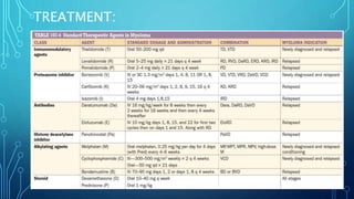
MULTIPLE MYELOMA
DR BIPUL BORTHAKUR
PROF & HEAD
DEPT OF ORTHOPAEDICS
SILCHAR MEDICAL COLLEGE
DEFINITION:
• Multiple myeloma represents a malignant
proliferation of plasma cells derived from a
single clone.
• Most common primary malignancy of bone.
• Malignant B cell lymphoproliferative disorder
of the marrow with plasma cell
predominating,.
• Males> females
• Common in 5th to 7th decade
DEFINITION:
• The tumor, its products and the host response to it results in number of
dysfunctions and symptoms:
• Bone pain or fracture
• Renal failure
• Susceptibility to infection
• Anemia
• Hypercalcemia
• Occasional clotting abnormalities, neurological symptoms and
manifestations of hyperviscosity
ETIOLOGY:
• The cause of myeloma is not known.
• More common seen in farmers, wood workers, leather workers, and those
exposed to petroleum products.
• Chromosomal alterations in MM:
• Hyperdiploidy
• 13q14 deletions
• Translocations t(11;14) t(4;14) t(14;16)
• N-ras, K-ras, and B-raf mutations are common (occurring in >40%
patients)
PATHOGENESIS AND CLINICAL MANIFESTATIONS:
• MM cells bind to bone marrow stromal cells (BMSCs) and extracellular
matrix (ECM) via cell surface adhesion molecules
triggers
• MM cell growth, survival, drug resistance, and migration in bone marrow.
• These effects are due to:
• Direct MM cell-BMSC binding via adhesion molecules (Adhesion
mediated signaling)
• Induction of various cytokines ( Cytokine mediated signaling)
PATHOGENESIS AND CLINICAL
MANIFESTATIONS:
PATHOGENESIS AND CLINICAL MANIFESTATIONS:
• Most common symptom : bone pain (70% patients)
• Persistent localised pain usually signifies a pathological fracture.
• Bone lesions are lytic in nature and are caused by:
• Proliferation of tumor cells
• Activation of osteoclast
• Suppression of osteoblasts
PATHOGENESIS AND CLINICAL MANIFESTATIONS:
• Increased osteoclastic activity is mediated by osteoclast activating factor
(OAFs)
• OAFs is produced by myeloma cells which is mediated by:
• IL-1
• Lymphotoxin
• VEGF
• Receptor activator of NF-k B ligand
• Macrophage inhibitory factor (MIF)-1 alpha
• Tumor necrosis factor(TNF)
• This bony lysis leads to hypercalcemia.
Typical “Punched out” lesion of skull Multiple small lytic foci throughout
the pelvic bone
PET-CT showing multiple FDG avid lesions in skeleton
PATHOGENESIS AND CLINICAL MANIFESTATIONS:
• Vertebral collapse- cord compression, radicular pain, loss of bowel
bladder control
• Susceptibility to bacterial infection : 2nd Most common clinical
problem
• Most common infection-pneumonias and pyelonephritis
• > 75% patients will have a serious infection at some time in their
course
PATHOGENESIS AND CLINICAL MANIFESTATIONS:
• Reason for susceptibility to infection:
• Diffuse hypogammaglobulinemia: both decreased
production and increased destruction of antibodies
• Decreased Th1 response, increased Th17 cells
producing proinflammatory cytokines and aberrant T
rec cell function
• Complement function abnormality
PATHOGENESIS AND CLINICAL MANIFESTATIONS:
• Renal failure in >25% patients
• Most common cause of renal failure : hypercalcemia
• other causes: glomerular amyloid deposition, hyperuricemia, recurrent
infection, use of NSAIDs, iodinated contrast dye for imaging, bisphosphonate
use
• Tubular damage associated with excretion of light chain is almost always
present.
• Proteinuria is observed if glomerulus involved
PATHOGENESIS AND CLINICAL MANIFESTATIONS:
• Normocytic normochromic anemia- 80% patients
• Granulocytopenia and thrombocytopenia are rarely seen
• Clotting abnormalities- due to failure of antibody coated platelets to function
properly
• Deep vein thrombosis- observed with use of thalidomide
• Hyperviscosity syndrome- may lead to headache, shortness of breath, heart
failure, visual disturbances, ataxia, vertigo, retinopathy.
• Hypercalcemia- lethargy, weakness, depression, confusion
DIAGNOSIS AND STAGING:
• The diagnosis of myeloma requires:
• marrow plasmocytosis (>10%)
• A serum and/or urine M component
• At least one of the following myeloma defining event-
DIAGNOSIS AND STAGING:
• The most important differential diagnosis in patients with myeloma are:
• MGUS ( Monoclonal gammapathy of undetermined significance)
• Smoldering multiple myeloma ( Asymptomatic myeloma)
DIAGNOSIS AND STAGING:
• Standard investigative workup in Multiple Myeloma:
• Investigations to evaluate for clonal plasma cells:
• Bone marrow aspirate and biopsy
• Histology
• Clonality by kappa/lambda immunostaining by flow
cytometry or immunohistochemistry
DIAGNOSIS AND STAGING:
• Investigation to evaluate clonal paraprotein:
• Serum protein electrophoresis and immunofixation
• Quantitative serum immunoglobin levels (IgG, IgA and IgM)
• 24 hour urine protein electrophoresis and immunofixation
• Serum free light chain and ratio
• Immunofixation for IgD or IgE in select cases
DIAGNOSIS AND STAGING:
• Investigation to evaluate End-Organ damage:
• Hemogram for anemia, leukopenia, thrombocytopenia
• ESR- Raised (often >100)
• Chemistry panel for renal function and calcium
• Skeletal survey to evaluate bone lesions
• PET/CT or MRI
DIAGNOSIS AND STAGING:
• Investigation for Risk Stratification:
• Beta 2 microglobulin and serum albumin for ISS stage
• Fluorescent in situ hybridization for hyperdiploidy, del 17p,
t(4;14), t(14;16), amp1q34, and del 13
• LDH
• Specialized investigation in selected cases:
• Abdominal fat pad for amyloid
• Serum viscosity
DIAGNOSIS AND STAGING:
• Clinical examination:
• Careful physical examination for tender bone and masses
• Chest and bone x-rays- lytic lesion or diffuse osteopenia
PROGNOSIS:
• Serum beta-2 microglobulin is single most powerful predictor of
survival.
• Three stage international staging system (ISS):
TREATMENT:
• Patients with symptomatic and/or progressive myeloma require
therapeutic intervention.
• Such therapy has two purpose:
1. Systemic therapy to control myeloma
2. Supportive care to control symptoms of the disease, its
complications, and adverse effects of therapy
TREATMENT:
• Therapy includes an initial induction regimen followed by consolidation and
maintenance therapy.
• For newly diagnosed MM patients-
• Thalidomide + dexamethasone – response in 2/3rd of cases
• Lenalidomide + dexamethasone- response in >80% cases
• Bortezomib + dexamethasone- response in >80% cases
• Lenalidomide + bortezomib + dexamethasone- 100% response rate
• Bortezomib + thalidomide + dexamethasone- >90% response
• Bortezomib + cyclophosphamide + dexamethasone- >90% response
TREATMENT:
• Patients who are transplant candidates- alkylating agents
(melphalan) should be avoided (damage stem cells)
• High dose therapy (HDT) and maintenance are standard practice
in majority of eligible patients.
• Two successive HDTs (tandem transplantation) are more effective
than single HDT.
• Maintenance therapy prolongs remissions following standard dose
regimens as well as HDT.
TREATMENT:
• RELAPSED DISEASE:
• Almost all patients with MM who survive initial treatment will eventually
relapse
• Relapsed or refractory MM is usually identified on routine surveillance.
• Therapy options are:
• HDT
• A rechallenge of previous chemotherapy regimen
• A trial of a new regimen
TREATMENT:
TREATMENT:
Treatment algorithm for Multiple Myeloma
TREATMENT:
• The median overall survival of patients with myeloma is 8+ years, with
younger patients surviving >10 years.
• SUPPORTIVE CARE:
• Hypercalcemia: respond to bisphosphonates, glucocorticoids, hydration,
calcitonin.
• Kyphoplasty and vertebroplasty in painful collapsed vertebra
• Hyperviscosity syndrome- treatment of choice is plasmapheresis
TREATMENT:
• In patients in whom neurologic deficit is increasing- surgical
decompression may be necessary.
• Anemia: respond to erythropoietin along with hematinic (iron,
folate, cobalamin)
THANK YOU

Multiple myeloma

  • 1.
    MULTIPLE MYELOMA DR BIPULBORTHAKUR PROF & HEAD DEPT OF ORTHOPAEDICS SILCHAR MEDICAL COLLEGE
  • 2.
    DEFINITION: • Multiple myelomarepresents a malignant proliferation of plasma cells derived from a single clone. • Most common primary malignancy of bone. • Malignant B cell lymphoproliferative disorder of the marrow with plasma cell predominating,. • Males> females • Common in 5th to 7th decade
  • 3.
    DEFINITION: • The tumor,its products and the host response to it results in number of dysfunctions and symptoms: • Bone pain or fracture • Renal failure • Susceptibility to infection • Anemia • Hypercalcemia • Occasional clotting abnormalities, neurological symptoms and manifestations of hyperviscosity
  • 4.
    ETIOLOGY: • The causeof myeloma is not known. • More common seen in farmers, wood workers, leather workers, and those exposed to petroleum products. • Chromosomal alterations in MM: • Hyperdiploidy • 13q14 deletions • Translocations t(11;14) t(4;14) t(14;16) • N-ras, K-ras, and B-raf mutations are common (occurring in >40% patients)
  • 5.
    PATHOGENESIS AND CLINICALMANIFESTATIONS: • MM cells bind to bone marrow stromal cells (BMSCs) and extracellular matrix (ECM) via cell surface adhesion molecules triggers • MM cell growth, survival, drug resistance, and migration in bone marrow. • These effects are due to: • Direct MM cell-BMSC binding via adhesion molecules (Adhesion mediated signaling) • Induction of various cytokines ( Cytokine mediated signaling)
  • 6.
  • 7.
    PATHOGENESIS AND CLINICALMANIFESTATIONS: • Most common symptom : bone pain (70% patients) • Persistent localised pain usually signifies a pathological fracture. • Bone lesions are lytic in nature and are caused by: • Proliferation of tumor cells • Activation of osteoclast • Suppression of osteoblasts
  • 8.
    PATHOGENESIS AND CLINICALMANIFESTATIONS: • Increased osteoclastic activity is mediated by osteoclast activating factor (OAFs) • OAFs is produced by myeloma cells which is mediated by: • IL-1 • Lymphotoxin • VEGF • Receptor activator of NF-k B ligand • Macrophage inhibitory factor (MIF)-1 alpha • Tumor necrosis factor(TNF) • This bony lysis leads to hypercalcemia.
  • 9.
    Typical “Punched out”lesion of skull Multiple small lytic foci throughout the pelvic bone
  • 10.
    PET-CT showing multipleFDG avid lesions in skeleton
  • 11.
    PATHOGENESIS AND CLINICALMANIFESTATIONS: • Vertebral collapse- cord compression, radicular pain, loss of bowel bladder control • Susceptibility to bacterial infection : 2nd Most common clinical problem • Most common infection-pneumonias and pyelonephritis • > 75% patients will have a serious infection at some time in their course
  • 12.
    PATHOGENESIS AND CLINICALMANIFESTATIONS: • Reason for susceptibility to infection: • Diffuse hypogammaglobulinemia: both decreased production and increased destruction of antibodies • Decreased Th1 response, increased Th17 cells producing proinflammatory cytokines and aberrant T rec cell function • Complement function abnormality
  • 13.
    PATHOGENESIS AND CLINICALMANIFESTATIONS: • Renal failure in >25% patients • Most common cause of renal failure : hypercalcemia • other causes: glomerular amyloid deposition, hyperuricemia, recurrent infection, use of NSAIDs, iodinated contrast dye for imaging, bisphosphonate use • Tubular damage associated with excretion of light chain is almost always present. • Proteinuria is observed if glomerulus involved
  • 14.
    PATHOGENESIS AND CLINICALMANIFESTATIONS: • Normocytic normochromic anemia- 80% patients • Granulocytopenia and thrombocytopenia are rarely seen • Clotting abnormalities- due to failure of antibody coated platelets to function properly • Deep vein thrombosis- observed with use of thalidomide • Hyperviscosity syndrome- may lead to headache, shortness of breath, heart failure, visual disturbances, ataxia, vertigo, retinopathy. • Hypercalcemia- lethargy, weakness, depression, confusion
  • 15.
    DIAGNOSIS AND STAGING: •The diagnosis of myeloma requires: • marrow plasmocytosis (>10%) • A serum and/or urine M component • At least one of the following myeloma defining event-
  • 16.
    DIAGNOSIS AND STAGING: •The most important differential diagnosis in patients with myeloma are: • MGUS ( Monoclonal gammapathy of undetermined significance) • Smoldering multiple myeloma ( Asymptomatic myeloma)
  • 17.
    DIAGNOSIS AND STAGING: •Standard investigative workup in Multiple Myeloma: • Investigations to evaluate for clonal plasma cells: • Bone marrow aspirate and biopsy • Histology • Clonality by kappa/lambda immunostaining by flow cytometry or immunohistochemistry
  • 18.
  • 19.
    DIAGNOSIS AND STAGING: •Investigation to evaluate clonal paraprotein: • Serum protein electrophoresis and immunofixation • Quantitative serum immunoglobin levels (IgG, IgA and IgM) • 24 hour urine protein electrophoresis and immunofixation • Serum free light chain and ratio • Immunofixation for IgD or IgE in select cases
  • 20.
    DIAGNOSIS AND STAGING: •Investigation to evaluate End-Organ damage: • Hemogram for anemia, leukopenia, thrombocytopenia • ESR- Raised (often >100) • Chemistry panel for renal function and calcium • Skeletal survey to evaluate bone lesions • PET/CT or MRI
  • 21.
    DIAGNOSIS AND STAGING: •Investigation for Risk Stratification: • Beta 2 microglobulin and serum albumin for ISS stage • Fluorescent in situ hybridization for hyperdiploidy, del 17p, t(4;14), t(14;16), amp1q34, and del 13 • LDH • Specialized investigation in selected cases: • Abdominal fat pad for amyloid • Serum viscosity
  • 22.
    DIAGNOSIS AND STAGING: •Clinical examination: • Careful physical examination for tender bone and masses • Chest and bone x-rays- lytic lesion or diffuse osteopenia
  • 23.
    PROGNOSIS: • Serum beta-2microglobulin is single most powerful predictor of survival. • Three stage international staging system (ISS):
  • 24.
    TREATMENT: • Patients withsymptomatic and/or progressive myeloma require therapeutic intervention. • Such therapy has two purpose: 1. Systemic therapy to control myeloma 2. Supportive care to control symptoms of the disease, its complications, and adverse effects of therapy
  • 25.
    TREATMENT: • Therapy includesan initial induction regimen followed by consolidation and maintenance therapy. • For newly diagnosed MM patients- • Thalidomide + dexamethasone – response in 2/3rd of cases • Lenalidomide + dexamethasone- response in >80% cases • Bortezomib + dexamethasone- response in >80% cases • Lenalidomide + bortezomib + dexamethasone- 100% response rate • Bortezomib + thalidomide + dexamethasone- >90% response • Bortezomib + cyclophosphamide + dexamethasone- >90% response
  • 26.
    TREATMENT: • Patients whoare transplant candidates- alkylating agents (melphalan) should be avoided (damage stem cells) • High dose therapy (HDT) and maintenance are standard practice in majority of eligible patients. • Two successive HDTs (tandem transplantation) are more effective than single HDT. • Maintenance therapy prolongs remissions following standard dose regimens as well as HDT.
  • 27.
    TREATMENT: • RELAPSED DISEASE: •Almost all patients with MM who survive initial treatment will eventually relapse • Relapsed or refractory MM is usually identified on routine surveillance. • Therapy options are: • HDT • A rechallenge of previous chemotherapy regimen • A trial of a new regimen
  • 28.
  • 29.
  • 30.
    TREATMENT: • The medianoverall survival of patients with myeloma is 8+ years, with younger patients surviving >10 years. • SUPPORTIVE CARE: • Hypercalcemia: respond to bisphosphonates, glucocorticoids, hydration, calcitonin. • Kyphoplasty and vertebroplasty in painful collapsed vertebra • Hyperviscosity syndrome- treatment of choice is plasmapheresis
  • 31.
    TREATMENT: • In patientsin whom neurologic deficit is increasing- surgical decompression may be necessary. • Anemia: respond to erythropoietin along with hematinic (iron, folate, cobalamin)
  • 32.