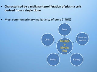
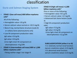
MULTIPLE
MYELOMA
Dr. Kiran Bikkad
• Characterised by a malignant proliferation of plasma cells
derived from a single clone
• Most common primary malignancy of bone (~40%)
Multipl
e
Myelo
ma
Bone
Nervous
system
KidneyBlood
Chest
Incidence
• Age group – more common in 4th to 6th decade
• More in african-americans than caucasians
• Exact cause not known
• Risk factors –
• radiation
• exposure to petroleum products
• t(11,14), t(4,14), t( 14,16), del13q14
Clinical features
• Early stages  Silent
• Bone pain- increases on movements – m/c symptom (70%)
• Pathological fractures
• Symptoms of anaemia Normocytic Normochromic (80%)
• Renal failure(25%)
• Recurrent infections (25%)(respiratory & urinary)
• Hyperviscosity  headache, SOB, Heart failure, Visual
disturbances
• Neurological involvement (sensory)
Recurrent Infections
• M/C : Pneumonia & Pyelonephritis
• Staph, strepto, E.Coli, Klebsiella
• Bcoz diffuse hypogammaglobulinemia except M-component
• Decreased production & increased destruction of normal
antibodies
• Fractional catabolic rate of antibody increased in MM.
• Very poor Ab. Response to antigen specially to
polysaccharisde antigen.
Recurrent Infections
• CD4+ cell decreased
• Granulocyte : lysosome component is low & migration is also
slow.
• Complement functioning abnormality +
• Dexamethasone suppresses immune response & bortezomib
predisposes herpes infection
Renal Failure
• Amyloid deposit
• Hyperuricemia
• Recurrent infections
• NSAIDs use for pain relief
• Bisphosphonates
• Iodinated contrast dyes
• Light chain mediated tubular damage (Type 2 proximal RTA i.e.
Adult Fanconi Syndrome)
• Proteinuria is not asso with HTN  bcoz protein lost is mainly
light chain protein  bcoz glomerular function is normal,
when it is damaged then non selective proteinuria starts.
Anion gap & Pseudohyponatremia
• Anion gap is decreased
• Because M – component is cationic & retains Cl- ions.
• Artificial Pseudohyponatremia
• Each volume of serum has less water because of increased
protein
Anaemia
• Normocytic Normochromic
• Replacement of normal marrow by expanding plasma cells
• Decreased EPO by damaged kidney
• Thrombocytopenia indused bleed
• Bleed : may be due to
1. Failure of antibody coated platelet to function
2. Interaction of M component to clotting factors 1,2,5,7,8
3. Amyloid mediated damage to endothelium.
• DVT observed with use of Thalidomide with Dexamethasone
• Raynouds Phenomenon bcoz M protein forms cryoglobulins
& may cause Hyperviscocity syndrome.
• Neurologic Symptoms :
• Hypercalcemia: Lethargy, weakness, depression & confusion
• Hyperviscocity : Ataxia, somnolence, Vertigo, retinopathy,
coma
• Bony damage : cord compression
• Amyloid deposition of peripheral nerves. E.g. Carpal Tunnel S.
Lab findings
• Anemia, leukopenia, thrombocytopenia
• ALb, reversed A:G ratio
• serum urea, creat, uric acid
• Abnormal coagulation
• Serum Ca+2
• Proteinuria i.e. light chain (selective) and cast.
• ESR
• LOW to NORMAL ALKALINE PHOSPHATASE
• Red cells show rouleaux formation
• BENCE-JONES PROTEIN in urine in 30% (24 hr urine protein)
Lab findings
• Serum electrophoresis- screening method for detection of Pl.
cell disorders.
• It reveals monoclonal component (narrow band peak:
“church spike”)
• found in 98% of patients, in serum, urine or both
“M” spike or
church spike on
electrophoresis
*Microscopic
appearance – -
Eccentric nucleus with
nucleolus
Coarsely clumped
chromatin in spoke-of-
wheel fashion
Basophilic cytoplasm
Perinuclear halo
Radiology
Multiple, punched-out,
sharply demarceted,
purely lytic lesion
without surrounding
reactive sclerosis
Diagnosis
Diagnostic features of active or
symptomatic myeloma
Organ damage classified as “CRAB”
• C – calcium elevation (>10 mg/L)
• R – renal dysfunction (creatinine >2 mg/dL)
• A – anemia (hemoglobin <10 g/dL or ≥2 g/dL decrease
from patient’s normal)
• B – bone disease (lytic lesions or osteoporosis)
*ONE OR MORE required for diagnosis of SYMPTOMATIC
MYELOMA.
Other less common features can also be criteria for an
individual patient, including:
• Recurrent severe infections
• Neuropathy linked to myeloma
• Amyloidosis or M-component deposition
• Other unique features
• Other Pl cell disorders-
• Waldenstrom`s disease
• Bone metastasis – breast, prostatic Ca
• Hyperparathyroidism
• Other reasons for renal failure - ex. chronic glomerulo-
nephritis
classification
• STAGE I (low cell mass) 600 billion myeloma
cells*
All of the following:
• Hemoglobin value >10 g/dL
• Serum calcium value normal or <10.5 mg/dL
• Bone X-ray, normal bone structure (scale 0)
or solitary bone plasmacytoma only
• Low M-component production rates
IgG value <5.0 g/dL
IgA value <3.0 g/dL
Urine light chain M-component on
electrophoresis <4 g/24h
• STAGE II (intermediate cell mass) 600 to 1,200
billion myeloma cells*
Fitting neither stage I nor stage III
SUBCLASSIFICATION (either A or B)
• A: relatively normal renal function
(serum creatinine value) <2.0 mg/dL
• B: abnormal renal function
(serum creatinine value) >2.0 mg/dL
STAGE III (high cell mass) >1,200
billion myeloma cells*
One or more of the following:
• Hemoglobin value <8.5 g/dL
• Serum calcium value >12 mg/dL
• Advanced lytic bone lesions (scale
3)
• High M-component production
rates
IgG value >7.0 g/dL
IgA value >5.0 g/dL
Urine light chain M-component on
electrophoresis >12 g/24h
Durie and Salmon Staging System
International staging system (ISS)
SURVIVAL
STAGE 1 β2M <3.5 62 MONTHS
ALB ≥3.5
STAGE 2 β2M <3.5
ALB <3.5 or 44 MONTHS
β2M 3.5 – 5.5
STAGE3 β2M >5.5 29 MONTHS
β2M = Serum β2 microglobulin in mg/L
ALB = Serum albumin in g/dL
MANAGEMENT
1. Chemotherapy
2. Hematopoietic stem cell transplant
3. Radiation
4. Maintenance therapy
5. Supportive care
6. Management of drug-resistant or refractory
disease
7. New and emerging treatments
In asymptomatic myeloma or MGUS
Supportive treatment including
• Erythropoietin
• Pain medication
• Bisphosphonates
• Growth factors
• Antibiotics
Systemic anti-myeloma treatment
• Thalidomide 200mg daily + dexamethasone
• Lenalidomide25mg/day on day1-25 every monthly
(DVT prophylaxis)
• Bortezomib 1.3mg/m2 on day 1,4,8,11 every 3 week +
Dexamethasone 40mg once weekly
• Combination of Lenalidomide, Bortezomib & Dexa
shown close to 100% response rate
• Melphalan with prednisone used for older patients
>70yrs
– If stem cell transplantation is NOT planned
• Relapse Myeloma T/t: Lenalidomide and/or
Bortezomib
• High dose Melphalan with Stem Cell Transplant in
Refractory cases
• Bisphosphonate
– Pamidronate 90mg or Zoledronate 4mg/month
• Plasmapheresis for Hyperviscocity Syndrome
• Cord compression treated with local radiarion therapy
with glucocorticoids or Vertebroplasty
THANK YOU

Multiple myeloma

  • 1.
  • 2.
    • Characterised bya malignant proliferation of plasma cells derived from a single clone • Most common primary malignancy of bone (~40%) Multipl e Myelo ma Bone Nervous system KidneyBlood Chest
  • 3.
    Incidence • Age group– more common in 4th to 6th decade • More in african-americans than caucasians • Exact cause not known • Risk factors – • radiation • exposure to petroleum products • t(11,14), t(4,14), t( 14,16), del13q14
  • 5.
    Clinical features • Earlystages  Silent • Bone pain- increases on movements – m/c symptom (70%) • Pathological fractures • Symptoms of anaemia Normocytic Normochromic (80%) • Renal failure(25%) • Recurrent infections (25%)(respiratory & urinary) • Hyperviscosity  headache, SOB, Heart failure, Visual disturbances • Neurological involvement (sensory)
  • 6.
    Recurrent Infections • M/C: Pneumonia & Pyelonephritis • Staph, strepto, E.Coli, Klebsiella • Bcoz diffuse hypogammaglobulinemia except M-component • Decreased production & increased destruction of normal antibodies • Fractional catabolic rate of antibody increased in MM. • Very poor Ab. Response to antigen specially to polysaccharisde antigen.
  • 7.
    Recurrent Infections • CD4+cell decreased • Granulocyte : lysosome component is low & migration is also slow. • Complement functioning abnormality + • Dexamethasone suppresses immune response & bortezomib predisposes herpes infection
  • 8.
    Renal Failure • Amyloiddeposit • Hyperuricemia • Recurrent infections • NSAIDs use for pain relief • Bisphosphonates • Iodinated contrast dyes • Light chain mediated tubular damage (Type 2 proximal RTA i.e. Adult Fanconi Syndrome) • Proteinuria is not asso with HTN  bcoz protein lost is mainly light chain protein  bcoz glomerular function is normal, when it is damaged then non selective proteinuria starts.
  • 9.
    Anion gap &Pseudohyponatremia • Anion gap is decreased • Because M – component is cationic & retains Cl- ions. • Artificial Pseudohyponatremia • Each volume of serum has less water because of increased protein
  • 10.
    Anaemia • Normocytic Normochromic •Replacement of normal marrow by expanding plasma cells • Decreased EPO by damaged kidney • Thrombocytopenia indused bleed • Bleed : may be due to 1. Failure of antibody coated platelet to function 2. Interaction of M component to clotting factors 1,2,5,7,8 3. Amyloid mediated damage to endothelium.
  • 11.
    • DVT observedwith use of Thalidomide with Dexamethasone • Raynouds Phenomenon bcoz M protein forms cryoglobulins & may cause Hyperviscocity syndrome. • Neurologic Symptoms : • Hypercalcemia: Lethargy, weakness, depression & confusion • Hyperviscocity : Ataxia, somnolence, Vertigo, retinopathy, coma • Bony damage : cord compression • Amyloid deposition of peripheral nerves. E.g. Carpal Tunnel S.
  • 12.
    Lab findings • Anemia,leukopenia, thrombocytopenia • ALb, reversed A:G ratio • serum urea, creat, uric acid • Abnormal coagulation • Serum Ca+2 • Proteinuria i.e. light chain (selective) and cast. • ESR • LOW to NORMAL ALKALINE PHOSPHATASE • Red cells show rouleaux formation • BENCE-JONES PROTEIN in urine in 30% (24 hr urine protein)
  • 13.
    Lab findings • Serumelectrophoresis- screening method for detection of Pl. cell disorders. • It reveals monoclonal component (narrow band peak: “church spike”) • found in 98% of patients, in serum, urine or both “M” spike or church spike on electrophoresis
  • 14.
    *Microscopic appearance – - Eccentricnucleus with nucleolus Coarsely clumped chromatin in spoke-of- wheel fashion Basophilic cytoplasm Perinuclear halo
  • 15.
    Radiology Multiple, punched-out, sharply demarceted, purelylytic lesion without surrounding reactive sclerosis
  • 16.
  • 18.
    Diagnostic features ofactive or symptomatic myeloma Organ damage classified as “CRAB” • C – calcium elevation (>10 mg/L) • R – renal dysfunction (creatinine >2 mg/dL) • A – anemia (hemoglobin <10 g/dL or ≥2 g/dL decrease from patient’s normal) • B – bone disease (lytic lesions or osteoporosis) *ONE OR MORE required for diagnosis of SYMPTOMATIC MYELOMA. Other less common features can also be criteria for an individual patient, including: • Recurrent severe infections • Neuropathy linked to myeloma • Amyloidosis or M-component deposition • Other unique features
  • 19.
    • Other Plcell disorders- • Waldenstrom`s disease • Bone metastasis – breast, prostatic Ca • Hyperparathyroidism • Other reasons for renal failure - ex. chronic glomerulo- nephritis
  • 20.
    classification • STAGE I(low cell mass) 600 billion myeloma cells* All of the following: • Hemoglobin value >10 g/dL • Serum calcium value normal or <10.5 mg/dL • Bone X-ray, normal bone structure (scale 0) or solitary bone plasmacytoma only • Low M-component production rates IgG value <5.0 g/dL IgA value <3.0 g/dL Urine light chain M-component on electrophoresis <4 g/24h • STAGE II (intermediate cell mass) 600 to 1,200 billion myeloma cells* Fitting neither stage I nor stage III SUBCLASSIFICATION (either A or B) • A: relatively normal renal function (serum creatinine value) <2.0 mg/dL • B: abnormal renal function (serum creatinine value) >2.0 mg/dL STAGE III (high cell mass) >1,200 billion myeloma cells* One or more of the following: • Hemoglobin value <8.5 g/dL • Serum calcium value >12 mg/dL • Advanced lytic bone lesions (scale 3) • High M-component production rates IgG value >7.0 g/dL IgA value >5.0 g/dL Urine light chain M-component on electrophoresis >12 g/24h Durie and Salmon Staging System
  • 21.
    International staging system(ISS) SURVIVAL STAGE 1 β2M <3.5 62 MONTHS ALB ≥3.5 STAGE 2 β2M <3.5 ALB <3.5 or 44 MONTHS β2M 3.5 – 5.5 STAGE3 β2M >5.5 29 MONTHS β2M = Serum β2 microglobulin in mg/L ALB = Serum albumin in g/dL
  • 22.
    MANAGEMENT 1. Chemotherapy 2. Hematopoieticstem cell transplant 3. Radiation 4. Maintenance therapy 5. Supportive care 6. Management of drug-resistant or refractory disease 7. New and emerging treatments
  • 23.
    In asymptomatic myelomaor MGUS Supportive treatment including • Erythropoietin • Pain medication • Bisphosphonates • Growth factors • Antibiotics
  • 24.
    Systemic anti-myeloma treatment •Thalidomide 200mg daily + dexamethasone • Lenalidomide25mg/day on day1-25 every monthly (DVT prophylaxis) • Bortezomib 1.3mg/m2 on day 1,4,8,11 every 3 week + Dexamethasone 40mg once weekly • Combination of Lenalidomide, Bortezomib & Dexa shown close to 100% response rate • Melphalan with prednisone used for older patients >70yrs – If stem cell transplantation is NOT planned
  • 25.
    • Relapse MyelomaT/t: Lenalidomide and/or Bortezomib • High dose Melphalan with Stem Cell Transplant in Refractory cases • Bisphosphonate – Pamidronate 90mg or Zoledronate 4mg/month • Plasmapheresis for Hyperviscocity Syndrome • Cord compression treated with local radiarion therapy with glucocorticoids or Vertebroplasty
  • 26.

Editor's Notes

  • #6 Recurrent Infections: Pneumonia & Pyelonephritis, Bcoz diffuse hypogammaglobulinemia except M-component, decreased production & increased destruction of normal antibodies, Fractional catabolic rate of antibody increased in MM. V.poor Ab. Response to antigen specially to polysaccharisde antigen. CD4+ cell decreased, Granulocyte lysosome component is low & migration is also slow. Complement functioning abnormality & on top Dexamethasone suppresses immune response & bortezomib predisposes herpes infection Renal Failure : Amyloid deposit, Hyperuricemia, Recurrent infections, NSAIDs use for pain relief, Bisphosphonates, Iodinated contrast dyes, Light chain mediated tubular damage (Type 2 proximal RTA i.e. Adult Fanconi Syndrome), proteinuria is not asso with HTN bcoz protein lost is mainly light chain protein bcoz glomerular function is normal, when it is damaged then non selective proteinuria starts. Anion gap is decreased, bcz M – component is cationic & retains Cl- ions. Artificial Pseudohyponatremia (each volume of serum has less water because of increased protein) Anemia : NN, bcoz replacement of normal marrow by expanding plasma cells, decreased EPO by damaged kidney, , thrombocytopenia indused bleed may add on, *bleed may b due to failure of antibody coated platelet to function & interaction of M component to clotting factors 1,2,5,7,8 & Amyloid mediated damage to endothelium. DVT observed with use of Thalidomide with Dexamethasone Raynouds Phenomenon bcoz M protein forms cryoglobulins & may cause Hyperviscocity syndrome. Neurologic Symptoms : Hypercalcemia: Lethargy, weakness, depression & confusion
  • #13 (ALP : if raised bad prognosis s/o active bony lysis)