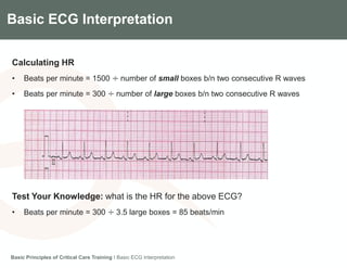
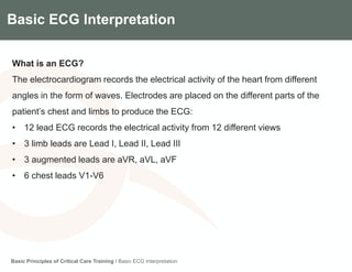
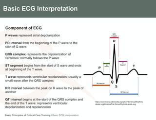
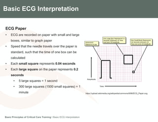
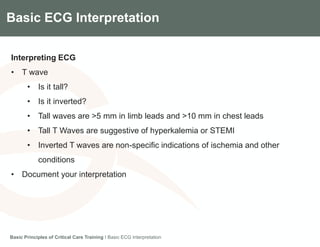
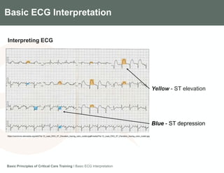
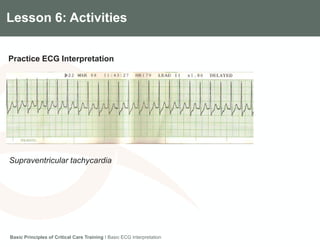
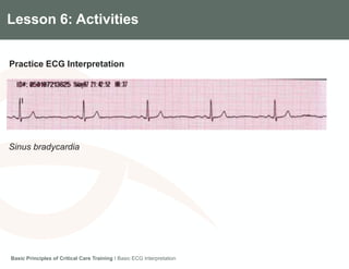
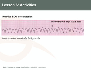
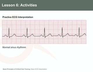
BASIC ECG INTERPRETATION
Gradian Health Systems
Basic Principles of Critical Care
CARDIOVASCULAR SYSTEM
Disclaimer
Basic Principles of Critical Care Training I Basic ECG Interpretation
Disclaimer: Gradian Health Systems cannot provide formal recommendations or indications
regarding medical care and clinical service delivery. The tables, checklists, and other clinical
documents referenced in this training have not been validated in all settings. These documents are
intended to serve as examples only. We recognize that all clinical training content and activities
must be customized to meet the needs of each facility and its clinical staff, factoring in available
resources, practitioner skill level, and other environmental considerations.
For any questions regarding the contents or applications of this training,
please contact Gradian Health Systems:
40 W 25th St, 6th Floor
New York, NY 10010 USA
+1 212-537-0340
training@gradianhealth.org
Module 3
Gradian Health Systems
Basics Principles of Critical Care
Cardiovascular System
Module 3: Cardiovascular System
MODULE OVERVIEW
Lesson 1 I Cardiovascular System Anatomy and Physiology
Lesson 2 I Hemodynamic Monitoring
Lesson 3 I Shock
Lesson 4 I Fluid Replacement
Lesson 5 I Vasoactive Medications
Lesson 6 I Basic ECG Interpretation
Basic Principles of Critical Care Training I Basic ECG Interpretation
Components of the Gradian CCV SystemLesson 6: Basic ECG Interpretation
Lesson Objectives
• Describe the basics of ECGs
• Interpret basic ECGs
Basic Principles of Critical Care Training I Basic ECG Interpretation
Components of the Gradian CCV SystemLesson 6: Basic ECG Interpretation
Key Concepts
• Rate
• Rhythm
• Waves
• Segments
• Clinical interpretation
Basic Principles of Critical Care Training I Basic ECG Interpretation
Components of the Gradian CCV SystemBasic ECG Interpretation
What is an ECG?
The electrocardiogram records the electrical activity of the heart from different
angles in the form of waves. Electrodes are placed on the different parts of the
patient’s chest and limbs to produce the ECG:
• 12 lead ECG records the electrical activity from 12 different views
• 3 limb leads are Lead I, Lead II, Lead III
• 3 augmented leads are aVR, aVL, aVF
• 6 chest leads V1-V6
Basic Principles of Critical Care Training I Basic ECG Interpretation
Components of the Gradian CCV SystemBasic ECG Interpretation
Limb Electrode Placement
Basic Principles of Critical Care Training I Basic ECG Interpretation
https://commons.wikimedia.org/wiki/File:Limb_leads.svg#/media/File:Limb_leads.svg
Components of the Gradian CCV SystemBasic ECG Interpretation
Chest Electrode Placement
Basic Principles of Critical Care Training I Basic ECG Interpretation
https://commons.wikimedia.org/wiki/File:Precordial_leads_i
n_ECG.png#/media/File:Precordial_leads_in_ECG.png
Components of the Gradian CCV SystemBasic ECG Interpretation
ECG Waves
A normal 12 lead ECG strip
Note: the waves have a certain
morphology based on where the
electrode is placed
Basic Principles of Critical Care Training I Basic ECG Interpretation
https://commons.wikimedia.org/wiki/File:Normal_12_lead_EKG.jpg#/media/File:Normal_12_lead_EKG.jpg
Components of the Gradian CCV SystemBasic ECG Interpretation
Component of ECG
Basic Principles of Critical Care Training I Basic ECG Interpretation
P waves represent atrial depolarization
PR interval from the beginning of the P wave to the
start of Q wave
QRS complex represents the depolarization of
ventricles; normally follows the P wave
ST segment begins from the start of S wave and ends
at beginning of the T wave.
T wave represents ventricular repolarization; usually a
small wave after the QRS complex
RR interval between the peak on R wave to the peak of
another
QT interval begins at the start of the QRS complex and
the end of the T wave; represents ventricular
depolarization and repolarization
https://commons.wikimedia.org/wiki/File:SinusRhythmL
abels.svg#/media/File:SinusRhythmLabels.svg
Components of the Gradian CCV SystemBasic ECG Interpretation
ECG Paper
• ECG are recorded on paper with small and large
boxes, similar to graph paper
• Speed that the needle travels over the paper is
standard, such that the time of one box can be
calculated
• Each small square represents 0.04 seconds
• Each large square on the paper represents 0.2
seconds
• 5 large squares = 1 second
• 300 large squares (1500 small squares) = 1
minute
Basic Principles of Critical Care Training I Basic ECG Interpretation
https://upload.wikimedia.org/wikipedia/commons/8/86/ECG_Paper.svg
Components of the Gradian CCV SystemBasic ECG Interpretation
Calculating HR
• Beats per minute = 1500 ÷ number of small boxes b/n two consecutive R waves
• Beats per minute = 300 ÷ number of large boxes b/n two consecutive R waves
Basic Principles of Critical Care Training I Basic ECG Interpretation
Test Your Knowledge: what is the HR for the above ECG?
• Beats per minute = 300 ÷ 3.5 large boxes = 85 beats/min
Components of the Gradian CCV SystemBasic ECG Interpretation
Interpreting ECG
• Confirm patient particulars
• Calculate the HR
• HR < 50/min is bradycardia
• HR > 100/min is tachycardia
• Determine if the heart rhythm is regular or irregular
• Drawn from the pattern of the waves
• Do the R waves occur at regular intervals (same number of boxes)?
• P wave
• Are they present?
• Do they follow the QRS complex?
Basic Principles of Critical Care Training I Basic ECG Interpretation
Components of the Gradian CCV SystemBasic ECG Interpretation
Interpreting ECG
• PR interval
• Normal is 0.12-0.2 secs (3-5 small boxes)
• Is it normal or prolonged (>5 small boxes)?
• This is suggestive of a conduction problem (heart block)
• QRS complex – width and morphology
• Width can be narrow (<0.12 seconds) or broad (>0.12 seconds)
Basic Principles of Critical Care Training I Basic ECG Interpretation
Components of the Gradian CCV SystemBasic ECG Interpretation
Interpreting ECG
• ST segment
• Should be an isoelectric line in normal patients
• Can be elevated or depressed in sick patients
• ST elevation is significant if >1mm (height of 1 small square) in 2 or more
contiguous limb leads
• ST elevation is a sign of acute myocardial infarction (STEMI - ST
Elevated Myocardial Ischemia)
• ST depression is ≥ 0.5mm in 2 or more contiguous leads and indicates
myocardial ischemia
Basic Principles of Critical Care Training I Basic ECG Interpretation
Components of the Gradian CCV SystemBasic ECG Interpretation
Interpreting ECG
• T wave
• Is it tall?
• Is it inverted?
• Tall waves are >5 mm in limb leads and >10 mm in chest leads
• Tall T Waves are suggestive of hyperkalemia or STEMI
• Inverted T waves are non-specific indications of ischemia and other
conditions
• Document your interpretation
Basic Principles of Critical Care Training I Basic ECG Interpretation
Components of the Gradian CCV SystemBasic ECG Interpretation
Interpreting ECG
Basic Principles of Critical Care Training I Basic ECG Interpretation
https://commons.wikimedia.org/wiki/File:12_Lead_EKG_ST_Elevation_tracing_color_coded.jpg#/media/File:12_Lead_EKG_ST_Elevation_tracing_color_coded.jpg
Yellow - ST elevation
Blue - ST depression
Components of the Gradian CCV SystemLesson 6: Summary
ECG interpretation takes practice. Follow the steps indicated during this lesson
and confirm findings with an expert.
Basic Principles of Critical Care Training I Basic ECG Interpretation
Components of the Gradian CCV SystemLesson 6: Activities
Practice ECG Interpretation
Remember the steps
• Confirm patient particulars
• Calculate HR
• Determine if the heart rhythm is regular or irregular
• P wave – are they present? Do they follow the QRS complex?
• PR interval – is it normal or prolonged?
• QRS complex – narrow or wide?
• ST segment – elevated or depressed?
• T wave – tall or inverted?.
• Document your interpretation
Basic Principles of Critical Care Training I Basic ECG Interpretation
Components of the Gradian CCV SystemLesson 6: Activities
Practice ECG Interpretation
Basic Principles of Critical Care Training I Basic ECG Interpretation
Supraventricular tachycardia
Components of the Gradian CCV SystemLesson 6: Activities
Practice ECG Interpretation
Basic Principles of Critical Care Training I Basic ECG Interpretation
Sinus bradycardia
Components of the Gradian CCV SystemLesson 6: Activities
Practice ECG Interpretation
Basic Principles of Critical Care Training I Basic ECG Interpretation
Monomorphic ventricular tachycardia
Components of the Gradian CCV SystemLesson 6: Activities
Practice ECG Interpretation
Basic Principles of Critical Care Training I Basic ECG Interpretation
Normal sinus rhythmm

Module 3.6 Basic ECG Interpretation

  • 1.
    BASIC ECG INTERPRETATION GradianHealth Systems Basic Principles of Critical Care CARDIOVASCULAR SYSTEM
  • 2.
    Disclaimer Basic Principles ofCritical Care Training I Basic ECG Interpretation Disclaimer: Gradian Health Systems cannot provide formal recommendations or indications regarding medical care and clinical service delivery. The tables, checklists, and other clinical documents referenced in this training have not been validated in all settings. These documents are intended to serve as examples only. We recognize that all clinical training content and activities must be customized to meet the needs of each facility and its clinical staff, factoring in available resources, practitioner skill level, and other environmental considerations. For any questions regarding the contents or applications of this training, please contact Gradian Health Systems: 40 W 25th St, 6th Floor New York, NY 10010 USA +1 212-537-0340 training@gradianhealth.org
  • 3.
    Module 3 Gradian HealthSystems Basics Principles of Critical Care Cardiovascular System
  • 4.
    Module 3: CardiovascularSystem MODULE OVERVIEW Lesson 1 I Cardiovascular System Anatomy and Physiology Lesson 2 I Hemodynamic Monitoring Lesson 3 I Shock Lesson 4 I Fluid Replacement Lesson 5 I Vasoactive Medications Lesson 6 I Basic ECG Interpretation Basic Principles of Critical Care Training I Basic ECG Interpretation
  • 5.
    Components of theGradian CCV SystemLesson 6: Basic ECG Interpretation Lesson Objectives • Describe the basics of ECGs • Interpret basic ECGs Basic Principles of Critical Care Training I Basic ECG Interpretation
  • 6.
    Components of theGradian CCV SystemLesson 6: Basic ECG Interpretation Key Concepts • Rate • Rhythm • Waves • Segments • Clinical interpretation Basic Principles of Critical Care Training I Basic ECG Interpretation
  • 7.
    Components of theGradian CCV SystemBasic ECG Interpretation What is an ECG? The electrocardiogram records the electrical activity of the heart from different angles in the form of waves. Electrodes are placed on the different parts of the patient’s chest and limbs to produce the ECG: • 12 lead ECG records the electrical activity from 12 different views • 3 limb leads are Lead I, Lead II, Lead III • 3 augmented leads are aVR, aVL, aVF • 6 chest leads V1-V6 Basic Principles of Critical Care Training I Basic ECG Interpretation
  • 8.
    Components of theGradian CCV SystemBasic ECG Interpretation Limb Electrode Placement Basic Principles of Critical Care Training I Basic ECG Interpretation https://commons.wikimedia.org/wiki/File:Limb_leads.svg#/media/File:Limb_leads.svg
  • 9.
    Components of theGradian CCV SystemBasic ECG Interpretation Chest Electrode Placement Basic Principles of Critical Care Training I Basic ECG Interpretation https://commons.wikimedia.org/wiki/File:Precordial_leads_i n_ECG.png#/media/File:Precordial_leads_in_ECG.png
  • 10.
    Components of theGradian CCV SystemBasic ECG Interpretation ECG Waves A normal 12 lead ECG strip Note: the waves have a certain morphology based on where the electrode is placed Basic Principles of Critical Care Training I Basic ECG Interpretation https://commons.wikimedia.org/wiki/File:Normal_12_lead_EKG.jpg#/media/File:Normal_12_lead_EKG.jpg
  • 11.
    Components of theGradian CCV SystemBasic ECG Interpretation Component of ECG Basic Principles of Critical Care Training I Basic ECG Interpretation P waves represent atrial depolarization PR interval from the beginning of the P wave to the start of Q wave QRS complex represents the depolarization of ventricles; normally follows the P wave ST segment begins from the start of S wave and ends at beginning of the T wave. T wave represents ventricular repolarization; usually a small wave after the QRS complex RR interval between the peak on R wave to the peak of another QT interval begins at the start of the QRS complex and the end of the T wave; represents ventricular depolarization and repolarization https://commons.wikimedia.org/wiki/File:SinusRhythmL abels.svg#/media/File:SinusRhythmLabels.svg
  • 12.
    Components of theGradian CCV SystemBasic ECG Interpretation ECG Paper • ECG are recorded on paper with small and large boxes, similar to graph paper • Speed that the needle travels over the paper is standard, such that the time of one box can be calculated • Each small square represents 0.04 seconds • Each large square on the paper represents 0.2 seconds • 5 large squares = 1 second • 300 large squares (1500 small squares) = 1 minute Basic Principles of Critical Care Training I Basic ECG Interpretation https://upload.wikimedia.org/wikipedia/commons/8/86/ECG_Paper.svg
  • 13.
    Components of theGradian CCV SystemBasic ECG Interpretation Calculating HR • Beats per minute = 1500 ÷ number of small boxes b/n two consecutive R waves • Beats per minute = 300 ÷ number of large boxes b/n two consecutive R waves Basic Principles of Critical Care Training I Basic ECG Interpretation Test Your Knowledge: what is the HR for the above ECG? • Beats per minute = 300 ÷ 3.5 large boxes = 85 beats/min
  • 14.
    Components of theGradian CCV SystemBasic ECG Interpretation Interpreting ECG • Confirm patient particulars • Calculate the HR • HR < 50/min is bradycardia • HR > 100/min is tachycardia • Determine if the heart rhythm is regular or irregular • Drawn from the pattern of the waves • Do the R waves occur at regular intervals (same number of boxes)? • P wave • Are they present? • Do they follow the QRS complex? Basic Principles of Critical Care Training I Basic ECG Interpretation
  • 15.
    Components of theGradian CCV SystemBasic ECG Interpretation Interpreting ECG • PR interval • Normal is 0.12-0.2 secs (3-5 small boxes) • Is it normal or prolonged (>5 small boxes)? • This is suggestive of a conduction problem (heart block) • QRS complex – width and morphology • Width can be narrow (<0.12 seconds) or broad (>0.12 seconds) Basic Principles of Critical Care Training I Basic ECG Interpretation
  • 16.
    Components of theGradian CCV SystemBasic ECG Interpretation Interpreting ECG • ST segment • Should be an isoelectric line in normal patients • Can be elevated or depressed in sick patients • ST elevation is significant if >1mm (height of 1 small square) in 2 or more contiguous limb leads • ST elevation is a sign of acute myocardial infarction (STEMI - ST Elevated Myocardial Ischemia) • ST depression is ≥ 0.5mm in 2 or more contiguous leads and indicates myocardial ischemia Basic Principles of Critical Care Training I Basic ECG Interpretation
  • 17.
    Components of theGradian CCV SystemBasic ECG Interpretation Interpreting ECG • T wave • Is it tall? • Is it inverted? • Tall waves are >5 mm in limb leads and >10 mm in chest leads • Tall T Waves are suggestive of hyperkalemia or STEMI • Inverted T waves are non-specific indications of ischemia and other conditions • Document your interpretation Basic Principles of Critical Care Training I Basic ECG Interpretation
  • 18.
    Components of theGradian CCV SystemBasic ECG Interpretation Interpreting ECG Basic Principles of Critical Care Training I Basic ECG Interpretation https://commons.wikimedia.org/wiki/File:12_Lead_EKG_ST_Elevation_tracing_color_coded.jpg#/media/File:12_Lead_EKG_ST_Elevation_tracing_color_coded.jpg Yellow - ST elevation Blue - ST depression
  • 19.
    Components of theGradian CCV SystemLesson 6: Summary ECG interpretation takes practice. Follow the steps indicated during this lesson and confirm findings with an expert. Basic Principles of Critical Care Training I Basic ECG Interpretation
  • 20.
    Components of theGradian CCV SystemLesson 6: Activities Practice ECG Interpretation Remember the steps • Confirm patient particulars • Calculate HR • Determine if the heart rhythm is regular or irregular • P wave – are they present? Do they follow the QRS complex? • PR interval – is it normal or prolonged? • QRS complex – narrow or wide? • ST segment – elevated or depressed? • T wave – tall or inverted?. • Document your interpretation Basic Principles of Critical Care Training I Basic ECG Interpretation
  • 21.
    Components of theGradian CCV SystemLesson 6: Activities Practice ECG Interpretation Basic Principles of Critical Care Training I Basic ECG Interpretation Supraventricular tachycardia
  • 22.
    Components of theGradian CCV SystemLesson 6: Activities Practice ECG Interpretation Basic Principles of Critical Care Training I Basic ECG Interpretation Sinus bradycardia
  • 23.
    Components of theGradian CCV SystemLesson 6: Activities Practice ECG Interpretation Basic Principles of Critical Care Training I Basic ECG Interpretation Monomorphic ventricular tachycardia
  • 24.
    Components of theGradian CCV SystemLesson 6: Activities Practice ECG Interpretation Basic Principles of Critical Care Training I Basic ECG Interpretation Normal sinus rhythmm