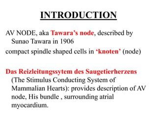
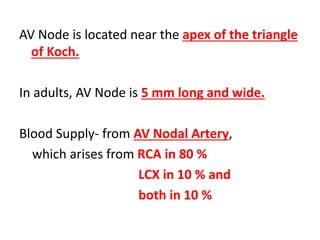
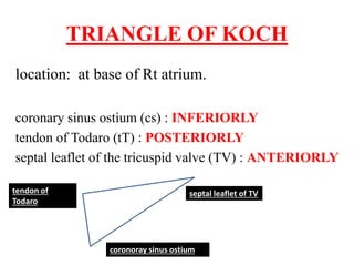
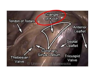
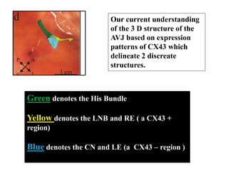
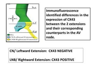
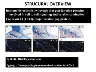
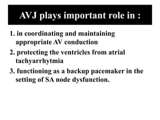
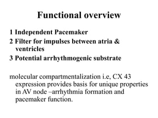
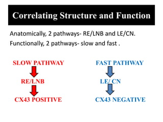
ANATOMYAND
ELECTROPHYSIOLOGY OF
THE HUMAN AV NODE
GUIDE- Dr A UDYAVAR
BY- Dr SUMEDH
INTRODUCTION
AV NODE, aka Tawara’s node, described by
Sunao Tawara in 1906
compact spindle shaped cells in ‘knoten’ (node)
Das Reizleitungssytem des Saugetierherzens
(The Stimulus Conducting System of
Mammalian Hearts): provides description of AV
node, His bundle , surrounding atrial
myocardium.
AV Node is located near the apex of the triangle
of Koch.
In adults, AV Node is 5 mm long and wide.
Blood Supply- from AV Nodal Artery,
which arises from RCA in 80 %
LCX in 10 % and
both in 10 %
TRIANGLE OF KOCH
location: at base of Rt atrium.
coronary sinus ostium (cs) : INFERIORLY
tendon of Todaro (tT) : POSTERIORLY
septal leaflet of the tricuspid valve (TV) : ANTERIORLY
septal leaflet of TVtendon of
Todaro
coronoray sinus ostium
using central fibrous body (CFB) as demarcation
between His bundle and AV node, as Tawara
suggested,
AV node divided into----------
Lower Nodal Bundle (LNB)
and Compact Node (CN)
LNB:
cells are longer and arranged more parallel to
one another
CN:
cells are small and spindle shaped with no clear
orientation.
extending proximally from LNB towards CS is
“inferior nodal extension/ rightward nodal
extension”
“second nodal extension/ leftward nodal
extension” , only in humans, extends from CN
towards the CS, is usually shorter than right
one.
Our current understanding
of the 3 D structure of the
AVJ based on expression
patterns of CX43 which
delineate 2 discreate
structures.
Green denotes the His Bundle
Yellow denotes the LNB and RE ( a CX43 +
region)
Blue denotes the CN and LE (a CX43 – region )
Immunofluoroscence
identified differences in the
expression of CX43
between the 2 extensions
and their corresponding
counterparts in the AV
node.
CN/ Leftward Extension: CX43 NEGATIVE
LNB/ Rightward Extension: CX43 POSITIVE
STRUCURAL OVERVIEW
Immunohistochemistry reveals that gap junction proteins
involved in cell to cell signaling and cardiac conduction.
Connexin 43 (Cx43), major cardiac gap protein.
fig (b-d) : Histological sections
fig (e-g) : Corresponding immunostained sections for CX43.
AVJ plays important role in :
1. in coordinating and maintaining
appropriate AV conduction
2. protecting the ventricles from atrial
tachyarrhytmia
3. functioning as a backup pacemaker in the
setting of SA node dysfunction.
Functional overview
1 Independent Pacemaker
2 Filter for impulses between atria &
ventricles
3 Potential arrhythmogenic substrate
molecular compartmentalization i.e, CX 43
expression provides basis for unique properties
in AV node –arrhythmia formation and
pacemaker function.
INE inferior nodal extension/Rightward
extension – considered as leading pacemaker
location in the absence of SA nodal impulses
in rabbit hearts.
This same location in another study was found to
have high levels of HCN4- which is the major
cardiac pacemaker current responsible for If.
In other study in human hearts, leading
pacemaker was found to be located in CN area
Functional longitudinal dissociation
between fast and slow pathway
A spontaneous reentrant beat originates in CN
area, similar to where the intrinsic junctional
rhythm originates, progresses in retrograde
fashion through the fast compartment, returns
anterogradely through the slow pathway.
This displays functional compartmentalization
within AVJ between Cx43(+) & Cx43(-)
regions.
AV nodal dual pathway
A standard S1-S2 protocol was applied to
unmask dual pathway electrophysiology.
During a routine S1 train @ 1000ms, conduction
consistently occurred through fast pathway
Then, decreased to S2 of 600 ms, conduction
block occurred in fast pathway d/t long
refractory period
Correlating Structure and Function
Anatomically, 2 pathways- RE/LNB and LE/CN.
Functionally, 2 pathways- slow and fast .
SLOW PATHWAY FAST PATHWAY
RE/LNB LE/ CN
CX43 POSITIVE CX43 NEGATIVE
Substrate for the slow pathway involves
structures in isthmus of myocardium located
along tricuspid annulus below coronary sinus.
Evidence exists- RE is the anatomical substrate
of slow pathway.
Fast pathway is less well defined.
The probable anatomical substrate of this is
transitional cell layers located around the CN
at the interface between CN and transiional
cells, which expresses Cx43 at levels similar to
the interatrial septum.
Cx43(-)- >as the fast pathway compartment
Cx43(+)->as the slow pathway compartment
We dont believe that Cx43 expression is directly
responsible for slow pathway conduction, nor
the absence of it responsible for fast pathway
conduction.
However, Cx43 expression provides a starting
point for characterizing the region from a
molecular standpoint.
Clinical Implications
AVJ pacemaker located within region of slow
pathway opens the possibility of genetic &
cellular manipulation , results in modification
of existing native AVJ pacemaker function.
Proteins/ encoding genes involved in modulating
the pacemaker (HCN4, calcium handling etc)
and conduction could be delivered.
Role of ANS in regulating the AVJ
Bianchi, et al implanted the atrial lead in the
postero septal right atrium (slow pathway) in
pt with AF receiving defibrillators, with help
of high frequency stimulation at this particular
pacing, they were able to induce conduction
block at level of AVJ.
This may be used in rate control of rapid
ventricular response in atrial fibrillation.
THANK
YOU

Av node

  • 2.
    ANATOMYAND ELECTROPHYSIOLOGY OF THE HUMANAV NODE GUIDE- Dr A UDYAVAR BY- Dr SUMEDH
  • 3.
    INTRODUCTION AV NODE, akaTawara’s node, described by Sunao Tawara in 1906 compact spindle shaped cells in ‘knoten’ (node) Das Reizleitungssytem des Saugetierherzens (The Stimulus Conducting System of Mammalian Hearts): provides description of AV node, His bundle , surrounding atrial myocardium.
  • 4.
    AV Node islocated near the apex of the triangle of Koch. In adults, AV Node is 5 mm long and wide. Blood Supply- from AV Nodal Artery, which arises from RCA in 80 % LCX in 10 % and both in 10 %
  • 5.
    TRIANGLE OF KOCH location:at base of Rt atrium. coronary sinus ostium (cs) : INFERIORLY tendon of Todaro (tT) : POSTERIORLY septal leaflet of the tricuspid valve (TV) : ANTERIORLY septal leaflet of TVtendon of Todaro coronoray sinus ostium
  • 7.
    using central fibrousbody (CFB) as demarcation between His bundle and AV node, as Tawara suggested, AV node divided into---------- Lower Nodal Bundle (LNB) and Compact Node (CN)
  • 8.
    LNB: cells are longerand arranged more parallel to one another CN: cells are small and spindle shaped with no clear orientation.
  • 9.
    extending proximally fromLNB towards CS is “inferior nodal extension/ rightward nodal extension” “second nodal extension/ leftward nodal extension” , only in humans, extends from CN towards the CS, is usually shorter than right one.
  • 10.
    Our current understanding ofthe 3 D structure of the AVJ based on expression patterns of CX43 which delineate 2 discreate structures. Green denotes the His Bundle Yellow denotes the LNB and RE ( a CX43 + region) Blue denotes the CN and LE (a CX43 – region )
  • 11.
    Immunofluoroscence identified differences inthe expression of CX43 between the 2 extensions and their corresponding counterparts in the AV node. CN/ Leftward Extension: CX43 NEGATIVE LNB/ Rightward Extension: CX43 POSITIVE
  • 12.
    STRUCURAL OVERVIEW Immunohistochemistry revealsthat gap junction proteins involved in cell to cell signaling and cardiac conduction. Connexin 43 (Cx43), major cardiac gap protein. fig (b-d) : Histological sections fig (e-g) : Corresponding immunostained sections for CX43.
  • 13.
    AVJ plays importantrole in : 1. in coordinating and maintaining appropriate AV conduction 2. protecting the ventricles from atrial tachyarrhytmia 3. functioning as a backup pacemaker in the setting of SA node dysfunction.
  • 14.
    Functional overview 1 IndependentPacemaker 2 Filter for impulses between atria & ventricles 3 Potential arrhythmogenic substrate molecular compartmentalization i.e, CX 43 expression provides basis for unique properties in AV node –arrhythmia formation and pacemaker function.
  • 15.
    INE inferior nodalextension/Rightward extension – considered as leading pacemaker location in the absence of SA nodal impulses in rabbit hearts. This same location in another study was found to have high levels of HCN4- which is the major cardiac pacemaker current responsible for If. In other study in human hearts, leading pacemaker was found to be located in CN area
  • 16.
    Functional longitudinal dissociation betweenfast and slow pathway A spontaneous reentrant beat originates in CN area, similar to where the intrinsic junctional rhythm originates, progresses in retrograde fashion through the fast compartment, returns anterogradely through the slow pathway. This displays functional compartmentalization within AVJ between Cx43(+) & Cx43(-) regions.
  • 17.
    AV nodal dualpathway A standard S1-S2 protocol was applied to unmask dual pathway electrophysiology. During a routine S1 train @ 1000ms, conduction consistently occurred through fast pathway Then, decreased to S2 of 600 ms, conduction block occurred in fast pathway d/t long refractory period
  • 18.
    Correlating Structure andFunction Anatomically, 2 pathways- RE/LNB and LE/CN. Functionally, 2 pathways- slow and fast . SLOW PATHWAY FAST PATHWAY RE/LNB LE/ CN CX43 POSITIVE CX43 NEGATIVE
  • 19.
    Substrate for theslow pathway involves structures in isthmus of myocardium located along tricuspid annulus below coronary sinus. Evidence exists- RE is the anatomical substrate of slow pathway.
  • 20.
    Fast pathway isless well defined. The probable anatomical substrate of this is transitional cell layers located around the CN at the interface between CN and transiional cells, which expresses Cx43 at levels similar to the interatrial septum.
  • 21.
    Cx43(-)- >as thefast pathway compartment Cx43(+)->as the slow pathway compartment We dont believe that Cx43 expression is directly responsible for slow pathway conduction, nor the absence of it responsible for fast pathway conduction. However, Cx43 expression provides a starting point for characterizing the region from a molecular standpoint.
  • 22.
    Clinical Implications AVJ pacemakerlocated within region of slow pathway opens the possibility of genetic & cellular manipulation , results in modification of existing native AVJ pacemaker function. Proteins/ encoding genes involved in modulating the pacemaker (HCN4, calcium handling etc) and conduction could be delivered.
  • 23.
    Role of ANSin regulating the AVJ Bianchi, et al implanted the atrial lead in the postero septal right atrium (slow pathway) in pt with AF receiving defibrillators, with help of high frequency stimulation at this particular pacing, they were able to induce conduction block at level of AVJ. This may be used in rate control of rapid ventricular response in atrial fibrillation.
  • 24.